Table of contents
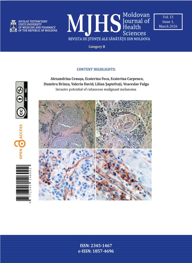
24 papers · pp. 3–159Invasive potential of cutaneous malignant melanoma
Introduction
Cutaneous malignant melanoma is the most aggressive skin cancer, with a high mortality rate despite advances in therapy. This study aimed to evaluate the relationship between lymphovascular and perineural invasion and key clinicopathological parameters in superficial spreading melanoma, in order to assess their potential prognostic significance.
Materials and methods
A retrospective analysis was conducted on 47 cases of superficial spreading melanoma obtained from the Oncology Institute in Chisinau. All cases were histologically confirmed and reviewed for tumor thickness, Clark level, ulceration, mitotic activity, microsatellitosis, pigmentation, and lymph node involvement. Lymphovascular and perineural invasion were assessed using hematoxylin–eosin staining and, where available, immunohistochemistry. Correlations between invasion patterns and clinicopathologic features were analyzed using Pearson correlation coefficients, with statistical significance set at p < 0.05.
Results
Lymphovascular invasion was positive and significantly correlated with tumor thickness (r = 0.54, p< 0.001), Clark level (r = 0.46, p < 0.001), microsatellitosis (r = 0.50, p < 0.001), tumor stage (r = 0.33, p = 0.01), and lymph node involvement (r = 0.29, p = 0.02). A negative correlation was observed with pigmentation (r = –0.26, p = 0.04). Perineural invasion was less frequent, but correlated positively with lymphovascular invasion (r = 0.28, p = 0.03) and showed a trend toward association with amelanotic tumors (r = –0.24, p = 0.05). No significant relationships were found with ulceration or mitotic activity.
Conclusions
Lymphovascular invasion represents a significant indicator of aggressive biological behavior in superficial spreading melanoma, closely associated with established prognostic factors. Perineural invasion occurs less frequently, but may further reflect invasive potential, particularly in amelanotic variants. Routine histopathologic assessment of both invasion patterns is recommended to improve prognostic evaluation.
Contemporary insights into diagnosis and treatment of gastrointestinal non-Hodgkin lymphomas
Introduction
The gastrointestinal tract (GIT) is the most common site of extranodal primary non-Hodgkin lymphoma (NHL), accounting for 20% to 40% of all extranodal lymphomas. The advanced stages at diagnosis and complications remain significant issues in NHL management, imposing a substantial disease burden on patients and healthcare systems.
Material and methods
We performed a descriptive cross-sectional and cohort study of patients with gastrointestinal NHL and a narrative review of the literature in the Discussion section. This study included 50 prospective and retrospective patients with NHL treated between 2015-2024 in the Institute of Oncology in Moldova. A bibliographic search was conducted using databases such as PubMed, Hinari, SpringerLink, the National Center for Biotechnology Information, and Medline. The final bibliography included 18 relevant sources deemed to be representative of the literature published on the topic of this article.
Results
According to the International Clinical Classification, most patients (22, 44.0%) were diagnosed with clinical stage IV. B symptoms occurred in 38 (76.0%) patients. The overwhelming predominance of diffuse large B-cell lymphomas (46 cases - 90.2%) was observed. The complete blood count, bone marrow aspiration and biopsy of the iliac crest did not detect any specific changes in cases without bone marrow involvement, with the exception of a decrease in hemoglobin and erythrocyte counts observed in cases of posthemorrhagic anemia. The overall survival (OS) of all patients with gastrointestinal NHL was 78.1% at 1 year, 59.4% at 3 years, and 35.9% at 5 years. In patients with stage IE NHL, the 1-, 3-, and ≥5-year OS was 93.4%, 76.5%, and 69.9%, respectively. In patients with stage IIE, the OS was 91.2% at 1 year, 71.4% at 3 years, and 63.8% at ≥5 years. In patients with stage IIIE-IV, the OS was 75.1% at 1 year, 54.8% at 3 years, and 28.5% at ≥5 years with combined chemotherapy.
Conclusions
Our study demonstrated that non-Hodgkin lymphomas with primary involvement of the gastrointestinal tract exhibited distinct histopathological, clinical-evolutionary and hematological features, which influenced treatment outcomes. The aggressive histological types and the advanced stages IIIE and IV prevailed within the structure of non-Hodgkin lymphomas with primary gastrointestinal involvement, and, thus, negatively impacted the survival and prognosis.
From prolonged premature rupture of membranes to bronchopulmonary dysplasia: the role of chorioamnionitis in the respiratory outcomes of preterm infants
Introduction
Prolonged premature rupture of membranes predisposes to intrauterine infection and chorioamnionitis, both of which have significant implications for neonatal outcomes. While chorioamnionitis has been linked to accelerated surfactant production and reduced respiratory distress syndrome, it is also associated with long-term pulmonary injury, including bronchopulmonary dysplasia and pulmonary hypertension. The objective of the study is to investigate the association between prolonged premature rupture of membranes, chorioamnionitis, and respiratory outcomes among preterm infants ≤34 weeks of gestation.
Material and methods
A prospective cohort of 108 preterm infants admitted to the Neonatal Intensive Care Unit of the Mother and Child Institute, Chișinău, between October 2023 and July 2024, was divided into two groups: infants born to mothers with clinical/histological chorioamnionitis (n = 54) and controls (n = 54). Maternal risk factors, incidence of prolonged premature rupture of membranes incidence, Apgar scores, type and duration of respiratory support, and pulmonary complications were analyzed. Statistical significance was tested using chi-square and logistic regression.
Results
Prolonged premature rupture of membranes was significantly more frequent in chorioamnionitis group (67% vs. 22%, p<0.001). Infants exposed to chorioamnionitis had lower 1-minute Apgar scores, greater need for invasive ventilation (5.9 ± 10.6 vs. 2.2 ± 4.8 days, p<0.05), and prolonged hospitalization. BPD incidence was higher in the chorioamnionitis group (25.9% vs. 3.7%, p<0.05). Mortality did not differ significantly between groups (27.8% vs. 22.2%).
Conclusions
Prolonged premature rupture of membranes is strongly associated with chorioamnionitis, which in turn significantly increases the risk of long-term pulmonary complications in preterm infants. Early recognition of prolonged premature rupture of membranes, antibiotic prophylaxis, antenatal corticosteroids, and interdisciplinary obstetric–neonatal management are essential to reduce the burden of bronchopulmonary dysplasia.
Exploring the clinical spectrum of DiGeorge syndrome
Introduction
DiGeorge syndrome, known also as 22q11.2 deletion syndrome, is a rare multisystemic disorder characterized by a wide range of clinical features and may include thymic aplasia and subsequent immunodeficiency, conotruncal cardiac anomalies, typical facial features, palatal abnormalities, and hypocalcemia due to hypoparathyroidism.
Material and methods
Data were collected for 10 patients genetically confirmed with DiGeorge syndrome at the Institute of Mother and Child. This included general information, laboratory results, and clinical features.
Results
The mean age at diagnosis was 74.6 months (3 months – 28 years). Most cases were sporadic, with only 2 patients having a history of DGS (n=1), or close relatives with cardiac malformations (n=1). The most common symptoms that led to diagnosis were congenital heart defects (90%), and facial dysmorphism (90%). Common clinical features included recurrent infections (40%) and ENT disorders (20%). Weight was within normal percentiles for the entire group, but a delay in height growth was noted. Regarding the immunological characteristics: lymphopenia was recorded in 20% of patients, and thrombocytopenia in 2 patients.
Conclusions
Given the diverse array of symptoms associated with DiGeorge syndrome, physicians should be knowledgeable about both typical and less common characteristics of the syndrome to facilitate optimal treatment and potentially enable early diagnosis.
Development and testing of a questionnaire: assessment of occupational risk factors in surgeons
Introduction
Surgery exposes professionals to significant physical and psychological risks, including intense exertion, prolonged static postures, and repetitive gestures, often leading to musculoskeletal pain. International studies report prevalence rates above 90%, linked to long procedures and poor ergonomics, alongside exposure to biological and chemical hazards, radiation, toxic smoke, and chronic stress. In the Republic of Moldova, occupational health in the medical sector is underexplored, with no tools tailored to surgeons. This study aims to develop and validate the first nationally standardized questionnaire to assess these risks and support public health policies.
Material and methods
The study used a sequential mixed-methods design: a qualitative phase to identify domains and indicators, followed by a quantitative phase for psychometric testing. Four domains were defined: working conditions, occupational factors, general health, and psycho-emotional state. The questionnaire was refined from 70 to 47 items after expert review and validation, with internal consistency (Cronbach’s α) and content validity (I-CVI, modified Kappa) assessed. It was pre-tested on 52 surgeons, and data were analyzed using SPSS 27 (p<0.05).
Results
Content validation revealed S-CVI/Ave values ranging from 0.934 to 1.00 and S-CVI/UA values from 0.738 to 1.00, with all domains exceeding the accepted threshold for relevance and clarity, except for one domain, which fell slightly below the recommended level for unanimous agreement. I-CVI values ranged from 0.857 to 1.00 (relevance) and 0.847 to 1.00 (clarity), while κ* indices were all rated as "excellent". Based on expert feedback, 24 questions were revised and 15 were removed. Overall internal consistency was very good (α = 0.808), with section values ranging from 0.769 to 0.864, the highest being for "Psycho-emotional state" (α = 0.864). The pre-test sample comprised 52 surgeons, mostly male (57.7%), with a mean age of 44.92 years, predominantly from urban areas (92.3%). Respondents generally found the questions clear but noted some lengthy formulations, repetitiveness, and sensitive items. These observations contributed to optimizing the final version of the instrument.
Conclusions
The final questionnaire meets the initial theoretical dimensions and shows strong psychometric properties, with high validity and internal consistency. The tool is comparable to established instruments and suitable for assessing surgeons’ health and occupational risks, with potential for wider use.
Evolution of maxillary expansion in patients with cleft lip and palate
Introduction
The craniofacial orthodontist who is part of the multidisciplinary team evaluating children with cleft lip and palate plays an important role in examining the development of dental occlusion. Early surgical interventions in children with cleft lip and palate frequently cause occlusion disorders with a prevalence of anterior crossbite in 62%. The expansion of the maxilla is important for normalizing the morphology and correct symmetrical tooth eruption. The aim is to evaluate the evolution of maxillary expansion in patients with cleft lip and palate.
Material and methods
This study included 20 patients with cleft lip and palate, including 8 girls and 12 boys aged 6-12 years, with a mean age of 9.35 years. Unilateral cleft lip and palate was present in 16 patients, bilateral cleft lip and palate – 2 patients, and clefts of the hard and soft palate – 2 patients. The study models were scanned, and the maxillary dimensions were examined using a 3D Dolphin Imaging program before and after maxillary slow expansion over an average of 12.2 months.
Results
Clinical evaluation of patients with cleft lip and palate showed crossbite occlusion. Of these patients, anterior and posterior crossbite occlusion was present in 11 (55%) patients, anterior crossbite occlusion in 3 (15%) patients, unilateral posterior crossbite occlusion in 3 (15%) patients and bilateral in 3 (15%) patients. In the transverse plane, the size of the upper jaw increased statistically significantly (p = 0.002), but in the sagittal plane we found a statistically significant elongation of the upper jaw (p < 0.05) following slow expansion treatment with a removable orthodontic appliance over an average of 12.2 months in patients with cleft lip and palate.
Conclusions
Slow maxillary expansion treatment during the mixed dentition period in patients with cleft lip and palate was found to be more effective in the sagittal plane than in the transverse plane.
The role of odontogenic infection in the onset and evolution of focal disease
Introduction
The focal disease is a pathological condition characterized by a wide variety of functional disorders and organic tissue alterations, due to chronic foci of infection, from which various microbes, microbial toxins, and toxic products of septic tissue disintegration originate. According to the percentage distribution, 90% of the foci of the body are located in the cephalic region, and 10% in the rest of the body [17]. Research has shown that on the list of foci of infection, those in the oral cavity are in first place, with 93% of active foci being caused by teeth and their pathologies. Important clinical criteria that mark this fundamental difference of the odonto-periodontal focal infection include the profile of local inflammation, the level of the tissue hypersensitivity process, the level of the microbial load in the focus and, no less importantly, the reactivity of the body.
Material and methods
The present study is a retrospective clinical observational study and included 87 patients with foci of odontogenic infection, classified according to the frequency of pathologies encountered and the virulence of microbial pathogens as follows: with periodontal disease - 35 patients (gingivitis - 11 patients and 24 patients with different stages of periodontitis); with endodontic pathologies - 27 patients (pulpitis - 5 patients and periapical lesions - periodontitis (Pt) - 22 patients); with dental caries of varying severity - 19 patients, and 6 patients with oral mucosa pathologies.
Results
Through clinical observations, it was found that with the removal of the foci of infection, the “vegetative alarm symptoms” begin to disappear, some of them even very quickly, such as causeless fatigue. At the same time, symptoms that have been present for a longer period, such as long-term depressive states and memory disorders in patients with periodontal disease–particularly severe periodontitis with a major microbial load–and lesions of the oral mucosa, decreased more slowly, over a period of 1-3 months. The 100% disappearance of symptoms in the case of carious lesions and oral mucosa lesions demonstrates the direct relationship between the foci of infection and the patient's general health. In relation to periodontal disease and periapical lesions (over 90% of symptoms have subsided), the remaining clinical signs are related to the increased bacterial load and the virulence of the pathogens.
Conclusions
The identification, evaluation, and elimination of foci of odontogenic infection play an important role in aggravating already existing systemic conditions, thus triggering focal disease. The role of the dentist in the prophylaxis of focal disease is primary in the detection and elimination of foci of odontogenic infection. In the prophylaxis of focal disease, doctor-patient cooperation is very important, and no less important is collaboration with general medicine specialists.
Developing and validating a questionnaire on knowledge and attitudes in health research ethics
Introduction
Research ethics and integrity are fundamental for safeguarding human participants and ensuring trustworthy scientific practices. Understanding researchers’ knowledge, attitudes, and perceptions regarding ethical standards is important for all health researchers, but particularly relevant for early-career researchers. While several international instruments exist to evaluate specific aspects of research ethics, such as plagiarism, organizational climate, or responsible conduct of research, a multidimensional and contextually relevant tool is required.
Materials and methods
A comprehensive questionnaire was developed to assess ethical knowledge, attitudes toward research resources and institutional integrity measures, and self-perceived ethical competencies among doctoral students in the health sciences. Item formulation was guided by international standards, including the Declaration of Helsinki, the European Code of Conduct for Research Integrity, and Good Clinical Practice guidelines. The development process included a content validity assessment by 10 experts and a psychometric evaluation of the collected data from 274 doctoral students. Exploratory factor analysis (EFA) was applied to determine the latent structure of the questionnaire, and internal consistency was assessed using Cronbach’s alpha.
Results
EFA revealed a six-factor structure explaining 64.5% of the total variance. The factors measured: (I) perceived importance of research resources, (II) self-perceived ethical competencies, (III) implemented institutional measures for research integrity, (IV) ethical principles and moral responsibilities, (V) perceived accessibility of research resources, and (VI) importance of institutional integrity measures. Factor loadings were generally high, and internal consistency was good to excellent, with Cronbach’s alpha values ranging from 0.738 to 0.989. These findings indicate that the questionnaire captures multidimensional aspects of research ethics and integrity.
Conclusions
The developed questionnaire represents a robust, valid, and reliable instrument for assessing ethical knowledge and attitudes among researchers in health sciences. It can serve as an internal audit tool to evaluate research integrity climate, researcher satisfaction with available resources, and implementation of institutional policies. Moreover, it provides a foundation for designing targeted training programs and professional development initiatives aimed at improving ethical competencies.
The iCREATE registry: a model for strengthening injury surveillance in the Republic of Moldova
Introduction
Injuries represent a major public health issue, causing approximately 16,000 deaths globally each day (10% of all deaths), which is 32% more than the combined total caused by malaria, tuberculosis, and HIV/AIDS. Over the past 15 years, the WHO and regional initiatives have supported the piloting of trauma registries in low- and middle-income countries as essential tools for monitoring, planning, and prevention.
Objective
This article aims to assess the feasibility and utility of implementing a national trauma registry in the Republic of Moldova, in order to improve injury surveillance and emergency service planning.
Materials and methods
In 2018, the pilot iCREATE trauma registry was tested for the first time in three countries: Moldova, Armenia, and Georgia. The data collection instrument was developed based on WHO recommendations, ICD-10, and IDB-JAMIE standards under the guidance of partners from the University of Iowa and Babeș-Bolyai University, Cluj-Napoca. All trauma cases from the Institute of Emergency Medicine and the Valentin Ignatenco Municipal Clinical Children’s Hospital in Chișinău were included in the registry.
Results
The analyzed sample consists of 7,942 individuals, predominantly male (57.3%). The most represented age groups were 19-29 years (17.8%) and 30-39 years (17.6%), while individuals aged 70 and above accounted for 11% of the total. Most incidents occurred in urban areas (76.9%). Of the total patients, 52.4% were treated and discharged, while 37.5% required hospitalization. Injuries occurred primarily at home (55.4%) and on public roads (24.7%). The leading mechanism of injury was falls (68.2%), followed by other causes (12.2%) and cut/pierce injuries (10.0%). The most frequently affected body regions were the head/skull (12.5%) and knee (11.8%), followed by the hip (6.7%) and wrist (6.4%). Fractures were the most common injury type (34.5%), followed by contusions (23.0%) and open wounds (14.7%). Several gaps in data collection and reporting were identified and should be considered in future efforts to enhance trauma surveillance.
Conclusions
The data highlight the need to develop a national trauma registry as an essential tool for monitoring, prevention, and effective intervention, alongside health promotion campaigns targeting vulnerable groups and the involvement of relevant stakeholders.
Emergency care for trauma patients in the red zone: clinical experience from 2024
Introduction
The traumatized patient with signs of shock remains a major cause of morbidity and mortality, requiring rapid diagnosis and multidisciplinary management. This study evaluates clinical, paraclinical, and therapeutic aspects of trauma patients admitted with shock signs in the red zone of the Emergency Department of the Institute of Emergency Medicine during 2024.
Material and methods
A prospective analysis of 60 polytrauma patients in shock was performed. Demographic data, trauma mechanisms, Glasgow Coma Score, vital signs, imaging, emergency interventions, and outcomes were evaluated. The patients were managed according to an experimental protocol developed at the institutional level, which provides for a multidisciplinary approach to the traumatized patient. This protocol was developed based on the international protocol for Advanced Trauma Life Support. It will later be implemented at the institutional level.
Results
The cohort included 79% males, with a mean age of 49.7 ± 15.2 years; 75% urban residents. Grade III and IV shock were diagnosed in 40% and 28.3%, respectively. Causes of trauma included physical aggression (29.5%), road traffic accidents (28.5%), and accidental trauma (21.0%), falls from height (21.0%). Radiological lesions were detected in 63.3%, positive findings on Focused Assessment with Sonography for Trauma in 40%, and cranial lesions identified by computed tomography in 60%. Intraglotic intubation was required in 80% of patients, vasopressor support was administered in 50%, antibiotic therapy in 95%, and emergency surgery was performed in 30%. Mortality was 1.7%.
Conclusions
Protocol-driven multidisciplinary care facilitated effective stabilization of traumatized patients presenting with shock. Early diagnosis, the use of advanced imaging techniques, and timely therapeutic interventions significantly contributed to improved patient outcomes. These findings underscore the critical role of structured trauma life support protocols in emergency medical settings.
Micronutrient needs and supplementation strategies during pregnancy
Introduction.
icronutrient deficiencies during pregnancy remain a major global public health concern, with implications for maternal health, fetal development, and long-term child outcomes. International organizations such as the WHO and FIGO have issued evidence-based guidelines on micronutrient supplementation, which are increasingly being adapted into national protocols.
Material and methods
This narrative review was based on literature searches in PubMed, Scopus, Web of Science, and Google Scholar, covering January 2000 to May 2025. Peer-reviewed studies, systematic reviews, and clinical guidelines from WHO, FIGO, and the Moldovan Ministry of Health were included. The review focused on iron, folic acid, iodine, calcium, vitamin D, and selected trace elements.
Results
Iron and folic acid emerged as the most consistently recommended supplements across guidelines, with proven efficacy in reducing maternal anemia and neural tube defects. Iodine and calcium are also emphasized, particularly in regions with documented dietary insufficiency. Moldova’s antenatal care protocol largely aligns with WHO and FIGO recommendations, prioritizing targeted over universal supplementation for nutrients beyond iron and folate. Evidence on routine supplementation with multivitamin complexes remains inconclusive.
Conclusions
Evidence-based micronutrient supplementation is essential to optimizing pregnancy outcomes. Universal iron and folic acid supplementation remain the cornerstone of antenatal nutrition strategies. Context-specific approaches, as exemplified by the Moldovan model, can enhance implementation in resource-limited settings.
Thromboprophylaxis in pregnancy, delivery and puerperium: a review of literature and current guidelines
Introduction
Venous thromboembolism is one of the leading causes of maternal morbidity and mortality. Pregnancy induces a hypercoagulable state as an adaptive mechanism to prevent hemorrhage during childbirth. These physiological changes significantly increase the risk of venous thromboembolism – by up to six-fold during pregnancy and up to 10-fold during the postpartum period compared to the non-pregnant population. Given these risks, proper identification of patients who may benefit from thromboprophylaxis is essential to improve maternal outcomes.
Material and methods
This manuscript reviews medical articles and current clinical guidelines on thromboprophylaxis in pregnancy and the puerperium, highlighting similarities, differences, and practical considerations in the management of at-risk patients. International guidelines developed to aid clinicians in venous thromboembolism risk stratification and prevention, including the Royal College of Obstetricians and Gynecologists, the American College of Obstetricians and Gynecologists, the American Society of Hematology, and the National Institute for Health and Care Excellence were analyzed.
Results
The incidence of Venous thromboembolism ranges from 1 to 2 per 1,000, with up to 80% attributed to deep vein thrombosis cases occurring during the antepartum period, and 20%-25% being pulmonary embolism cases. In contrast, the incidence of pulmonary embolism is significantly higher after childbirth, with 40% to 60% of all pulmonary embolism cases occurring during the postpartum period. The impact of venous thromboembolism is not limited to mortality. Acute venous thromboembolism and the need for long-term anticoagulant therapy are associated with a significant clinical and psychological burden, while potential long-term sequelae, such as pulmonary hypertension and post-thrombotic syndrome, can have lifelong consequences. Ensuring thromboprophylaxis is essential, and the primary responsibility lies with the obstetrician. Ultimately, effective thromboprophylaxis is about balancing efficacy and safety between the need to prevent a potentially life-threatening event and the cost of an increased risk of bleeding.
Conclusions
Effective thromboprophylaxis during pregnancy and the puerperium remains a critical component of maternal care. The national protocol aims to assist healthcare professionals in identifying women at increased risk of venous thromboembolism during pregnancy, childbirth, and the postpartum period, and in making evidence-based decisions regarding the use of thromboprophylaxis and anticoagulant agents.
Pregnancy-related inflammatory markers and their association with perinatal mental illness: a systematic review
Introduction
This study investigates the relationship between immune dysregulation and perinatal mental disorders by analyzing clinical data and biomarker profiles in pregnant individuals with varying severity of psychiatric symptoms. Understanding these associations may support the development of early screening tools and targeted interventions to improve maternal and infant mental health outcomes.
Material and methods
A comprehensive literature review was conducted using PubMed, MEDLINE, and Scopus, covering studies published through 2025. Key proinflammatory and anti-inflammatory cytokines, including IL-6, TNF-α, IL-1β, CRP, IL-8, and IL-10, were extracted from peer-reviewed articles. When numerical values were unavailable, data were estimated from published figures using digitization tools. Extracted data were standardized and analyzed using Python (Pandas, Matplotlib). Statistical procedures included correlation analysis, ROC curve modelling, and ANOVA testing to assess group differences and diagnostic performance of biomarkers.
Results
Analysis revealed strong associations between cytokine levels and perinatal depressive symptoms. In one dataset, nine cytokines were inversely correlated with postpartum depression severity (Pearson r = –0.79, p = 0.004; Spearman rₛ = –0.87, p = 0.00085; Kendall τ = –0.72, p = 0.0031), and ANOVA confirmed significant group differences (F = 5.8, p = 0.022). Other studies reported elevated IL-6 and TNF-α levels in postpartum depression (p < 0.05). Co-expression of IL-2, IL-6, IL-8, and TNF-α was very high (r = 0.9991, p = 0.00006), likely reflecting cytokine collinearity and limited sample size, with ANOVA indicating significant elevation in affected individuals (F = 45.42, p = 0.0151). ROC analyses identified IL-8, IL-6, CRP, and TNF-α as reliable markers of perinatal depression and psychosis. Tryptophan metabolites and MCP-1 were more specific for psychosis, while IFN-γ showed a regulatory rather than a diagnostic function.
Conclusions
Perinatal mental disorders are associated with significant immune alterations. IL-6, IL-8, IL-2, and TNF-α appear to play a central role in the pathophysiology of postpartum depression. The findings support the utility of cytokine profiling for early detection and differential diagnosis of perinatal psychiatric conditions.
Advances in disease-modifying therapies for multiple sclerosis: global updates and a regional comparison between the Republic of Moldova and Romania
Introduction
The therapeutic landscape of multiple sclerosis has undergone a remarkable transformation over the past two decades. The paradigm has shifted from reliance on moderate-efficacy, first-generation injectable therapies toward earlier adoption of high-efficacy disease-modifying treatments, particularly in relapsing forms of MS. This evolution reflects an increasing focus on early intensive treatment strategies aimed at preserving long-term neurological function and brain health.
Materials and methods
This narrative review synthesizes recent global evidence on progress in disease-modifying treatments across all multiple sclerosis phenotypes, drawing from randomized controlled trials, real-world studies, and expert consensus guidelines. In addition, it includes a comparative health policy analysis assessing DMT availability, access, and implementation in Romania and the Republic of Moldova, based on national formularies, reimbursement frameworks, and care delivery models.
Results
Globally, the MS treatment algorithm has been reoriented toward early intensive treatment, supported by emerging evidence favoring high-efficacy therapies in the early disease course. While many countries have aligned their protocols accordingly, regional discrepancies persist. Romania, as an EU member, has expanded patient access to 16 reimbursed therapies and biomarker-driven monitoring, and has developed a network of specialized Multiple Sclerosis centers. Conversely, the Republic of Moldova faces structural and economic barriers that restrict access to high-efficacy treatments, advanced diagnostics, and multidisciplinary care—factors contributing to delayed treatment and suboptimal outcomes.
Conclusions
Understanding both global innovations and regional realities is necessary to place current Multiple Sclerosis care in context. Further advancements in science, health policy, and infrastructure will ultimately determine how effectively different nations can convert therapeutic progress into actual improvements in patient outcomes.
Correlation between pathogenetic factors and vascular endothelial damage in patients with rheumatoid arthritis
Introduction
Systemic rheumatoid vasculitis accounts for 1 to 5% of complications seen in rheumatoid arthritis, while autopsy studies report an average of 23% incidence. This enormous difference in numbers emphasizes the rate of misdiagnosis or underdiagnosis of systemic rheumatoid vasculitis. It mainly affects people with a median age of 65 years. It is particularly noteworthy, as systemic rheumatoid vasculitis has a high mortality and relapse rate. Also, the multifactorial aetiology: cytokines/immune cells and other particles determines clinical complexity of this type of angiitis.
Materials and methods
A comprehensive literature search of articles published since 1996 was conducted using MEDLINE via PubMed and HINARI. The search included terms such as "rheumatoid vasculitis", "rheumatoid arthritis", and "endothelial dysfunction", focusing on mechanisms driving vascular damage. A total of 217 relevant sources were identified, including original studies, reviews, and book chapters. The study evaluated pathogenic factors like cytokines, immune complexes, and systemic inflammation, highlighting their roles in endothelial dysfunction and hypercoagulability.
Results
The main pathogenetic factors in systemic rheumatoid vasculitis were immune complexes, cytokines (IL-6/TNF-α and IL-17), immune and blood cells (CD20/TH17/Platelets) and others (microparticles, blood rheology modifications). Even though, taken separately, those factors appear to have little to no impact on vascular endothelium, their synergistic effect lead to significant endothelial damage.
Conclusions
To conclude, each disease has its own pathogenetic factors which determine the natural course of this pathology. Understanding these mechanisms plays an important role for clinicians helping them to diagnose and effective treatment. Taking into consideration the relationships between specific factors in rheumatoid angiitis, we can make more specific decisions for its diagnosis and treatment. We can also use the pathophysiology of systemic rheumatoid vasculitis as a foundation for developing prevention measures.
Rheumatoid arthritis worldwide: inequalities in epidemiology and care
Introduction
Rheumatoid arthritis (RA) is a chronic autoimmune disease affecting approximately 0.5% of the global population. It represents a major cause of disability, reduced quality of life, and healthcare burden. The prevalence of RA is rising, especially in older populations and in low-income regions.
Materials and methods
A systematic literature search was performed in PubMed, ScienceDirect and Google Scholar for articles published between 2000 and 2025. Search terms included “rheumatoid arthritis”, “epidemiology”, “risk factors”, “economic burden”, and “healthcare disparities”.
Results
RA prevalence ranges from 0.3% to 1.0%, with the highest values in Northern Europe (0.8–1.0%) and North America (0.7–0.9%), and the lowest in Africa (0.1–0.3%) and rural Asia (0.2–0.4%). Work incapacity has declined in several high-income countries, attributed to earlier diagnosis and the widespread use of disease-modifying antirheumatic drugs. Socio-economic status is a key factor for RA outcomes, with patients in the lowest income groups showing up to 50% higher disability rates. Other risk factors include female sex, HLA-DRB1 alleles, smoking, and environmental exposures. The economic burden is considerable, with direct and indirect costs disproportionately affecting low- and middle-income countries, where RA frequently results in early work disability.
Conclusions
RA causes disability and reduces quality of life. Its prevalence is rising worldwide, with higher detection and better outcomes in high-income countries, while low-income countries face underdiagnosis and limited access to modern therapies. Reducing these disparities requires stronger healthcare systems, early diagnosis, and affordable and accessible treatments.
Current concepts in the surgical management of chronic suppurative otitis media
Introduction
Chronic suppurative otitis media represents a major public health concern in both children and adults, particularly in developing countries. The condition poses not only a medical challenge requiring complex clinical management but also a public health issue with significant socioeconomic implications and costs.
Material and methods
A review of selected literature from the PubMed, Hinari, SpringerLink, National Center for Biotechnology Information, and Medline databases was conducted. Articles published between 2000 and 2025 were selected using the following keywords: “chronic otitis media” and “chronic suppurative otitis media,” combined in various ways with the terms “surgical treatment,” “mastoidectomy,” “canal wall-down,” “canal wall-up,” “canal wall-intact,” “canal wall reconstruction,” “tympanoplasty,” and “ossiculoplasty” to maximize search efficiency. Based on the search criteria, 325 full-text articles were initially identified. The final bibliography included 66 relevant sources considered representative of the published material related to the topic of this review article.
Results
There are four traditional surgical procedures for the treatment of chronic suppurative otitis media: 1) simple (cortical) mastoidectomy, 2) radical mastoidectomy, 3) modified radical mastoidectomy, and 4) mastoidectomy with tympanoplasty. These surgical interventions can be classified into two categories: open cavity (canal wall-down – CWD) and closed cavity (canal wall-up – CWU) or canal wall-intact (CWI). The CWD technique is the most effective method for cholesteatoma eradication, as it allows a wide-angle view of the mastoid and middle ear structures. Currently, radical CWD mastoidectomy is rarely performed but may be indicated when complete excision of the cholesteatoma is not possible. To prevent complications associated with the mastoid cavity following CWD, surgeons may opt for CWU mastoidectomy or CWD mastoidectomy with mastoid cavity obliteration and reconstruction of the external auditory canal wall.
Conclusions
The current concept of managing patients with chronic suppurative otitis media involves developing a personalized approach based on anatomical, biological, radiological, and social factors. The selection of the surgical technique should be tailored to each patient according to the location and extent of the cholesteatoma, defects of the posterior canal wall, associated lesions, presence of complications, degree of hearing loss, and the patient’s overall medical condition.
Surgical treatment of dysphagia lusoria caused by an aberrant right subclavian artery: a case report and literature review
Introduction
Dysphagia lusoria refers to dysphagia caused by an aberrant right subclavian artery (ARSA), a rare congenital anomaly of the aortic arch. This article presents a clinical case of symptomatic ARSA, accompanied by a comprehensive review of its anatomical, clinical, and therapeutic aspects.
Material and methods
We present the case of a 54-year-old female patient diagnosed with dysphagia lusoria due to ARSA, who underwent surgical intervention after clinical and imaging evaluations. The literature was reviewed through PubMed using the keywords: “aberrant right subclavian artery”, “dysphagia lusoria”, and “arteria lusoria”.
Results
The patient presented with progressive dysphagia, morning cough, fatigue, and weight loss. Imaging revealed ARSA compressing the esophagus, and the presence of a bicarotid trunk. Surgical correction involved right subclavian-to-carotid transposition, with complete resolution of symptoms. The literature review showed a modest grade of evidence regarding the management of patients with dysphagia lusoria, mostly including case reports and limited case series. Treatment is usually indicated in symptomatic patients, with options including open surgery or hybrid approaches.
Conclusions
ARSA can cause significant esophageal symptoms in adults, representing a diagnostic challenge. Appropriate imaging and timely surgical intervention are crucial in symptomatic cases. Awareness of aortic anatomical variants is essential for both diagnosis and treatment planning.
Psoriasis vulgaris and B-cell non-Hodgkin lymphoma: a complex case with rare cephalic localization
Introduction
Psoriasis is a chronic immune-mediated inflammatory condition and is considered a potential risk factor for the development of hematologic malignancies, particularly in the context of immunosuppressive therapy and T-cell dysfunction. B-cell non-Hodgkin lymphomas are neoplasms of the lymphatic system with variable clinical manifestations, most commonly presenting with peripheral lymphadenopathy. Primary localization in the soft tissues of the head, with bone invasion, is rare.
Case presentation
We report a rare case of cephalic aggressive NHL Not Otherwise Specified (NOS) in a 63-year-old patient with a history of psoriasis vulgaris and Clear cell carcinoma (T1N0M0, treated in 2021 at the Oncology Institute in Chișinău), who presented with a painless right temporo-parietal mass. MRI revealed a 48×19×50 mm lesion in the temporal soft tissues with extension into the frontal bone. Surgical biopsy and immunohistochemistry (CD20+, CD79a+, CD45+, BCL6-) confirmed the diagnosis of B-cell NHL NOS. In 2022, the patient received 8 induction cycles of immunochemotherapy followed by maintenance therapy with Rituximab. PET/CT evaluation showed a Deauville score of 3, indicating a partial favorable response. Associated comorbidities (psoriasis, type 2 diabetes mellitus, hypertension) required multidisciplinary monitoring.
Conclusions
This case illustrates an unusual cranial localization of aggressive B-cell lymphoma NOS and highlights the potential link between psoriasis and lymphoproliferative risk, as previously suggested in the medical literature.
Proteus syndrome in a young African woman: a clinically diagnosed ultra-rare mosaic overgrowth disorder
Introduction
Proteus syndrome is an extremely rare congenital multisystem disorder characterized by highly variable clinical manifestations. Its exact prevalence remains unknown, with fewer than 200 cases reported in the medical literature worldwide.
Clinical case description
We report the case of an 18-year-old African woman diagnosed with Proteus syndrome, presenting with marked asymmetrical overgrowth affecting the upper limbs, trunk, and lower limbs. The distribution of the deformities resembled the appearance of a tree trunk with multiple stumps. Additionally, the patient exhibited soft tissue tumor-like formations at the left oral commissure and in the left retroauricular region.
Conclusion
The detailed phenotypic description of individuals with Proteus syndrome is essential due to the extreme rarity and clinical complexity of this disorder. Improved awareness and diagnostic acumen among healthcare professionals are critical for the early identification and appropriate multidisciplinary management of affected patients. Given the high risk of life-threatening complications, such as the development of malignant tumors and thromboembolic events, a structured and dynamic follow-up protocol is required. Moreover, the psychosocial dimension of the disease must not be overlooked. Many patients experience profound emotional distress, stigmatization, and moral suffering, which can significantly impact their quality of life. An integrated approach that combines medical surveillance with psychological and social support is imperative for optimizing long-term outcomes in Proteus syndrome.
In the cold days of January, when the Soroca Fortress seems to have stood guard for centuries at the country’s border, we celebrate a Man of rare spiritual nobility who, for a lifetime, has watched over the health of the heart of his fellow men – Valeriu Revenco, University Professor, Habilitated Doctor of Medical Sciences. Born on January 16, 1956, in the city of Soroca, Professor Revenco is, for
MONOGRAPH REVIEW “Ankylosing spondylitis – an old disease with new perspectives” Author: Lia Chișlari, MD, PhD, associate professor Discipline of Rheumatology and Nephrology, Department of Internal Medicine, Nicolae Testemițanu State University of Medicine and Pharmacy, Republic of Moldova Monograph details: Chișlari L. Spondilita anchilozantă – boală veche, cu viziuni noi [Ankylosing spondy
MONOGRAPH REVIEW “Psoriatic arthritis – from pathogenetic mechanisms to pharmacological management” Author: Eugeniu Russu, MD, PhD, associate professor Discipline of Rheumatology and Nephrology, Department of Internal Medicine, Nicolae Testemițanu State University of Medicine and Pharmacy, Republic of Moldova Monograph details: Russu E. Artrita psoriazică – de la mecanisme patogenetic
Leadership in living organ transplantation: beyond deceased donor shortages in Turkey and South Korea
Dear Editor, Turkey and South Korea rank among the countries with the highest rates of living donor organ transplantation, particularly kidney and liver transplants, worldwide [1]. This phenomenon is often attributed solely to the scarcity of deceased donor organs. However, current data and the literature suggest that this pattern arises from a more complex, multi-layered, and structural set