Monoclonal antibodies to human kappa and lambda light chains suitable for staining of formalin-fixed, paraffin-embedded tissue
https://doi.org/10.52645/MJHS.2024.4.01
Introduction
The hallmark of most B-cell neoplasia is proliferation of B-cells and clonal rearrangement of the immunoglobulin gene, which is lambda or kappa light chain restricted. Since fresh tissue is not always available to determine clonality, immunohistochemical (IHC) staining for kappa and lambda light chains is often performed on formalin-fixed, paraffin-embedded (FFPE) tissue. Commercially available polyclonal antibodies typically produce high background staining and have a significant false negative rate, which makes it difficult to prove clonality. We developed a series of monoclonal antibodies to human kappa and lambda light chain constant regions by immunizing mice with purified kappa and lambda immunoglobulin light chains.
Materials and methods
Screening of the hybridoma populations and clone selection was performed in two steps. Primary screening was done by ELISA in plates coated with purified human kappa and lambda light chains fixed with 10% formalin. The positive populations were then selected by IHC staining of FFPE containing B-cell neoplasms with known immunophenotype.
Results
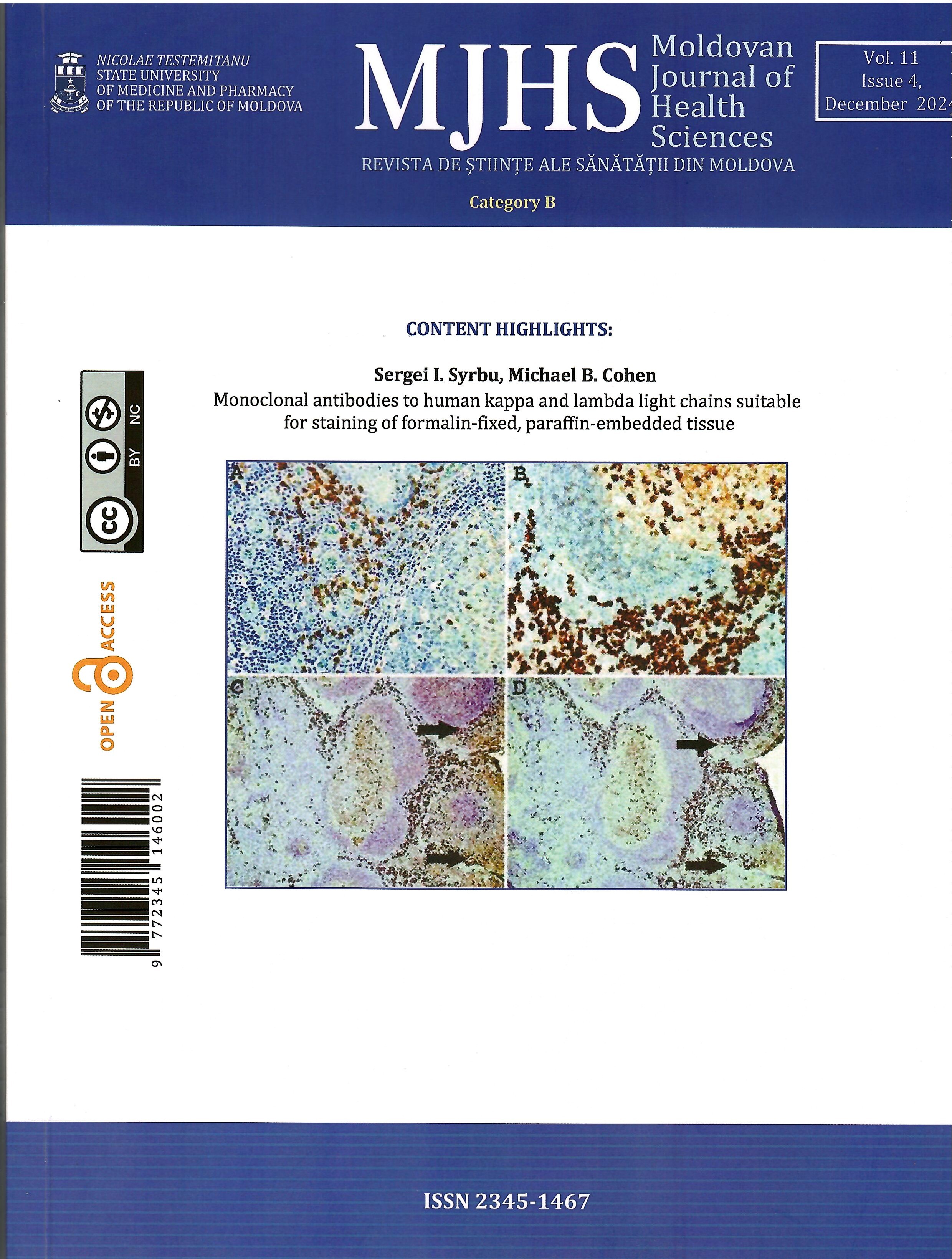
We selected eight hybridomas that produced highly specific monoclonal antibodies (mAbs) directed to the constant regions of kappa (four clones) and lambda light chains (four clones). The antibodies were used for IHC staining and direct immunofluorescence microscopy on FFPE tissues. Plasma cell neoplasms could be stained without antigen retrieval. For uniform and reproducible staining of diffuse large B-cell lymphomas and some marginal zone B-cell lymphomas with plasmacytoid differentiation, a heat-based antigen retrieval procedure was necessary. Compared with commercial polyclonal antibodies to kappa and lambda light chains these mAbs demonstrated lower background staining and produced fewer false negative results.