Table of contents
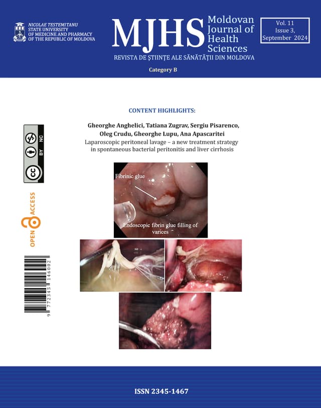
9 papers · pp. 3–64Laparoscopic peritoneal lavage – a new treatment strategy in spontaneous bacterial peritonitis and liver cirrhosis
Introduction
Bacterial infections in cirrhotic patients represent a major clinical problem, occuring 4-5 times more frequently compared to the general population and increasing mortality by leading to acute on chronic liver failure, subsequent decompensation, and multiorgan failure. The study’s purpose is to determine the possibilities of laparoscopy in the treatment of spontaneous bacterial peritonitis with decompensated liver cirrhosis.
Materials and methods
A retrospective descriptive study was conducted on 82 patients diagnosed with liver cirrhosis and spontaneous bacterial peritonitis, who were admitted to the Constantin Tibirna Surgery department No2, Holy Trinity Municipal Clinical Hospital and the Scientific Research Laboratory of Hepatic Surgery, Nicolae Testemițanu State University of Medicine and Pharmacy, Chisinau, Republic of Moldova, between January 2012 and December 2021. Patients who underwent surgical drainage of the abdominal cavity by laparoscopy with postoperative peritoneal lavage with antibiotics were selected. All patients received standard treatment for the correction of liver function and complications of portal hypertension. The data were extracted from the medical records of the hospital archive, and the patient database was compiled. Data analysis was performed using simple statistical calculations.
Results
Positive ascitic fluid bacterial culture was in 29.2% (24 patients), while 70.7% (58 patients) had culture-negative ascites and peritonitis. The most frequent bacterial species was E. coli, present in 54.1% (13 patients). Mortality was 8.5% (7 patients) due to progressive liver failure. Recurrence of ascites and peritonitis at 1 month was 6.0% (5 patients).
Conclusions
The laparoscopic approach in spontaneous bacterial peritonitis in patients with decompensated liver cirrhosis allows for better sanitation of the abdominal cavity, improves peritoneal absorption, and deserves establishment as clinical practice for patients with ascites and peritonitis and cirrhosis.
Childbirth at advanced reproductive age: the impact of biopsychosocial factors on the mode of delivery
Introduction
Contemporary society reflects a clear trend towards delayed motherhood, raising significant concerns in the management of pregnancy and childbirth in women of advanced reproductive age. In this context, the mode of delivery and associated risks for this age category require increased attention. Birth methods have been thoroughly examined to identify risks and influencing factors within this specific cohort.
Material and methods
A descriptive cross-sectional study was conducted on a sample of 528 women. Data were collected using a pre-tested semi-structured questionnaire, and respondents were divided into three groups based on the mode of delivery: vaginal delivery, planned cesarean section, and emergency cesarean section. Sociodemographic, anthropometric, medical, and obstetric characteristics were analyzed using linear regression. Statistical analyses included descriptive and inferential statistics (Chi-square), with a 95.0% confidence interval.
Results
The analysis revealed statistically significant variations in the mode of delivery based on maternal age (p=0.013) and paternal age (p=0.001), with an increased rate of cesarean sections at more advanced ages. Significant variations were also found in relation to area of residence (p=0.003), education level (p=0.001), nature of work (p=0.028), GP appointments (p=0.020), number of GP appointments(p<0.001), number of obstetrician appointments (p=0.032), time of informing on risk factors (p=0.005), parity (p<0.001), multiple pregnancies (p=0.016), mode of first delivery (p<0.001), pregnancy complications (p=0.003), delivery complications (p<0.001), gestational age at birth (p=0.017), Apgar scores at 1 and 5 minutes (p<0.001).
Conclusions
Advanced reproductive age has been associated with a higher risk of cesarean section compared to vaginal delivery. The influence of age is modulated by various sociodemographic, medical, and obstetric characteristics, including area of residence, education level, history of cesarean section, parity, pregnancy and delivery complications, pre-existing chronic conditions, antenatal care and provision of information on risk factors.
Deep endometriosis – impact on infertility, endometriosis fertility index, and reproductive prognosis (comparative study)
Introduction
Deep infiltrating endometriosis (DIE) is the most severe form of endometriosis, contributes to pelvic pain syndrome, extragenital symptoms, fertility problems, and diminished reproductive prognosis for affected individuals. It is recommended to use the Endometriosis Fertility Index (EFI) to assess reproductive prognosis and conduct clinical research to compare reproductive prognosis in different forms of endometriosis.
Objective
Comparison of the impact of the DIE and others forms of endometriosis on EFI, patients' fertility and on reproductive prognosis to understand the management approach.
Materials and methods
A cohort study included 190 reproductive-age women, divided according to the #Enzian classification: the main group - 85 patients with DIE, the control group - 105 women with other forms of endometriosis. The EFI was utilized for reproductive prognosis. Pain was evaluated with Visual Analog Scale (VAS). Statistical analyses were performed using SPSS, with calculation of the Mann-Whitney U and Pearson's chi-square test (χ²).
Results
In main group, the frequency of infertility was 83.5% compared to the control group's 71.4% (p > 0.05). The EFI in main group was 7.18±0.25 points vs the control group's 7.13±0.28 points (p = 0.852). Patients in the main group suffered from intense pelvic pain (>7 points by the VAS, p < 0.01), including severe dyspareunia (7.85±0.33 points vs 2.18±0.46 points in control group, p < 0.01).
Conclusions
Our results suggest that infertility in women with DIE may be more often associated with sexual abstinence due to significant dyspareunia rather than organic impairments. Thus, EFI in patients with DIE does not reflect all aspects of infertility and has reservations, and consideration of both physical symptoms and sexual health is crucial in managing DIE to optimize fertility outcomes. These findings open the way to the feasibility of surgical treatment of DIE to improve sexual quality of life, which will reduce the need for IVF and increase the chances of spontaneous pregnancy in patients, but this conclusion requires further investigation in randomized clinical trials.
Postoperative urinary retention - prevalence and risk factors: prospective, cohort study
Introduction
Ensuring perioperative urination maintenance can often be challenging, as postoperative urinary retention is frequently overlooked in favor of more clearly defined goals such as successful surgery, comprehensive postoperative pain control, reducing the risk of postoperative cardiorespiratory complications and shortening the patient's overall hospital stay. However, the inability to initiate urination and empty the bladder in the early postoperative period may negatively affect each of the listed success criteria.
Material and methods
A single-center, prospective, observational, cohort study was conducted, enrolling elderly patients without severe comorbidities. A total of 127 complete datasets were analyzed. Anthropometric parameters, type of surgery, duration of anesthesia and surgery; and several parameters previously reported as risk factors for postoperative urinary retention were recorded. The main objective wasto identify the prevalence of postoperative urinary retention in a surgical group in the Republic of Moldova. The secondary objective was to test the predictive value of a series of parameters (modifiable and non-modifiable) related to the patient or surgical treatment received as risk factors for urinary retention in the first 24 hours postoperatively. Statistical software used: Social Science Statistics (https://www.socscistatistics.com).
Results
The studied surgical population was homogeneous in terms of body mass, height, duration of surgery and anesthesia; heterogeneous by gender (62.2% male) and type of anesthesia (64% general anesthesia). Depending on the definition criteria, the prevalence of postoperative urinary retention varied between 5.5% and 7.9%. The preoperative unmodifiable risk factors for postoperative urinary retention: positive history for hypertension OR = 9.0 (X2 (1, N = 127) = 5.6, p = 0.017), diabetes mellitus OR = 5.1 (X2 (1, N = 127) = 5.36, p = 0.021) and stroke OR = 4.83 (X2 (1, N = 127) = 2.098, p = 0.148).
Conclusions
The prevalence of postoperative urinary retention in a single-center surgical population from the Republic of Moldova varies between 5.5% and 7.9%, depending on the criteria for postoperative urinary retention applied. This variation highlights the need for a consensus on diagnostic criteria for postoperative urinary retention is needed. Patients with hypertension and diabetes mellitus were more likely to develop postoperative urinary retention. Patients with pre-existing neurological disorders such as positive history for stroke and diabetic polyneuropathy were more susceptible for postoperative urinary retention.
The impact of ozone therapy on the progression of COVID-19 patients
Introduction
Ozone therapy can be used as a monotherapy or as an adjunctive treatment to standard COVID-19 treatment protocols. Current evidence indicates that this approach may improve clinical outcomes, paraclinical markers, and reduce radiological signs of inflammation, with no side effects.
Material and methods
he study included 100 consecutive patients aged 18 and older with COVID-19, admitted to the Intensive Care Unit at the Institute of Emergency Medicine. Patients were randomly divided into two groups: 50 patients underwent treatment according to the National Clinical Protocol along with major ozonated autohemotherapy (the study group), while the other 50 patients were treated only according to the National Clinical Protocol (the control group).
Results
Although the initial oxygenation index (PaO2/FiO2) values were similar in both study groups, a dynamic analysis revealed a clear efficacy of ozone therapy. By the end of the first-week treatment, the mean oxygenation index in the ozone-treated group was significantly higher than in the standard treatment group: 296.8±105.1 mm Hg versus 232.8±110.6 mm Hg (p<0.01). The use of oxygen therapy (70.0% vs. 78.0%), non-invasive ventilation (70.0% vs. 76.0%), and invasive mechanical ventilation (22.0% vs. 38.0%) tended to be lower in the ozone group, though this difference was not statistically significant (p>0.05). Both treatment groups showed a significant clinical improvement, with 54.0% of COVID-19 patients in the ozone group and 50.0% in the conventional treatment group achieving a two-point reduction in clinical severity score (p>0.05).
Conclusions
The mean oxygenation index significantly increased in the study patient group (246.86±30.3 mm Hg on day 1 and 296.75±105.1 mm Hg on day 7 of treatment; p<0.01) and remained unchanged in the control group (235.86±33.4 mm Hg on day 1 and 232.82±110.6 mm Hg on day 7 of treatment; p>0.05). Although the mortality rate was lower among COVID-19 patients treated with ozone therapy (24.0%) compared to those receiving standard treatment (34.0%), this difference did not achieve any statistical significance.
Comparative analysis of imaging data in sensory and motor disorders in lumbar neurocompressive syndrome
Introduction
Lumbar neurocompressive syndrome is a condition characterized by radicular pain, motor, sensory and reflex changes, as well as paresthesia or numbness in the lower limb. These symptoms can be triggered by positions and/or movements of the spine. In lumbar radiculopathy, both mechanical and inflammatory factors play significant roles.
Material and methods
The study included 102 patients with signs of lumbar neurocompressive syndrome. Of these, 51 (group I) patients were examined using MRI and the other 51 patients (group II) were examined using conventional radiographic investigation of the lumbosacral region of the spine.
Results
By analyzing the magnetic resonance imaging data of the lumbar spine, a threshold of statistical significance was determined (of 10%, p < 0.10) for patients with sensory disturbances in the lower limb in cases of stenosis of the lumbar spinal canal, and in patients with motor disorders in the lower limb, in the case of disc protrusions. The analysis of the magnetic resonance imaging data determined a significance threshold (of 5%, p < 0.05) in patients with sensory disorders (in the case of disc sequestrations and in the case of disc extrusions) as well as for motor disorders (in the case of disc extrusions, disc sequestrations and static disorders of the spine). The analysis of standard radiographs of the lumbar spine allowed the determination of the threshold of statistical significance (of 5%, p < 0.05) in patients with sensory disorders in the lower limb in cases of coxofemoral osteoarthritis and Schmorl’s hernias. For patients with motor disorders at the level of the lower limb, conventional radiography was informative in the presence of calcification of the intervertebral discs and in coxofemoral osteoarthritis.
Conclusion
MRI can be considered the first-choice imaging technique for diagnosis of the lumbar spine pathologies characterized by sensory and motor changes in the lower limbs.
Temporary splinting in periodontally mobile teeth: review before application
Introduction
Temporary splinting of mobile, periodontally compromised teeth is an important stage in the complex treatment of periodontitis, used to improve mastication, aesthetics, and the prognosis for teeth with pathological mobility. It helps prevent this phenomenon and consolidate the success of conservative and surgical treatment, which also has a positive effect on the patient's psycho-emotional state.
Material and methods
12 patients (6 female and 6 male), aged 26–60 years, with pathological teeth mobility due to chronic generalized periodontitis, were included in the study. Temporary splinting was performed using fiberglass threads braided with polyester microfibers, polyethylene fibers, and aramid fibers, and in all clinical cases, it was secured with photo-composite material designed for teeth splinting.
Results
Temporary splinting is a very important step in periodontitis treatment, and dynamic monitoring is necessary throughout the entire period of splinting. The interval between visits is determined based on the type of splinting, the risk of complications, the severity of periodontitis, systemic and local factors, the level of individual oral hygiene, and the patient's motivation. The interval can be 6 months, 4 months, or, in aggressive forms of periodontitis, 2 months.
Conclusions
Temporary splinting contributes to the effectiveness of pathogenetic and symptomatic therapy.
Management of diabetic retinopathy in pregnancy
Introduction
The onset and development of diabetic retinopathy are more common during pregnancy. Pregnancy has no long-term effect on diabetic retinopathy; however, in 50-70% of cases, changes in retinopathy continues. The probability of worsening is highest in the second trimester and up to one year postpartum. Additional factors that have been associated with disease progression include duration of diabetes, the degree of retinopathy at the time of conception, management of hyperglycemia, anemia, and development of associated hypertension. In cases of severe non-proliferative retinopathy, it is recommended to promptly initiate laser photocoagulation rather than wait for early proliferative changes. Maintaining good diabetic control before and during pregnancy can help prevent disease progression and serious vision loss.
Material and methods
Diabetic retinopathy management in pregnancy was the subject of a comprehensive review of the scientific and medical literature. A structured search was performed in the PubMed, Scopus and HINARI databases, considering relevant articles published in the last 10 years. The search terms used (in English) were: „Diabetic retinopathy”; „pregnancy”; „laser photocoagulation”; „intravitreal steroids”; „anti-vascular endothelial growth factor”.
Results
It is suggested that women with diabetes receive pre-conception and post-pregnancy counselling from a multidisciplinary team including an ophthalmologist, endocrinologist, and perinatologist, as diabetic retinopathy may worsen during pregnancy. The risk of progression of the disease and the importance of appropriate metabolic control before and during pregnancy should be clearly explained to the patient. Careful monitoring is required in patients with advanced gestation, significant retinopathy, concomitant hypertension, and nephropathy.
Conclusion
The risk of retinopathy development may increase during pregnancy. Serious effects can arise for both the mother and the fetus, even though retinopathy is not common during pregnancy. It is possible to avoid significant retinopathy by carefully planning a young diabetic woman's pregnancy and proceeding promptly to laser photocoagulate in cases of severe non-proliferative retinopathy. A tendency for regress is frequently seen in diabetic retinopathy during the post-natal period. Subsequent pregnancies do not significantly increase the risk of progression if the retinopathy is stable before pregnancy.
Acute autoimmune hemolytic anemia in a patient with systemic lupus erythematosus
Introduction
Autoimmune hemolytic anemia occurs due to the accelerated destruction of erythrocytes as a result of the dysfunction of immune system cells, which produce antibodies against the normal antigens of the membrane of hematopoietic cells. One of its causes is systemic lupus erythematosus.
Materials and methods
We present a case of a 20-year-old patient who was hospitalized with acute autoimmune hemolytic anemia, having been diagnosed with SLE at the age of 18 years. At the onset of the disease, hemolytic anemia was a differential diagnostic challenge.
Results
The differential diagnosis between primary and secondary autoimmune hemolytic anemia (AIHA) was an important step. The presence of antinuclear antibodies (ANA Hep2, Anti-dsDNA, Anti-cardiolipin, Anti-phospholipids, anti-Ro, Anti-Sm B) were important arguments in making the diagnosis. The relapse of AIHA was caused by inadequate treatment, due to a lack of compliance. Pulse therapy combined with methylprednisolone and cyclophosphamide successfully resolved the AIHA.
Conclusions
Hematological abnormalities are commonly seen in SLE patients, but hemolytic autoimmune anemia is a rare condition. A timely diagnosis of the cause of hemolytic anemia and proper treatment of lupus by correcting autoimmune disorders are crucial in disease management. Pulse therapy combined with corticosteroids and immunosuppressants is effective in acute relapses of hemolytic anemia.