MJHS
Navigation
Journal Navigation
Article Navigation
opened article
Volum 10, Issue 3
September 2023
Cover Image

Article contents

opened journal
Volum 10, Issue 3
September 2023
Cover Image

Abstract

Introduction

The clinical concept that would argue that the activity of the lateral pterygoid muscle, being disturbed, would play an important role as an etiological factor in temporomandibular joint dysfunctions is still widely accepted, being also a decisive factor in the correct choice of the treatment plan. However, because of the fact that very few research and clear evidence were conducted and presented to support completely that concept, it continues to remain a very controversial one.

Materials and methods

For this literature review were considered and studied scientific articles published between 2000 and 2023, in the following electronic databases: PubMed, MEDLINE, Google Scholar, BIR Publications, ScienceDirect. Research methods – analysis, synthesis, systematization, and description.

Results

Patients presenting temporomandibular joint dysfunction complain about pain in the temporomandibular joint or/and in masticatory muscles, limitation and sounds during mandibular activity. Temporomandibular dysfunction is a non-specific collective term, used to describe a heterogeneous group of pathological conditions located in the territory of the stomatognathic system. These are considered musculoskeletal conditions that cause pain while performing the function (mastication, speech, swallowing), with increased sensitivity in the masticatory muscles and/or the temporomandibular joint, with possible limitations of the range of motion, the appearance of joint noises and otological symptoms. One of the theories claims that in temporomandibular dysfunction, the lateral pterygoid muscle becomes hyperactive, hypoactive or that there is a lack of coordination between the superior and inferior branches of the muscle, or that there is a disturbance during the performance of the role of the muscle to control and stabilize the temporomandibular joint. However, the in-depth study of the specialized literature indicates that no scientific evidence is yet available that the function of the lateral pterygoid muscles in temporomandibular dysfunction is somehow disturbed. Moreover, the muscle's role during the execution of its normal function has also been questioned and remains a matter of controversy.

Conclusions

The lateral pterygoid muscle obviously plays an important role in the development of temporomandibular dysfunction through the prism of its anatomical and functional particularities, referring to the superior fascicle responsible for the correct anatomical maintenance of the articular disc during function.

Key Messages

What is not yet known about the issue addressed in the submitted manuscript

The literature study shows that there is still no clear scientific evidence that the function of the lateral pterygoid muscles in temporomandibular dysfunction is somehow disturbed. Additionally, their involvement in the development and evolution of temporomandibular dysfunction is uncertain.

The research hypothesis

Examining the lateral pterygoid muscle requires a deep knowledge of its anatomy and function, as both palpation and improperly or superficially performed functional manipulation can provide us with erroneous data for the diagnosis and treatment of temporomandibular dysfunction.

The novelty added by the manuscript to the already published scientific literature

A systematic study of the specialized literature was conducted regarding the role of the lateral pterygoid muscle in the development of temporomandibular joint dysfunctions, the evaluation of the anatomical aspects of the lateral pterygoid muscle and its interrelationship with the occurrence of temporomandibular joint dysfunction, as well as the elucidation of  methods for examining the lateral pterygoid muscle and the importance of its role in the evolution of temporomandibular dysfunction.

Introduction

The clinical concept that argues that the activity of the lateral pterygoid muscle, being disturbed, plays an important role as an etiological factor in temporomandibular joint (TMJ) dysfunctions is still widely accepted. It also remains a decisive factor in the correct choice of the treatment plan. However, due to the fact that few rigorous studies and clear evidence have been conducted and presented to fully support this concept, it continues to be a very controversial one [1, 2]. Patients with temporomandibular disorders (TMD) complain of pain in the TMJ and/or masticatory muscles, limitations and sounds during mandibular movements. Temporomandibular disorders are a non-specific collective term used to describe a heterogeneous group of pathological conditions located in the territory of the stomatognathic/masticatory system. These are considered musculoskeletal disorders, which cause pain during the performance of the function (mastication, speech, swallowing), with increased sensitivity at the level of masticatory muscles and/or the TMJ, along with possible limitations of the range of motion and the occurrence of joint noises and otological symptoms [1, 3-6]. Current data demonstrate that TMD is one of the most commonly diagnosed forms of musculoskeletal pain in all age groups. Ethnicity, age, geographic location, and the time of assessment influence the level of prevalence, causes, factors, and spectrum of clinical manifestation of the pathology [7, 8]. In adolescents, the prevalence is a controversial subject, considering that adults are more frequently affected by TMD, but at the same time, a significantly increased incidence is noted in subjects with mixed dentition (worldwide incidence – 25%, and in the developed countries – between 2% and 6%) [1, 3-5, 9]. One theory claims that in TMD, the lateral pterygoid muscle becomes hyperactive, hypoactive, or that there is a miscoordination between the superior and inferior branches of the muscle, or that there is a disturbance during the execution of the role of the muscle to control and stabilize the TMJ [1, 10]. However, a rigorous review of the literature indicates that no clear scientific evidence is yet available to suggest that the function of the lateral pterygoid muscles is somehow disrupted in TMD. Moreover, the muscle's role during the execution of its normal function has also been questioned and remains a matter of controversy. 

Materials and methods

For this literature review, scientific articles published between 2000 and 2023 were considered and studied using the following electronic databases: PubMed, MEDLINE, Google Scholar, BIR Publications, ScienceDirect. Keywords such as „lateral pterygoid muscle”, “temporomandibular joint”, and “TMJ dysfunction” were used. A total of 137 articles were studied, out of which 12 were duplicate articles, 45 articles presented studies performed on cadavers, 21 articles did not provide sufficient data, and 19 articles were conducted before the year 2000. The exclusion criteria included studies performed on cadavers, studies conducted before the year 2000, and studies that did not present trustworthy information on the topic under study.

Results and discussions

Anatomical variations of the lateral pterygoid muscle. In recent years, several studies have been carried out with the purpose of proving a theory that there may be a third fascicle of the lateral pterygoid muscle or that the two fascicles may be anatomically inserted differently at the level of the disc and condyle among different individuals. To study the anatomical variations of the lateral pterygoid muscle and its insertion variations, analyzes were performed using magnetic resonance imaging (MRI). In a 2013 study by Valenzuela et al., 698 patients underwent MRI analysis of the TMJ. Three types of muscle insertion of the superior fascicle of the muscle have been described (Table 1). The first type of muscle insertion involved muscle fibers inserting onto the articular disc. The second type of muscle insertion was at the level of the articular disc and condyle, and the third type was at the level between the articular disc and the articular capsule (Figure 1) [11, 12]. In another similar study conducted in 2016 by Eberhard et al., in which 382 patients with articular disc dislocation were evaluated by MRI, the results showed that the prevailing type of muscle insertion of the superior fascicle among subjects was type 2, at 67%, and the one with the lowest prevalence was type 1, at 7.6%. MRI images were taken in the position of maximum intercuspation (MI) and full opening of the oral cavity [13].

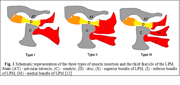
Table 1. Types of muscle insertion of the lateral pterygoid muscle. Litko classification [13]

Type of insertion

Bundle

Insertion

Type 1

Superior

Inferior

Disc

Condyle

Type 2*

Superior

Inferior

Disc and condyle

Condyle

Type 3

Superior

Medial

Inferior

Disc

Condyle

Condyle

Note: (*) – the highest prevalence with 76% of subjects according to Eberhard et al. studies.

Function of the lateral pterygoid muscles. The international theory about the anatomy of the lateral pterygoid muscle says that it is a masticatory muscle, which is part of the dento-maxillary system. It is considered to be an important masticatory component, in some sources even the main one, either structurally or functionally, due to its direct insertion at the level of the components of the temporomandibular joint. In some temporomandibular pathologies or dysfunctions, the muscle would be described as being directly or indirectly involved. Directly, it is described as the muscle that specifically causes TMD. The dysfunction involves the alteration of the movement of the joint and causes pain in neighboring structures, which are nervous, vascular or bone [11, 14, 15]. Pathologically, it is described that at the level of the joint there would be an impediment or an inconsistency between the elements, but the most affected component would be the articular disc, which is directly associated with the lateral pterygoid muscle that causes the anterior dislocation of the disc [11]. Mechanically, this joint dysfunction produces an anterior disc dislocation, which, in turn, compresses with the bony tissues, causing a bony stop for the articular condyle, reducing joint space and range of motion at the joint, producing combined clinical and mechanical symptoms [11, 12].

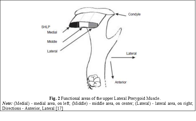
The functions of the lateral pterygoid muscle were defined as follows: the superior bundle is responsible for closing, retropulsion, and ipsilateral movements of the mandible, while the inferior bundle is active in opening, propulsion, and contralateral movements of the mandible. However, recent studies suggest that some fibers of the superior bundle may also be involved in opening, propulsion, and contralateral mandibular movements, and that it may consist of three mediolaterally arranged functional areas. This indicates that the concept that the occurrence of clicking, crackles in the TMJ is due to uncoordinated movements between both bundles of the lateral pterygoid muscle needs to be re-evaluated [10, 16]. Both fascicles of the lateral pterygoid muscle, in some recent reports, have been shown to be inactive during electromyography in the mandibular posture position. An important role of the lateral pterygoid muscle demonstrated electromyographically is that it generates laterality and propulsive movements of the mandible. This was observed when the activity of the inferior fasciculus changed when the direction of the horizontal force of the mandible oscillated from side to side [1, 10]. Murray et al. claim that the cause that would lead to different conclusions and results regarding the function of the lateral pterygoid muscle would be the incorrect placement of the electromyographic electrodes at the level of the muscle [17]. The results obtained could belong to other adjacent muscles such as the temporalis muscle or medial pterygoid muscle, or they could belong to the lateral pterygoid muscle (LPM) but be incorrectly assigned to a specific fascicle. With the help of computed tomography, they analyzed the correct placement of the electrodes at the level of the lateral pterygoid muscle and confirmed the classical notion that the inferior LPM would be involved in opening, protrusion, and contralateral mandibular movements. Studying the functional unit of the muscle, they concluded that the supero-medial part of the inferior LPM has an important role in initiating contralateral movements of the mandible, while the infero-medial part has a role in controlling fine movements [17]. The activity of the superior lateral pterygoid muscle was also confirmed by the correct placement of the electrodes, and it turned out to be much more complex than originally thought [17]. In contrast to the idea that the superior LPM is active only in closure, retrusion, and ipsilateral movements of the mandible, most muscle fibers of the superior LPM are at least active in opening, protrusion, and contralateral movements. Moreover, the pattern of activity varies depending on the location of the muscle fibers in the muscle and has been classified into three functional areas (Figure 2) [17]. The medial area produces patterns of activity similar to those produced by the inferior LPM. The lateral area may be equally active in closure, ipsilateral movements, and retrusion, while the central area generates different patterns of activity. These obtained functional data play an important role in understanding the role of the lateral pterygoid muscle in TMD [17]. 

The relationship between the lateral pterygoid muscle and the elements of the temporomandibular joint. J. Okeson described the functional importance of the superior lateral pterygoid muscle, namely: it becomes evident when the unilateral masticatory function is exercised. When an individual unilaterally bites a hard substance, both temporomandibular joints are functionally loaded differently. This occurs because the force is not applied to the joint but to the hard substance. Intra-articular pressure increases contralaterally to the biting area, and decreases at the ipsilateral articular level. This fact can lead to a separation between the articular surfaces, resulting in a dislocation of the articular disc on the same side. To prevent this, the superior lateral pterygoid muscle becomes active during biting, rotating the disc forward on the condylar surface so that the thicker edge of the disc remains in permanent contact with the articular surface [5].

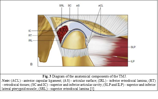
The articular disc is composed of several layers of collagen fibers, each oriented in different directions to resist the shearing phenomenon that could occur during the sliding of the condyle. The disc is attached to the medial and lateral surfaces of the condyle, which allows it to move in unison with the articular condyle. The positioning of the disc is controlled by a combination of elastic fibers attached to the posterior surface of the disc, thereby holding the disc in tension against the action of the superior fasciculus of the lateral pterygoid muscle, which is attached to the anterior surface of the disc. When the disc ligaments pull the disc along with the movement of the condyle, the rotation of the disc on the condyle occurs due to the force of contraction or relaxation of the superior fascicle of the muscle (Figure 3) [1, 4, 18].

All the tissues attached to the disc have the role of preventing its anterior dislocation. However, the question arises as to how its anterior dislocation occurs, which is very common in patients with temporomandibular dysfunction. The only force that is directed anteriorly and can produce articular disc dislocation is the muscle that is attached to the anterior surface of the disc, namely the superior fascicle of the lateral pterygoid muscle. This muscle, together with the elastic collagen fibers attached posteriorly to the disc, controls the position of the disc on the surface of the condyle and is always aligned with the direction of the force when the condyle descends the slope of the tubercle downward [4]. If the articular condyle is located in centric relation, the articular disc is located in the most anterior position that the posterior ligaments allow. In this position, the condylar forces are directed upward toward the medial third of the disc and forward toward the anterior surface of the condyle. When the inferior fascicle of the lateral pterygoid muscle (+) begins to propel the condyle anteriorly, the superior fascicle of the muscle (-) relaxes its contraction to allow the elastic fibers to pull the disc over the condyle (Figure 4) [4].

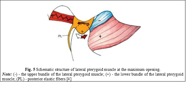
At the maximum opening of the oral cavity, when the condyle reaches the most inclined portion of the articular tubercle, the disc should be directly located above the articular condyle, as the forces are directed superiorly. At this point, the elastic fibers rotate the disc posteriorly because the superior fasciculus of the lateral pterygoid muscle is in a controlled relaxation (Figure 5) [4].

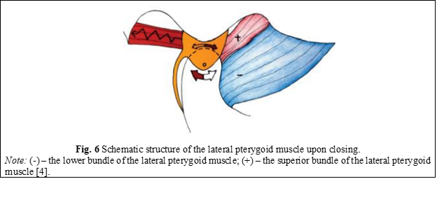
Upon closing the oral cavity, the condyle moves posteriorly and superiorly on the slope of the articular tubercle, so that the disc is again positioned in front of the condyle. For this, the upper fascicle (+) begins to contract, while the lower fascicle (-) relaxes and allows the condyle to return to its original position (Figure 6) [4].

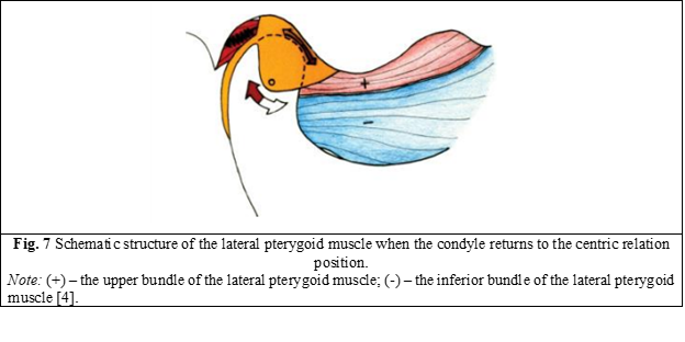
When the condyle returns to the centric relation position, the disc is located as anteriorly as possible, as far as the posterior ligament allows. If the ligament is intact and has not been stretched or torn, the disc is located in perfect alignment with the direction and position of the condyle. In the absence of occlusal interference, the inferior fascicle of the lateral pterygoid muscle is not active, even in the position of maximum intercuspation (MI). The superior fascicle remains contracted to keep the disc in the proper position (Figure 7) [4].

Correlation between anatomical variations of the lateral pterygoid muscle and temporomandibular dysfunctions.The correlation between the types of muscle insertions of the LPM and its anatomical variations is directly proven in studies of temporomandibular joint dysfunctions. Only two research studies from 2009 and 2012 (Dergin et al.; Matsunaga et al.) did not show any link between anatomical variations of the lateral pterygoid muscle and temporomandibular joint dysfunctions. This is because they represent studies carried out on corpses where only the morphological structure was studied and only the variations were observed [19, 20, 21]. The anatomical variation of muscle insertion type 2 according to the Litko classification [13] was the one that showed the closest connection with TMD, especially due to the insertion of the superior fasciculus of the LPM at the level of the articular disc and the articular condyle (Table 1). This variation can produce joint alterations or dysfunctions, characterized by local or loco-regional pain during closing or opening movements. However, studies have not shown any relationship between the type of insertion of the inferior fascicle of the LPM and TMD, both primary and secondary. The middle or accessory fascicle, which was described in 19% of cases in the analyzed studies, showed no connection between its presence and any TMD, primary or secondary, acute or chronic [21, 22-26]. Murray et al. state that observations made in recent years support the theory that the lack of coordination of movements between the upper and lower LPM could generate clicks, cracks, or articular blockages during the horizontal positioning of the disc in relation to the condyle. However, recent evidence indicates the possibility that fibers of the superior lateral pterygoid muscle that insert at the level of the articular disc could activate independently of other fibers of the same muscle, the superior LPM, and generate forces at the level of the disc that are not simultaneously generated with those inserted at the level of the condyle [17]. J. Okeson states that the exact percentage of insertion of the superior lateral pterygoid muscle at the level of the disc and at the level of the articular condyle is still under debate and is variable. However, it would be reasonable to assume that if the insertion of the muscle is predominant at the level of the neck of the articular condyle, and less at the level of the disc, the function of the superior muscle will have correspondingly less influence on the position of the articular disc. Moreover, vice versa, if the insertion of the superior muscle is predominant at the level of the articular disc, and less at the level of the condyle, the function of the muscle will greatly influence the position of the articular disc. This anatomical variation explains why, in some patients, the disc dislocates very quickly, without any antecedents or previous clinical features [5].

Conclusions

1. The lateral pterygoid muscle plays an obviously important role in the development of temporomandibular dysfunctions through the prism of its anatomical and functional particularities, specifically referring to the superior fascicle responsible for maintaining the correct anatomical position of the articular disc during function.

2. The anatomical variations of the muscle insertion according to various classifications are directly related to the development of TMJ dysfunctions, especially due to the insertion of the superior fascicle of the LPM at the level of the articular disc and the articular condyle. This variation can produce alterations at the level of the joint complex, characterized by the respective dysfunctional symptomatology. However, studies have not shown a clear link between the type of insertion of the inferior fascicle of the LPM and both primary and secondary TMJ dysfunction.

Competing interests 

None declared. 

Authors’ contribution 

All the authors have contributed equally at the results presentation in the paper, approved the „ready for print” version of the manuscript.

Author’s ORCID IDs

Vitalie Pântea – https://orcid.org/0000-0002-3489-030X

Felicia Tabără – https://orcid.org/0009-0000-9979-8371

Mariana Ceban – https://orcid.org/0000-0001-7203-358X

Veronica Burduja – https://orcid.org/0009-0007-8101-9043

Lilian Nistor – https://orcid.org/0009-0008-6282-9240

Olga Ursu –https://orcid.org/0000-0002-2923-5546

References

  1. Lai Y, Yap A, Turp J. Prevalence of temporomandibular disorders in patients seeking orthodontic treatment: a systematic review. J Oral Rehabil. 2020;47(2):270-280. doi: 10.1111/joor.12899.

  2. Murphy MK, MacBarb RF, Wong ME, Athanasiou KA. Temporomandibular disorders: a review of etiology, clinical management, and tissue engineering strategies. Int J Oral Maxillofac Implants. 2013 Nov-Dec;28(6):e393-414. doi: 10.11607/jomi.te20. 

  3. Bernal C, Gonzalez O, Moreno M. New anatomo-radiological findings of the lateral pterygoid muscle. Surg Radiol Anat. 2016;38(9):1033-1043. doi: 10.1007/s00276-016-1665-2.

  4. Dawson PE. Functional occlusion: from TMJ to smile design. St. Louis (Missouri): Mosby; 2007. 630 p.

  5. Okeson JP. Management of temporomandibular disorders and occlusion. 8th ed. St. Louis (Missouri): Elsevier; 2020. 514 p. 

  6. Li D, Leung Y. Temporomandibular disorders: current concepts and controversies in diagnosis and management. Diagnostics (Basel). 2021;11(3):459. doi: 10.3390/diagnostics11030459.

  7. Bordeniuc G. Indici clinico-fiziologici în disfuncția mușchilor masticatori [Clinical physiological index in masticator muscle disorder] [dissertation]. Chișinău: Nicolae Testemitanu State University of Medicine and Pharmacy; 2023. 159 p. Romanian.

  8. Fala V. Implementarea design-ului funcțional-estetic, conform conceptului ocluzal „Ocluzia consecutivă cu dominanta canină” în terapia restaurativă estetică, metoda directă [Implementation of functional design, based on the occlusal concept “Sequential guidance with canine dominance” in aesthetic restorative therapy, through the direct method]. Chișinău: Sirius; 2020. 168 p. Romanian.

  9. Sarlani E, Grace E, Reynolds M, Greenspan J. Evidence for up-regulated central nociceptive processing in patients with masticatory myofascial pain. J Orofac Pain. 2004;18(1):41-55. 

  10. Matsunaga K, Usui A, Yamaguchi K, Akita K. An anatomical study of the muscles that attach to the articular disc of the temporomandibular joint.  Clin Anat. 2009;22(8):932-940. doi: 10.1002/ca.20865.

  11. Antonopoulou M, Iatrou I, Paraschos A, Anagnostopoulou S. Variations of the attachment of the superior head of human lateral pterygoid muscle. J Craniomaxillofac Surg. 2013;41(6):91-97. doi: 10.1016/j.jcms.2012.11.021.

  12. Valenzuela JJ, Orellana M, Gold M, Garcia G, Santana A. Anatomy of the lateral pterygoid muscle and its relationship with temporomandibular disorders: a literature review. Eur J Anat. 2020;24(3):249-256.

  13. Litko M, Szkutnik J, Berger M, Rozyło-Kalinowska I. Correlation between the lateral pterygoid muscle attachment type and temporomandibular joint disc position in magnetic resonance imaging. Dentomaxillofac Radiol. 2016;45(8):20160229. doi: 101259/dmfr.20160229.

  14. Kawakami S, Kodama N, Maeda N, Sakamoto S, Oki K, Yanagi Y, Asaumi J, Maeda T, Minagi S. Mechanomyographic activity in the human lateral pterygoid muscle during mandibular movement. J Neurosci Methods. 2012;203(1):157-162. doi: 10.1016/j.jneumeth.2011.09.026.

  15. Omami G, Lurie A. Magnetic resonance imaging evaluation of discal attachement of superior head of lateral pterygoid muscle in individuals with symptomatic temporomanibular joint. Oral Surg Med Oral Pathol Oral Radiol. 2012; 114(5):650-657. doi.org/10.1016/j.oooo.2012.07.482.

  16. Ruangsri S, Whittle T, Wanigaratne K, Murray GM. Functional activity of superior head of human lateral pterygoid muscle and isometric force. J Dent Res. 2005;84(6):548-553. doi: 10.1177/154405910508400612.

  17. Murray GM, Phanachet I, Uchida S, Whittle T. The human lateral pterygoid muscle: a review of some experimental aspects and possible clinical relevance. Aust Dent J. 2004;49(1):2-8. doi: 10.1111/j.1834-7819.2004.tb00042.x.

  18. Sritara S, Tsutsumi M, Fukino K, Matsumoto Y, Ono T, Akita K. Evaluating the morphological features of the lateral pterygoid insertion into the medial surface of the condylar process. Clin Exp Dent Res. 2021;7(2):219-225. doi: 10.1002/cre2.353.

  19. Bhutada MK, Phanachet I, Whittle T, Peck CC, Murray GM. Regional properties of the superior head of human lateral pterygoid muscle. Eur J Oral Sci. 2008;116(6):518-524. doi: 10.1111/j.1600-0722.2008.00582.x.

  20. Stöckle M, Fanghänel J, Knüttel H, Alamanos C, Behr M. The morphological variations of the lateral pterygoid muscle: a systematic review. Ann Anat. 2019;222:79-87. doi: 10.1016/j.aanat.2018.10.006.

  21. Eberhard L, Giannakopoulos NN, Rohde S, Schmitter M. Temporomandibular joint (TMJ) disc position in patients with TMJ pain assessed by coronal MRI. Dentomaxillofac Radiol. 2013;42(6):20120199.  doi: 10.1259/dmfr.20120199.

  22. Dergin G, Kilic C, Gozneli R. Evaluating the correlation between the lateral pterygoid muscle attachment type and internal derangement of the temporomandibular joint with an emphasis on MR imaging findings. J Craniomaxillofac Surg. 2012;40(5):459-463. doi: 10.1016/j.jcms.2011.08.002.

  23. Palla S, Farella M. Masticatory muscle pain. In: Mense S, Gerwin RD, editors. Muscle pain: diagnosis and treatment. Heidelberg: Springer; 2010. p. 193-227.

  24. Phanachet I, Whittle T, Wanigaratne K, Murray GM. Functional properties of single motor units in inferior head of human lateral pterygoid muscle: task relations and thresholds. J Neurophysiol. 2001;86(5):2204-2218. doi: 10.1152/jn.2001.86.5.2204.

  25. Phanachet I, Whittle T, Wanigaratne K, Murray GM. Functional properties of single motor units in the inferior head of human lateral pterygoid muscle: task firing rates. J Neurophysiol. 2002;88(2):751-760. doi: 10.1152/jn.2002.88.2.751. 

  26. Șcerbatiuc D, Iovu G. Disfuncțiile articulației temporo-mandibulare [Temporomandibular joint dysfunction]. Med Stomatol (Chisinau). 2014;31(2):13-19. Romanian.

More articles

Research Harmfulness of prooxidants in bronchopulmonary dysplasia in preterm children
Mariana Ceahlău1*, Rodica Selevestru1,2, Olga Tagadiuc3, Svetlana Șciuca1,2
https://doi.org/10.52645/MJHS.2023.3.01
Oxidative stress can be defined as the imbalance of the redox state of a certain system including living one (organelle, cell, organ/tissue), which excessively produces reactive oxygen and/or reactive nitrogen species (ROS/RNS) that exceed the capacity of the antioxidant defense system, which have the ability to slow down or even prevent the oxidative damage of macromolecules. Oxidative stress is a pathogenic mechanism of a large variety of diseases, including pulmonary one.
Research Predicting sympathovagal balance using parameters of breathing patterns in abdominal breathing
Abdominal breathing is utilized as a non-pharmacological treatment method for various stress-related conditions and autonomic dysfunctions. The objective of the study was to determine the predictors in the modulation of sympathovagal balance, as indicated by the ratio of low frequency to high frequency power of heart rate variability, by utilizing the respiratory pattern parameters recorded during the abdominal breathing model.
Research The impact of imuheptin and imupurin on cytokine profile and antioxidant status in rat model of inflammation
Ina Guțu1*, Nicolae Bacinschi1, Valentin Gudumac2
https://doi.org/10.52645/MJHS.2023.3.03
Insects, throughout evolution, have developed a huge arsenal of active compounds, which they use to defend themselves against enemies and diseases, at the same time in recent years insects have shown great interest as a source of food rich in biologically active substances. Research in recent decades has shown that insects produce a variety of proteins and peptides with antibacterial, antifungal, antiviral, immunomodulatory, anti-inflammatory, antioxidant, antitumor, hepatoprotective, antithrombotic, antihypertensive and detoxifying activity during or after contact with the microbial agent or unfavourable factor.
Research Clinical effectiveness study of the new diagnostic score of acute appendicitis in the elderly
Acute appendicitis is among the three most frequent surgical diseases. The lifetime likelihood of developing acute appendicitis is around 7%. The incidence of acute appendicitis reduces with age after adolescence. Several studies evaluated the relevance of the current scores to the general population, mostly children, but a limited number of studies have studied the elderly population. This study aims to assess the clinical effectiveness of the new diagnostic score for the elderly population in comparison to both the Alvarado score and the non-standardized score.
Research Early results of endovascular treatment using percutaneous vacuum-assisted thromboaspiration in acute lower limb ischemia
Alexandru Predenciuc1*, Vasile Culiuc1,2, Dumitru Casian1,2
https://doi.org/10.52645/MJHS.2023.3.05
Open balloon thrombectomy and embolectomy remain the preferred initial option in the management of acute lower limb ischemia (ALI), but various endovascular techniques have become accessible and are growing in popularity. The aim of the study was to assess our early experience with percutaneous vacuum-assisted thromboaspiration using the Penumbra/Indigo® system for non-traumatic ALI.
Review Venous and arterial endothelium: markers of dysfunction and pathophysiological significance
Victor Ojog1*, Svetlana Lozovanu1
https://doi.org/10.52645/MJHS.2023.3.06
Endothelial dysfunction is a result of complex pathogenic interface involving inflammation, oxidative stress, disorders of endothelization and hemostasis etc., in both arteries and veins, leading to a lot of cardiovascular diseases. Identifying markers with high predictive value has an important diagnostic and prognostic significance.
Review Current affairs in the use of medical ozone. Biological effects. Mechanisms of action
Natalia Cernei1*, Serghei Șandru1, Serghei Cobîlețchi1, Ion Grabovschi2, Ivan Cîvîrjîc1, Ruslan Baltaga1,3
https://doi.org/10.52645/MJHS.2023.3.08
Oxygen-ozone therapy stands as a medically endorsed practice confirmed by numerous international clinical studies. Various authors have illustrated the beneficial clinical outcomes of ozone therapy in terms of its capacity to regulate redox balance, cellular inflammatory responses, and adaptation to ischemia/reperfusion processes. Ozone therapy extends to encompass a range of viral infections, inflammatory disorders, and degenerative ailments, used as both monotherapy and as an adjunct to unified conventional therapies.Introduction Ozone (O3), a gas discovered in the mid-19th century and composed of three oxygen atoms, represents a highly reactive allotropic form of oxygen. It exhibits high solubility in plasma, extracellular fluids, and water (approximately 10 times more soluble in water than conventional oxygen). At room temperature, it is unstable, causing rapid decomposition into ordinary diatomic oxygen. Notably, its half-life measures 25 minutes at 30°C, 40 minutes at 20°C, and 140 minutes at 0°C [1-10]. Medical ozone is a blend of oxygen and ozone derived from medical-grade oxygen through the utilization of a medical ozone generator. This medical ozone contains a concentration of 1-5% ozone and 90-95% pure medical oxygen, or 10-80 μg/mL (0.21-1.68 μmol/ml) of ozone per milliliter of blood. Ozone therapy stands as a current and significant avenue of research in contemporary medicine [1, 3-5, 7, 9, 10-15]. Oxygen-ozone therapy is a medically validated practice supported by numerous international clinical studies. Nowadays, many clinical trials have shown its beneficial effects on the modulation of the oxidoreduction balance, cellular inflammation state, and adaptation to ischemia/reperfusion processes. Ozonotherapy is an effective, safe, feasible, and easy-to-perform technique, which finds applications in various inflammatory, infectious, degenerative diseases, as well as in rehabilitation following acute cardiac and cerebral ischemic events. It demonstrates good efficacy both as an independent treatment and, notably, as an adjunct to conventional therapies [3-5, 7, 11, 16-22]. By incorporating this medical practice, patients can attain significant clinical benefits. When combined with standard therapies, it often leads to reduced medication dosages, complication rates, treatment duration, medication toxicity, and medical expenses. It also addresses the issue of bacterial resistance to antibiotics [2, 4, 18, 19, 21, 23]. In the context of the aforementioned, the purpose of this article is to present a synthesis of the most recent findings regarding ozone's mechanisms of action. Materials and methods To achieve the outlined purpose, an initial search of specialized scientific publications was conducted. These were identified through the Google Search engine, namely, PubMed, Hinari, SpringerLink, the National Center of Biotechnology Information, and Medline. The article selection criteria encompassed contemporary data regarding the mechanisms of action of ozone therapy, utilizing the following keywords: “ozone”, “ozone therapy”, “ozone mechanisms of action”, “biological effects of ozone”, “antioxidant effect”, “anti-inflammatory effect”, and “immunomodulatory effect.” These keywords were employed in various combinations to optimize search efficiency. For the advanced selection of bibliographic sources, the following filters were used: full-text articles, articles in English, articles published between 1990 and 2022. After a preliminary analysis of the titles, original articles, editorials, articles of narrative synthesis, taxonomy, and meta-analysis were selected, which contained up-to-date information and contemporary concepts regarding the mechanisms of ozone therapy. Furthermore, a search was conducted within the reference lists of the identified sources to highlight additional relevant publications that were not found during the initial database searches. The information from the publications included in the bibliography was gathered, organized, evaluated, and synthesized, showcasing the key aspects of the contemporary understanding of ozone's mechanisms of action, namely, its antioxidant capacity, vascular and hematological modulation, immune system activation, as well as its anti-inflammatory, bactericidal, virucidal, and fungicidal effects. To minimize the potential systematic errors (bias) in the study, a meticulous search was conducted within databases to identify a maximum number of relevant publications for the study's purpose. Only studies that satisfy validity criteria were evaluated, rigorous exclusion criteria for articles under consideration were applied, and a comprehensive review was conducted of both positive outcome studies and those that did not highlight the treatment's benefits. If necessary, additional sources of information were consulted to clarify some concepts. Duplicate publications and articles that did not meet the purpose of the article and were not available for full viewing were excluded from the list of publications generated by the search engine. Results Following the data processing, as identified by the Google Search engine and from databases such as PubMed, Hinari, SpringerLink, National Center of Biotechnology Information, and Medline, in accordance with the search criteria, a total 475 articles on the topic of ozone therapy were found. After a primary analysis of the titles, 59 articles were eventually deemed relevant for the given synthesis. Upon repeated review of these sources, a final selection of 52 relevant publications was ultimately made in alignment with the intended purpose. The final bibliography of this work comprises 52 articles that have been considered representative of the materials published on the subject of this synthesis article. Publications, the content of which did not reflect the relevant topic, despite being selected by the search program, as well as articles that were not accessible for open viewing through the HINARI (Health Internet Work Access to Research Initiative) database or available in the scientific medical library of the Nicolae Testemițanu State University of Medicine and Pharmacy, were subsequently removed from the list. Although ozone is the most potent natural oxidant, capable of oxidizing a wide range of organic and inorganic substances, and has the potential for cytotoxicity, researchers believe that under controlled conditions, it possesses numerous therapeutic effects. Moreover, the reactivity to ozone can be effectively mitigated by the blood and cellular antioxidant system [1, 2, 15, 24-28]. Initially used as an empirical approach, oxygen-ozone therapy has now evolved to a stage where the majority of ozone's biological mechanisms of action have been extensively studied and elucidated, these findings being found within the fields of biochemistry, physiology, and pharmacology [18, 22, 25, 27-29]. Ozone is not a pharmaceutical medicine but rather a regulatory molecule capable of generating bioactive mediators. The effects of ozone have been demonstrated to be consistent, safe, and associated with minimal preventable side effects [2, 30]. Chronic oxidative stress, chronic inflammatory processes, and immune overactivation are present and highly detrimental in a wide variety of diseases. The effectiveness of ozone therapy is determined by moderate oxidative stress, resulting from the interaction of ozone with the biological components of the body, triggering an endogenous cascade of biochemical reactions [31]. Ozone can function as an oxidant either directly, when it dissolves in plasma and other biological fluids, immediately reacting with polyunsaturated fatty acids, antioxidants, cysteine-rich proteins, and carbohydrates; or indirectly, by generating reactive oxygen species (ROS) and lipid oxidation products (LOP) [25, 28, 32-37]. At the onset of ozone therapy, an endogenous cascade is triggered, releasing bioactive substances in response to transient and moderately induced oxidative stress by ozone ('oxidative eustress'). Ozone can easily induce this oxidative stress due to its plasma solubility. Reacting with polyunsaturated fatty acids and water, ozone forms ROS in human fluids and tissues. The main molecule among ROS is hydrogen peroxide (H2O2) – a non-radical oxidant. Concurrently, ozone also gives rise to LOP – the lipoperoxide radical, hydroperoxides, malondialdehyde, isoprostanes, ozoneides, alkenes, and predominantly, 4-hydroxynonenal. ROS and LOP are the effector molecules responsible for modulating several biological and therapeutic effects in the body following ozone therapy [3, 8, 13, 27, 34, 37-39]. Having reacted with a number of biomolecules, ozone disappears, and hydrogen peroxide, the main molecule of ROS, and other mediators rapidly diffuse into cells, activating various metabolic pathways with numerous biological and therapeutic effects [3, 5, 27, 28, 35, 40]. Therefore, ROS and LOP are “biological messengers of ozone” and are responsible for the biological and therapeutic effects of ozone. ROSs are short-acting early messengers and are responsible for immediate biological effects, while LOPs are important late and long-term messengers [3, 5, 10, 13, 14, 17, 28, 36]. The formation of ROS in plasma occurs extremely quickly (less than a minute), accompanied by a moderate and transient decrease in the antioxidant capacity of the blood (from 5% to 25%). However, this antioxidant capacity returns to normal within 15-20 minutes [3, 9, 17, 28, 31, 40, 41]. Discussion Although not fully comprehended, the present article will delve into the mechanisms underlying the antioxidant, anti-inflammatory, immunomodulatory, antimicrobial, antiviral, and analgesic effects of ozone. The antioxidative capacity is considered one of the key impacts of ozone therapy. Moderate oxidative stress induced by ozone within the therapeutic range (10-80 μg/mL), most commonly 30-45 μg/mL (the 'physiological' dose of ozone), physiologically effective and recommended levels for systemic application, elicits controlled low doses of ROS acting primarily as signaling molecules, thereby stimulating the formation of LOP. ROS triggers the activation of nuclear erythroid factor 2 (Nrf2), well known as a pivotal regulator of manifold cytoprotective responses, responsible for upregulating antioxidant enzyme activity. In response to transient, moderate oxidative stress, the levels of antioxidant enzymes such as superoxide dismutase, glutathione peroxidase, glutathione S-transferase, catalase, and heme oxygenase-1 increase. Thus, moderate, transient, and repetitive oxidative stress causes an intense modulation of antioxidants in the body. A multitude of cells across various organs upregulate the synthesis of antioxidants, which are capable of significantly countering excessive ROS, thereby alleviating chronic oxidative stress, which is present and extremely harmful in a variety of diseases Consequently, ozone, either through oxidative preconditioning or adaptation to chronic oxidative stress, safeguards tissue integrity against ROS-induced damage, fostering a balance between antioxidant and pro-oxidant factors while preserving cellular redox balance [27, 30, 41-45]. Nrf2 and nuclear factor kappa Β (NF-κB) represent the primary signaling pathways through which ozone exerts its effects. Nrf2 activation regulates cell defense and maintains cellular homeostasis [36]. Furthermore, ozone therapy fosters adaptation to oxidative stress by gently triggering the immune system, releasing growth factors, and/or activating metabolic pathways that contribute to maintaining redox balance [38]. By activating Nrf2, LOP induces oxidative stress proteins, including heme-oxygenase-1 (HO-1), another inhibitor of the NF-κB pathway and one of the most crucial antioxidant defense enzymes. Through inhibiting the high expression level of hypoxia-inducible factor-1α (HIF-1α), it contributes to reducing the production of proinflammatory cytokines, directly activating anti-inflammatory cytokines, enhancing antioxidant protection, and consequently, safeguarding cellular integrity [8, 28, 30, 44-46]. Thus, ozone mimics acute oxidative stress which, when properly balanced, is not harmful, but can trigger several beneficial biochemical mechanisms. It can reactivate the intra- and extracellular antioxidant system, thereby reversing chronic oxidative stress in various inflammatory, degenerative processes, etc. During ozone treatment, cells throughout the body receive gradual and subtle impulses of LOP, significant long-term messengers that play a crucial role in up-regulating antioxidant enzymes in multiple cell types while rebalancing the oxidant/antioxidant system [46, 47]. Vascular and Hematological Modulation. Ozone serves as a catalyst for transmembrane oxygen flow. The increase in cellular oxygen levels resulting from ozone therapy enhances the efficiency of the mitochondrial respiratory chain. Moreover, ozone amplifies the production of prostacyclin, a widely acknowledged vasodilator [2, 6, 8, 26]. The effects of ozone on oxygen metabolism can be explained by promoting (1) changes in the rheological properties of blood (reversal of erythrocyte aggregation, increased flexibility and elasticity of red blood cells, favoring the transport and delivery of tissue oxygen), leading to enhanced blood flow in microcirculation; (2) increasing the speed of glycolysis in erythrocytes; and (3) the release of substances (adenosine triphosphate, nitric oxide, and prostaglandins) that may contribute to reducing peripheral vascular resistance and increasing oxygen supply to tissues [18, 25, 35, 37, 40, 48-50]. Hydrogen peroxide (H2O2) diffuses from the plasma into the cellular cytoplasm and serves as the triggering stimulus. Depending on the cell type, various biochemical pathways can be simultaneously activated in red blood cells, white blood cells, and platelets, leading to a multitude of biological effects [10, 28, 32]. The Impact of Ozone on Erythrocytes. Erythrocytes are the focus of ROS. During erythropoiesis, submicromolar concentrations of LOP positively regulate the synthesis of antioxidant enzymes. Consequently, ozone therapy increases the glycolytic rate by enhancing intracellular adenosine triphosphate production. This approach intensifies erythrocyte generation, yielding metabolically enhanced erythrocytes (super-endowed erythrocytes) capable of more effectively transporting and delivering oxygen to tissues, including ischemic tissues, thereby correcting hypoxia in vascular diseases [7, 12, 25, 27, 28, 39, 48]. Coupled with increased nitric oxide synthase activity, there is a significant increase in nitric oxide, an essential element in maintaining optimal levels of vasodilation and blood perfusion [1, 6, 8, 40]. Ozone therapy, through careful regulation of ozone dosage, stimulates the production of antioxidant enzymes within the system (catalase, glutathione peroxidase, and superoxide dismutase) while mitigating excessive formation of ROS, thereby reducing chronic oxidative stress [1, 6, 12, 14, 39, 43, 49]. The impact of ozone on leukocytes. Ozone acts as a mild cytokine and serves as a cytokine inducer by lymphocytes and monocytes, thereby enhancing the immune system's activity. This stimulation fosters intercellular matrix synthesis and contributes to the healing process [1, 12, 32, 35, 37, 39]. The Impact of ozone on platelets. Hydrogen Peroxide (H2O2) and other ROS generated through blood ozonation initiate a cascade of enzymatic reactions. These reactions gradually elevate intracellular Ca levels and trigger the release of prostaglandins (F2a and E2), leading to irreversible platelet aggregation. Increased levels of growth factors released from platelets, mobilization of endogenous stem cells, and stimulation of neoangiogenesis promote tissue regeneration, as well as healing of injuries and wounds [27, 49, 51]. Thus, the impact of ozone on oxygen metabolism is explained by how it alters the blood's rheological properties (reversing red blood cell clumping, enhancing the flexibility and elasticity of red blood cells, promoting the transport and delivery of oxygen to tissues). This, in turn, facilitates blood flow in the microcirculation, increases glycolysis in red blood cells, and triggers the production of substances (such as adenosine triphosphate, nitric oxide, and prostaglandins) that help reduce peripheral vascular resistance. Activation of the immune system. Ozone promotes an increase in the production of interferon-γ (IFN-γ) and some cytokines, with interleukin-2 (IL-2) being the primary one, subsequently triggering a whole cascade of immunological reactions [1, 2]. It has been shown that ROS, including H2O2 and LOP generated by ozone therapy, can easily diffuse into plasma cells and activate NF-κB, inducing the production of immunoactive cytokines in normal cells (IL-2, tumor necrosis factor alpha - TNF-a, IL-6 and IFN-γ), thereby enhancing the immune response [9, 13, 14, 26, 32, 40, 44]. Ozone indirectly activates the innate (non-specific) immune system by enhancing phagocytosis and promoting the synthesis of cytokines and interleukins in neutrophils and leukocytes. It also triggers the components of both cellular and humoral immunity [8, 26, 33, 39]. Within mononuclear cells, ozone stimulates immune responses by modulating the NF-kB transcription factor, thereby reactivating the suppressed immune system [27, 28]. Furthermore, ROSs trigger the activation of the immune system, which acts through monocytes and lymphocytes, promoting the production of a variety of cytokines (IL-1, IL-2, IL-6, IFN-β, IFN-γ, TNFα) [6, 49]. Thus, ozone induces mild immune system activation by stimulating neutrophils and initiating the synthesis of certain cytokines that trigger a whole cascade of immunological responses. Bactericidal, virucidal and fungicidal action of ozone. Ozone used in vitro acts directly on the membrane of bacterial cells (direct oxidative effect), disrupting and damaging the integrity of bacterial cell membranes, oxidizing phospholipids and lipoproteins, thereby impeding their enzymatic function. Additionally, ozone damages the viral capsids, disturbing their structure and interfering with the virus-cell interaction, leading to disruption in the reproductive cycle. When it comes to fungi, ozone inhibits cell growth by perturbing intracellular homeostasis, resulting from the compromised barrier properties of the plasma membrane [2, 6, 12, 16, 26, 44, 48, 50]. Although ozone is one of the most potent disinfectants, used in various ways, it cannot deactivate any pathogens (bacteria, viruses, and fungi) in vivo. This is because pathogens are well protected, especially within cells, by the cell's powerful antioxidant system. Consequently, ozone acts as a gentle enhancer of the immune system by activating neutrophils and stimulating the synthesis of certain cytokines [1, 10, 19, 22, 28, 39, 46]. The anti-inflammatory effect is revealed in ozone's ability to influence the inflammatory cascade by oxidizing biologically active substances (arachidonic acid and its derivatives - prostaglandins), which participate in the development and sustenance of the inflammatory process. Additionally, ozone significantly reduces the levels of pro-inflammatory cytokines (IL-1β, IL-6, IL-8, and TNF-α) without any signs of toxicity or recorded side effects [8, 26, 30, 31]. These cytokines induce the prostaglandin E2 pathway, which causes pain or increases the sensitivity of nerve roots to other algogenic substances (such as bradykinin) [31]. Severe oxidative stress, triggered by high concentrations of ozone, along with proinflammatory cytokines (IL-1β, IL-6, IL-8, TNF-α), activate NF-κB, a key regulator of the inflammatory response and muscle atrophy. This contributes to an increased inflammatory response and tissue damage, including the release of other inflammatory factors that enhance the migration of eosinophils and neutrophils [9, 13, 17, 47, 49]. On the contrary, mild oxidative stress induced by precise and small doses of ozone activates Nrf2. The latter indirectly inhibits the pro-inflammatory mechanism driven by the NF-kB pathway. As a result, there is a reduction in NF-κB activity along with a modification in the expression of inflammatory cytokines associated with NF-kB activity. This triggers an anti-inflammatory effect, leading to a decrease in IL-1, IL-2, IL-6, IL-7, and TNFα, as well as an increase in interleukins such as IL-4, IL-10, IL-13, and the transforming growth factor beta – TGF-β [11, 13, 19, 38, 43, 49, 50]. Nrf2 also plays an important role in intracellular inflammatory signaling pathways. Triggering the Nrf2-antioxidant signal can dampen NF-kB activity, leading to the downregulation of the inflammatory response by suppressing essential inflammatory mediators and cytokines (IL-6, IL-8, and TNF-a) [31, 38, 42, 50]. Moreover, a small amount of H2O2 stimulates the NF-kB pathway, which is typically balanced out by the Nrf2's blocking action, resulting in an immunomodulatory effect [11]. The analgesic effect of ozone is ensured by the oxidation of the byproducts of albuminolysis, known as algopeptides, which act on the nerve endings in the damaged tissue and determine the intensity of the pain response. Additionally, the analgesic effect is attributed to the restoration of the antioxidant system and, subsequently, the reduction of harmful molecular byproducts from lipid peroxidation [26]. Recent preclinical studies have elucidated the role of ROS in hyperalgesia by activating N-methyl-D-aspartate receptors [11]. Following ozone therapy, there has been a demonstrated increase in antioxidant molecules (serotonin and endogenous opioids), which induce pain relief by stimulating antinociceptive pathways [31, 39]. Data from scientific research acknowledge that the mechanisms of action of ozone are due to: (1) a decrease in the production of inflammatory mediators; (2) oxidation (inactivation) of metabolic mediators of pain; (3) improvement of local blood microcirculation leading to improved oxygen delivery to tissues; (4) elimination of toxins and resolution of physiological disorders that generate pain [42, 52]. Therefore, ozone exhibits pleiotropic properties, extending beyond its exclusive role as an antioxidant, anti-inflammatory, or immunomodulatory one. It also encompasses the capacity to employ ROS as a signaling molecule rather than merely as intracellular toxic substances. In existing experimental models and clinical studies, the anti-inflammatory, antioxidant, regenerative and immunomodulatory effects of ozone therapy have been associated with several molecular mechanisms, the main ones being the NF-kB/Nrf2 balance and IL-6 and IL-1β expression. NF-kB and Nrf2 are the most studied and important transcription factors and regulatory proteins that control the expression of a wide range of genes, encoding proteins involved in a multitude of vital biological functions, including those associated with redox status, immunity, and inflammatory responses. Additionally, indirectly through these pathways, LOP initiates the HIF-1α, HO-1, and NO/iNOS pathways. The main pharmacological effects of medical ozone through ozone-produced peroxides are as follows: (1) increased oxygen release by erythrocytes due to activated metabolism; (2) immunomodulation due to leukocyte activation; and (3) regulation of cellular antioxidants via Nrf2 signaling. Ozone therapy can elicit the following biological reactions: (a) improved blood circulation and oxygen delivery to ischemic tissue; (b) optimization of overall metabolism by improving oxygen delivery; (c) regulation of cellular antioxidant enzymes and induction of HO-1; (d) triggering a mild immune system activation and intensified release of growth factors; (e) providing a state of well-being in most patients, probably due to stimulation of the neuroendocrine system. Conclusions 1. Ozone induces both mild and moderate oxidative stress. When appropriately balanced, this stress poses no harm; instead, it can initiate several beneficial biochemical mechanisms. These mechanisms, in turn, reactivate the intracellular and extracellular antioxidant systems, effectively countering long-term oxidative stress in various inflammatory and degenerative processes, etc. Cells throughout the body receive small and gradual bursts of lipid oxidation products, important late and long-term messengers that are responsible for activating antioxidant enzymes in many cell types to rebalance the oxidant/antioxidant system. 2. The impact of ozone on oxygen metabolism is explained by changes in the blood's rheological properties. This involves reversing red blood cell aggregation, enhancing the flexibility and elasticity of hemoglobin, and promoting the efficient transport and delivery of oxygen to tissues. This process also facilitates blood flow within microcirculation, speeds up glycolysis within red blood cells, and triggers the release of substances like adenosine triphosphate, nitric oxide, and prostaglandins, which help to reduce peripheral vascular resistance. 3. Ozone triggers a slight activation of the immune system by up-regulating and activating neutrophils and promoting the synthesis of cytokines (IL-2, TNF-a, IL-6, and IFN-γ), setting off a chain reaction of immune responses. 4. Ozone therapy induces the following biological responses: enhanced blood circulation and oxygen delivery to ischemic tissue, regulation of cellular antioxidant enzymes, mild immune system activation, and intensified release of growth factors. 5. Ozone is an inherently toxic gas that should never be inhaled, cannot be stored, and must be handled with caution. Generally, no toxic effects were reported, and only the respiratory tract was found to be highly sensitive to inhaled ozone since the respiratory mucosal cells contain a minimal amount of antioxidants and are extremely susceptible to oxidation. 6. Although ozone ranks among the most potent disinfectants, being employed in various ways, it cannot neutralize any pathogens (bacteria, viruses, and fungi) in vivo, since pathogens are effectively shielded by the strong blood and cellular antioxidant system. Furthermore, elevated ozone concentrations induce severe oxidative stress, prompting increased inflammatory responses and tissue damage. Competing interests None declared. Authors’ contribution NC and RB conceived the study, participated in the study design and assisted in drafting the manuscript. SȘ and IC performed the analysis and data interpretation. IG drafted the manuscript. SC conceived the significant revision of the manuscript and provided significant intellectual involvement. The authors have read and approved the final version of the manuscript. Authors’ ORCID IDs Natalia Cernei – https://orcid.org/0000-0002-2031-5881 Serghei Șandru – https://orcid.org/0000-0002-2973-9154 Ion Grabovschi – https://orcid.org/0000-0002-7716-9926 Ivan Cîvîrjîc – https://orcid.org/0000-0002-1360-5485 Ruslan Baltaga – https://orcid.org/0000-0003-0659-4877 References 1. Cakir R. General aspects of ozone therapy [Internet]. In: Atroshi F, editor. Pharmacology and nutritional intervention in the treatment of disease. London: IntechOpen, 2014 [cited 2023 Apr 12]. Available from: http://dx.doi.org/10.5772/57470 2. Elvis A.M., Ekta J.S. Ozone therapy: a clinical review. J. Nat. Sci. Biol. Med., 2011 Jan; 2 (1): 66-70. doi: 10.4103/0976-9668.82319. 3. Bocci V. Ozone: a new medical drug. 2nd ed. New York: Springer, 2011; 315p. https://doi.org/10.1007/978-90-481-9234-2. 4. Allorto N. Oxygen-ozone therapy: an extra weapon for the general practitioners and their patients. Ozone Ther., 2019 July; 4 (2): 8424. doi: 10.4081/ozone.2019.8424. 5. Baeza-Noci J., Cabo-Soler J.R., Moraleda-Gómez M. et al. Revisión WFOT sobre Ozonoterapia Basada en Evidencias. Comité Científico Asesor de la WFOT – 2015 [WFOT Review on Evidence-Based Ozone Therapy. WFOT Scientific Advisory Committee – 2015]. Brescia: World Federation of Ozone Therapy, 2015; 117p. Spanish. https://semdor.es/wp-content/uploads/2021/10/WFOT-OZONE-2015-ESP.pdf. 6. Scassellati C., Ciani M., Galoforo A., Zanardini R., Bonvicini C., Geroldi C. Molecular mechanisms in cognitive frailty: potential therapeutic targets for oxygen-ozone treatment. Mech. Ageing Dev., 2020 Mar; 186: 111210. doi: 10.1016/j.mad.2020.111210. 7. Yousefi B., Banihashemian S., Feyzabadi Z., Hasanpour S., Kokhaei P., Abdolshahi A. et al. Potential therapeutic effect of oxygen-ozone in controlling of COVID-19 disease. Med. Gas Res., 2022 Apr-Jun; 12 (2): 33-40. doi: 10.4103/2045-9912.325989. 8. Smith N., Wilson A., Gandhi J., Vatsia S., Khan S. Ozone therapy: an overview of pharmacodynamics, current research, and clinical utility. Med. Gas Res., 2017 Oct 17; 7 (3): 212-9. doi: 10.4103/2045-9912.215752. 9. Sagai M., Bocci V. Mechanisms of action involved in ozone therapy: is healing induced via a mild oxidative stress? Med. Gas Res., 2011 Dec 20; 1:29. doi: 10.1186/2045-9912-1-29. 10. Bocci V., Borrelli E., Travagli V., Zanardi I. The ozone paradox: ozone is a strong oxidant as well as a medical drug. Med. Res. Rev., 2009 Jul; 29 (4): 646-82. doi: 10.1002/med.20150. 11. Hidalgo-Tallón F., Torres-Morera L., Baeza-Noci J., Carrillo-Izquierdo M., Pinto-Bonilla R. Updated review on ozone therapy in pain medicine. Front. Physiol., 2022 Feb 23; 13: 840623. doi: 10.3389/fphys.2022.840623. 12. Manjunath R., Singla D., Singh A. Ozone revisited. J. Adv. Oral Res., 2015 May-Aug; 6 (2): 5-9. https://doi.org/10.1177/222941122015020. 13. Cenci A., Macchia I., La Sorsa V., Sbarigia C., Di Donna V., Pietraforte D. Mechanisms of action of ozone therapy in emerging viral diseases: immunomodulatory effects and therapeutic advantages with reference to SARS-CoV-2. Front Microbiol., 2022 Apr 21; 13: 871645. doi: 10.3389/fmicb.2022.871645. 14. du Plessis L.H., van der Westhuizen F.H., Kotzé H.F. The protective effect of plasma antioxidants during ozone autohemotherapy. Afr. J. Biotechnol., 2008 Aug; 7 (14): 2472-7. doi: 10.4314/ajb.v7i14.59039. 15. Rowen R.J. Ozone and oxidation therapies as a solution to the emerging crisis in infectious disease management: a review of current knowledge and experience. Med. Gas Res., 2019 Oct-Dec; 9 (4): 232-7. doi: 10.4103/2045-9912.273962. 16. Tizaoui C. Ozone: a potential oxidant for COVID-19 virus (SARSCoV-2). Ozone Sci. Eng., 2020 Jul; 42 (5): 378-85. https://doi.org/10.1080/01919512.2020.1795614. 17. Zanardi I., Borrelli E., Valacchi G., Travagli V., Bocci V. Ozone: a multifaceted molecule with unexpected therapeutic activity. Curr. Med. Chem., 2016; 23 (4): 304-14. doi: 10.2174/0929867323666151221150420. 18. Radzimierska M., Śmigielski K. Ozone and products of ozonation in medical use. Biomed J. Sci. Tech. Res., 2019 Apr; 17 (1): 002959. doi: 10.26717/BJSTR.2019.17.002959. 19. de Sire A., Agostini F., Lippi L., Mangone M., Marchese S., Cisari C. et al. Oxygen-Ozone therapy in the rehabilitation field: state of the art on mechanisms of action, safety and effectiveness in patients with musculoskeletal disorders. Biomolecules, 2021 Feb 26; 11 (3): 356. doi: 10.3390/biom11030356. 20. Wen Q., Liu D., Wang X., Zhang Y., Fang S., Qiu X. et al. A systematic review of ozone therapy for treating chronically refractory wounds and ulcers. Int. Wound J., 2022 May; 19 (4): 853-70. doi: 10.1111/iwj.13687. 21. Mendes C., Cunha M., Ferreira A., Santos E. Ozone therapy as a coadjuvant in wound healing and pain reduction. Millenium, 2021 Sep; 2 (9): 131-8. https://doi.org/10.29352/mill029e.25237. 22. Smith A., Oertle J., Warren D., Pratoet D. Ozone therapy: a critical physiological and diverse clinical evaluation with regard to immune modulation, anti-infectious properties, anti-cancer potential, and impact on anti-oxidant enzymes. Open J. Mol. Integr. Physiol., 2015 Aug; 5 (3): 37-48. doi: 10.4236/ojmip.2015.53004. 23. León Fernández O.S., Oru G.T., Hansler R.V., Cabreja G.L., Espinosa I.S., Polo Vega J.P. et al. Medical Ozone: the pharmacological mechanisms accounting for its effectiveness against COVID-19/SARS-COV-2. J. Med. Clin. Res. Rev., 2021 Mar; 5 (3): 1-10. doi: 10.33425/2639-944X.1199. 24. Bocci V. Biological and clinical effects of ozone. Has ozone therapy a future in medicine? Br. J. Biomed. Sci., 1999 Apr; 56 (4): 270-9. 25. Di Mauro R., Cantarella G., Bernardini R., Di Rosa M., Barbagallo I., Distefano A. et al. The biochemical and pharmacological properties of Ozone: the smell of protection in acute and chronic diseases. Int. J. Mol. Sci., 2019 Feb 1; 20 (3): 634. doi: 10.3390/ijms20030634. 26. Maslennikov O.V., Kontorshchikova C.N., Gribkova I.A. Ozone therapy in practice. Health manual [Internet]. Nizhny Novgorod (Russia); 2008 [cited 2023 Feb 23]. 42 p. Available from: https://absoluteozone.com/wp-content/uploads/2022/05/Ozone-Therapy-in-Practice.pdf. 27. Tricarico G., Travagli V. The relationship between Ozone and human blood in the course of a well-controlled, mild, and transitory oxidative eustress. Antioxidants (Basel), 2021 Dec 4; 10 (12): 1946. doi: 10.3390/antiox10121946. 28. Borrelli E., Bocci V. Basic biological and therapeutic effects of ozone therapy in human medicine [Internet]. In: Munter R, editor. Ozone science and technology. Oxford, UK: Encyclopedia of Life Support Systems, 2010 [cited 2022 Apr 18]. Available from: https://www.eolss.net/sample-chapters/c07/E6-192-15-00.pdf 29. Inal M., Dokumacioglu A., Özcelik E., Ucar O. The effects of ozone therapy and coenzyme Q₁₀ combination on oxidative stress markers in healthy subjects. Ir. J. Med. Sci., 2011 Sep; 180 (3): 703-7. doi: 10.1007/s11845-011-0675-7. 30. Chirumbolo S., Valdenassi L., Simonetti V., Bertossi D., Ricevuti G., Franzini M. et al. Insights on the mechanisms of action of ozone in the medical therapy against COVID-19. Int. Immunopharmacol., 2021 Jul; 96: 107777. doi: 10.1016/j.intimp.2021.107777. 31. de Sire A., Marotta N., Ferrillo M., Agostini F., Sconza C., Lippi L. et al. Oxygen-ozone therapy for reducing pro-inflammatory cytokines serum levels in musculoskeletal and temporomandibular disorders: a comprehensive review. Int. J. Mol. Sci., 2022 Feb 25; 23 (5): 2528. doi: 10.3390/ijms23052528. 32. Bocci V., Zanardi I., Travagli V. Has oxygen-ozonetherapy a future in medicine? J. Exp. Integr. Med., 2011 Jan; 1: 5-11. doi: 10.5455/jeim.161210.ir.002. 33. Néri J., Lomba E., Karam A., Reis S., Marchionni A., Medrado A. Ozone therapy influence in the tissue repair process: a literature review. J. Oral Diagn., 2017 Jul; 2: e20170032. doi: 10.5935/2525-5711.20170032. 34. Bocci V., Valacchi G., Corradeschi F., Fanetti G. Studies on the biological effects of ozone: 8. Effects on the total antioxidant status and on interleukin-8 production. Mediators Inflamm., 1998 Jul; 7 (5): 313-7. doi: 10.1080/09629359890820. 35. Bocci V. Oxygen-ozone therapy: a critical evaluation. Dordrect: Kluwer Academic Publishers, 2002. 440 p. http://doi.org/10.1007/978-94-015-9952-8. 36. Viebahn-Haensler R., León Fernández O. Ozone in Medicine. The low-dose ozone concept and its basic biochemical mechanisms of action in chronic inflammatory diseases. Int. J. Mol. Sci., 2021 Jul 23; 22 (15): 7890. doi: 10.3390/ijms22157890. 37. Bocci V. Ozone as Janus: this controversial gas can be either toxic or medically useful. Mediators Inflamm., 2004 Feb; 13 (1): 3-11. doi: 10.1080/0962935062000197083. 38. Scassellati C., Galoforo A., Bonvicini C., Esposito C., Ricevuti G. Ozone: a natural bioactive molecule with antioxidant property as potential new strategy in aging and in neurodegenerative disorders. Ageing Res. Rev., 2020 Nov; 63: 101138. doi: 10.1016/j.arr.2020.101138. 39. Bocci V., Zanardi I., Travagli V. Ozone acting on human blood yields a hormetic dose-response relationship. J. Transl. Med., 2011 May 17; 9: 66. doi: 10.1186/1479-5876-9-66. 40. International Scientific Committee of Ozonetherapy (ISCO3). Madrid Declaration on Ozone Therapy [Internet]. 2nd ed. Madrid: ISCO3; 2012 [cited 2023 Feb 23]. 50 p. Available from: https://bornclinic.com/wp-content/uploads/2020/12/OZONE-THERAPY.pdf. 41. Thorp J., Thorp K., Thorp E., Viglione D. Ozone preconditioning in viral disease. Virol. Curr. Res. [Internet], 2022; 6 (S1): 001 [cited 2023 Feb 23]. Available from: https://www.hilarispublisher.com/open-access/ozone-preconditioning-in-viral-disease.pdf. 42. Chirumbolo S., Varesi A., Franzini M., Valdenassi L., Pandolfi S., Tirelli U. et al. The mito-hormetic mechanisms of ozone in the clearance of SARS-CoV2 and in the COVID-19 therapy. Biomedicines, 2022 Sep 12; 10 (9): 2258. doi: 10.3390/biomedicines10092258. 43. Pivotto A.P., Banhuk F.W., Staffen I.V., Daga M.A., Ayala T.S., Menolli R.A. Clinical uses and molecular aspects of ozone therapy: a review. OnLine J. Biol. Sci., 2020 Feb; 20 (1): 37-49. https://doi.org/10.3844/ojbsci.2020.37.49. 44. Obeid B.M. Ozone autohemotherapy: possible mechanisms of anti-viral action and anti oxidative. J. Infect. Dis. Epidemiol., 2020 Apr; 6: 117. doi: 10.23937/2474-3658/1510117. 45. Galiè M., Covi V., Tabaracci G., Malatesta M. The role of Nrf2 in the antioxidant cellular response to medical ozone exposure. Int. J. Mol. Sci., 2019 Aug 17; 20 (16): 4009. doi: 10.3390/ijms20164009. 46. Martínez-Sánchez G., Schwartz A., Donna V. Potential cytoprotective activity of ozone therapy in SARS-CoV-2/COVID-19. Antioxidants (Basel), 2020 May 6; 9 (5): 389. doi: 10.3390/antiox9050389. 47. Ranaldi G., Villani E., Franza L. Rationale for ozone-therapy as an adjuvant therapy in COVID-19: a narrative review. Med. Gas Res., 2020 Jul-Sep; 10 (3): 134-8. doi: 10.4103/2045-9912.289462. 48. Bodrug N., Barba D., Istrati V., Botezatu A. Eficacitatea terapiei cu ozon în medicină [Effectiveness of ozone therapy in medicine]. Chișinău: Medicina, 2012. 116 p. Romanian. 49. Dias E.N., Andrade K.F., Silveira R.S., Machado R.R.P. [The acting of ozoniotherapy in wounds, neuropathies, infections and inflammations: a systematic review]. Braz. J. Dev., 2021; 7 (5): 48604-29. Portuguese. https://doi.org/10.34117/bjdv.v7i5.29786. 50. Borges F., Meyer P., Jahara R., de Morais Carreiro E., Antonuzzo P., Picariello F. et al. Fundamentals of the use of ozone therapy in the treatment of aesthetic disorders: a review. J. Biosci. Med., 2021 Dec; 9: 40-70. doi: 10.4236/jbm.2021.912005. 51. Fitzpatrick E., Holland O., Vanderlelie J. Ozone therapy for the treatment of chronic wounds: a systematic review. Int. Wound J., 2018 Aug; 15 (4): 633-44. doi: 10.1111/iwj.12907. 52. Franzini M., Valdenassi L., Pandolfi S., Tirelli U., Ricevuti G., Simonetti V. et al. The biological activity of medical ozone in the hormetic range and the role of full expertise professionals. Front Public Health, 2022 Sep 16; 10: 979076. doi: 10.3389/fpubh.2022.979076.
Case study Laser ureteroscopic endopyelotomy efficacy in pyeloureteral junction stenosis
Vladimir Caraion1*, Eduard Pleșca2, Andrei Mezu2, Corneliu Maximciuc2
https://doi.org/10.52645/MJHS.2023.3.10
Pyeloureteral junction stenosis (PUJS) is a condition that affects urinary drainage at level of the renal pelvis and upper ureter. It is found in approximately 1 in 500 newborns, with a higher prevalence in males (2:1 ratio). PUJS is the main cause of congenital hydronephrosis and can also be caused by other specific pathologies. Endoscopic management is the primary treatment for PUJS, particularly in cases of aperistaltic and <2cm intrinsic ureteral stenosis without aberrant vessels.
Case study Treatment of deep carious lesions with mineral trioxide aggregate: clinical case report
Diana Trifan*, Diana Uncuța
https://doi.org/10.52645/MJHS.2023.3.11
Deep carious lesions are a dental disease widely spread among population of all ages. From clinical point of view, they have little symptoms and go unnoticed by the patients a long time, until they provoke dental pulp inflammations. If diagnosed and treated properly, the tooth can be treated conservatively with certain techniques of pulp vitality preservation. An important role in this process plays the innate capacity of regeneration of the pulp-dentine complex and the enhanced stimulating properties of new biomaterials used in dentistry. The aim of this clinical case report is to describe the clinical manifestations and the diagnostic algorithm used in deep caries and to establish a clinical guideline of treatment of deep carious lesion with a calcium silicate hydraulic cement.