MJHS
Navigation
Journal Navigation
Article Navigation
opened journal
Volume 11, Issue 1
March 2024
ISSN: 2345-1469
EISSN:
Cover Image

Volume 11, Issue 1, March 2024

Updated: 26.09.2024

Research

Parameters predicting non-invasive ventilation failure in COVID-19 patients

Ivan Cîvîrjic, Alina Nerpii, Natalia Stefantov, Ina Voleac, Natalia Cernei, Olga Gherasim, Serghei Șandru
https://doi.org/10.52645/MJHS.2024.1.01

Introduction

During COVID-19 pandemic, non-invasive ventilation (NIV) was widely used during COVID-19 Pandemic. The factors predicting NIV failure in COVID-19 patients remain debatable. The goal of this research is to identify the parameters that may correlate NIV failure.

Materials and methods

A retrospective analysis of COVID-19 patients’ data, who were admitted to ICU of the Institute of Emergency Medicine, Chisinau, during July-October 2020 and connected to NIV. The study analyzed the demographics, laboratory and respiratory parameters (at admission, at NIV initiation, 24-48h and 72-96h of NIV) and their relation with NIV failure. For continuous variables, the established confidence interval was 95%. The Kruskal-Wallis H test was used for continuous variables and the Fisher’s exact test or chi-squared test was used for category data.

Results

In study were included 154 patients. NIV failed in 52 patients. In NIV failure group were registered a higher rate of hypertension (88% vs 74%, p = 0.033), delirium (60% vs 20%, p=0.001) and need for sedation (83% vs 48, p=0.001). The urea levels were lower in NIV success group at admission, at NIV initiation and at 24-48h of NIV. The neutrophil/ lymphocyte ratio was higher in NIV failure group at NIV initiation; at 24-48h and 72-96h of NIV. NIV failure group had a higher level of WBC count and C-reactive protein at 24-48h and 72-96h as well as D-dimer at 72-96h of NIV. The ROX index was higher in NIV success group from NIV initiation and through 72h of NIV.

Conclusions

The presence of abnormal values of neutrophil/lymphocyte ratio, urea, lymphocytes, WBC count, C-reactive protein, D-dimer and ROX index during non-invasive ventilation, as well as association of delirium and need for sedation, can be suggestive and informative for high risk of NIV failure in COVID-19 patients. Continuous measurement of these parameters may help the clinicians to decide the optimal timing of conversion to invasive ventilation.
View Article

Manifestations of kidney involvement in COVID-19 patients and progression to chronic kidney disease

Tatiana Răzlog1,2*, Eugeniu Russu1,2, Zinaida Alexa2, Emil Ceban3, Costina Groza1,2, Liliana Groppa1,2
https://doi.org/10.52645/MJHS.2024.1.02

Introduction

COVID-19 was initially considered a predominantly respiratory infection, with mortality associated with progression of respiratory failure, but currently is recognized as a multisystem disease with a wide range of manifestations. One of the most common complications of COVID-19 is acute kidney injury (AKI), which predominantly evolved to chronic kidney disease (CKD). The objective of the study was to investigate the types of acute kidney injury and their prognostic value in hospitalized patients with COVID-19 with evolution to chronic kidney disease.

Material and methods

The study utilized a comprehensive database of patients admitted to general department of COVID-19 at Timofei Moșneaga Republican Clinical Hospital from 2020 to 2022 years (in total 1000 patients). The first part of the study was a retrospective study, focusing on patients with confirmed COVID-19 and lung injury was diagnosed by computer tomography. The second part was a prospective study assessing the prognostic value of inflammatory markers, renal functional status and kidney injury.

Results

AKI occurs in 29.6% of patients with COVID-19. The risk of AKI and CKD is higher in patients with more comorbidities, a more severe course of disease, elevated levels of ASAT/ALAT > 1.6 and hematuria at admission, which significantly increases the risk of progression to CKD. Patients with a history of CKD, and who had ASAT > 40 U/L, ASAT/ALAT > 1.6 and hematuria, experience the onset of AKI before hospitalization. Independent negative predictors of hospital-developed AKI include hypertension, Charlson Comorbidity Index > 4 points, respiratory failure, ASAT/ALAT > 1.6, D-dimers > 250 ng/ml, and hematuria. Hospital mortality in patients with COVID-19 was 20.8%, compared to 8.5% in patients without AKI, and this rate increased to 50% when AKI developed (p < 0.001).

Conclusions

Patients who developed AKI during admission had a higher incidence of negative outcomes compared to those with AKI prior to admission. Independent predictors of in-hospital mortality in COVID-19 patients were increased serum CRP. Death in hospitalized patients with COVID-19 and AKI was independently associated with factors such as age > 75 years, history of CKD, admission to Intensive Care Unit, leukocytosis, and ASAT/ALAT > 1.6.

View Article

The impact of family screening in patients with Wilson’s disease from the Republic of Moldova

Veronica Cumpata1,2*, Adela Turcanu1,2, Victoria Sacara3
https://doi.org/10.52645/MJHS.2024.1.03

Introduction

Wilson’s disease (WD) is a rare genetic disease with autosomal recessive transmission, thus screening of all family members of newly diagnosed patients is recommended. Therefore, we aimed to analyze the proband’s family members to detect asymptomatic cases and early treatment initiation.

Material and methods

There were retrospectively evaluated 12 families, between 2008 - 2023. The Leipzig Scoring System was used to assess the diagnosis. Genetic testing was performed in all cases by the Sanger sequencing method, examining exons with a high and moderate frequency of mutations.

Results

All patients were of Caucasian origin, and originally from Moldova. No patient reported consanguineous relationships. In 9 families, first-degree relatives were tested - parents and siblings, in the other 3 cases only their descendants were evaluated. In 6/12 cases: both parents were healthy carriers; in the other 3 families, one parent was a healthy carrier, but the other parent had not been tested. Among siblings, 4 healthy carriers and 2 healthy siblings were identified. 7 new family members with WD were identified in 5/12 families. 6 patients were asymptomatic, and 1 was symptomatic. The most frequent mutations detected were p.H1069Q and p.G1341D, both as compound heterozygous and homozygous recessive. A rare mutation has been detected.

Discussions

Genetic counseling is important for the family of the patient with Wilson’s disease, as the evaluation of first-degree relatives is recommended by all international guidelines. First-degree relatives include the proband’s siblings, as well as the proband’s offspring and parents. It is also important to assess distant relatives, especially in more isolated areas. Although it is an autosomal recessive disorder, systemic family screening is recommended, as cases of paradoxical transmission are recorded. The c.2292C>T variant, identified in one patient, represents a rare mutation that, when occurring in combination with another pathogenic mutation or a homozygous state, can cause WD.

Conclusions

Family screening greatly influences identifying asymptomatic members with Wilson’s disease. Genetic testing is very important in differentiating healthy carriers from asymptomatic members, especially when deciding treatment tactics.

View Article

Epidemiology of type 2 diabetes and prediabetes in the adult population of the Republic of Moldova (preliminary data)

Stela Vudu1,2, Ina Dușa1, Oleg Arnaut1, Aristia Șeremet1,2, Vlada Furdui1, Stela Bacinschi-Gheorghiță1,2, Tatiana Ambros1, Diana Munteanu1,2, Carolina Piterschi1,2, Lorina Vudu1,2*
https://doi.org/10.52645/MJHS.2024.1.04

Introduction

Type 2 DM accounts for over 95% of all diabetes cases worldwide and represents an important and independent cardio-metabolic risk factor. This is the first national study (Epidemiological study of widespread endocrine pathologies (diabetes, obesity) in the Republic of Moldova and its management strategy) that analyzes the prevalence of type 2 diabetes mellitus (DM), prediabetes, obesity, and dyslipidemia in the adult population of Moldova.

Material and methods

This is an epidemiological cross-sectional study with cluster random sampling. A face-to-face interview was conducted with the participants using a pre-tested semi-structured questionnaire. All biochemical tests were performed in a certified laboratory. Statistical analysis used Spearman’s correlation test, chi-square, and Wilcoxon tests. The Research Ethics Committee of Nicolae Testemițanu State University of Medicine and Pharmacy (Minutes 3 from December 28, 2020) approved this study.

Results

728 individuals were enrolled, of which 2.5% had unknown DM. Advanced age, obesity, and dyslipidemia were influencing factors for diabetes. 21.4% of participants had prediabetes, with a higher prevalence in men than in women (28.3% versus 18.9%). Only 23.2% of men and 30.4% of women had a BMI within the normal range. Abdominal circumference (AC) values greater than 102 cm and 88 cm in men and women, respectively, were determined in 39.4% of men and 53.8% of women.

Conclusions

Our study showed an increased prevalence of carbohydrate metabolism disorders, including prediabetes, as well as a high prevalence of abdominal obesity. Persons with unknown diabetes mellitus have been identified.

View Article

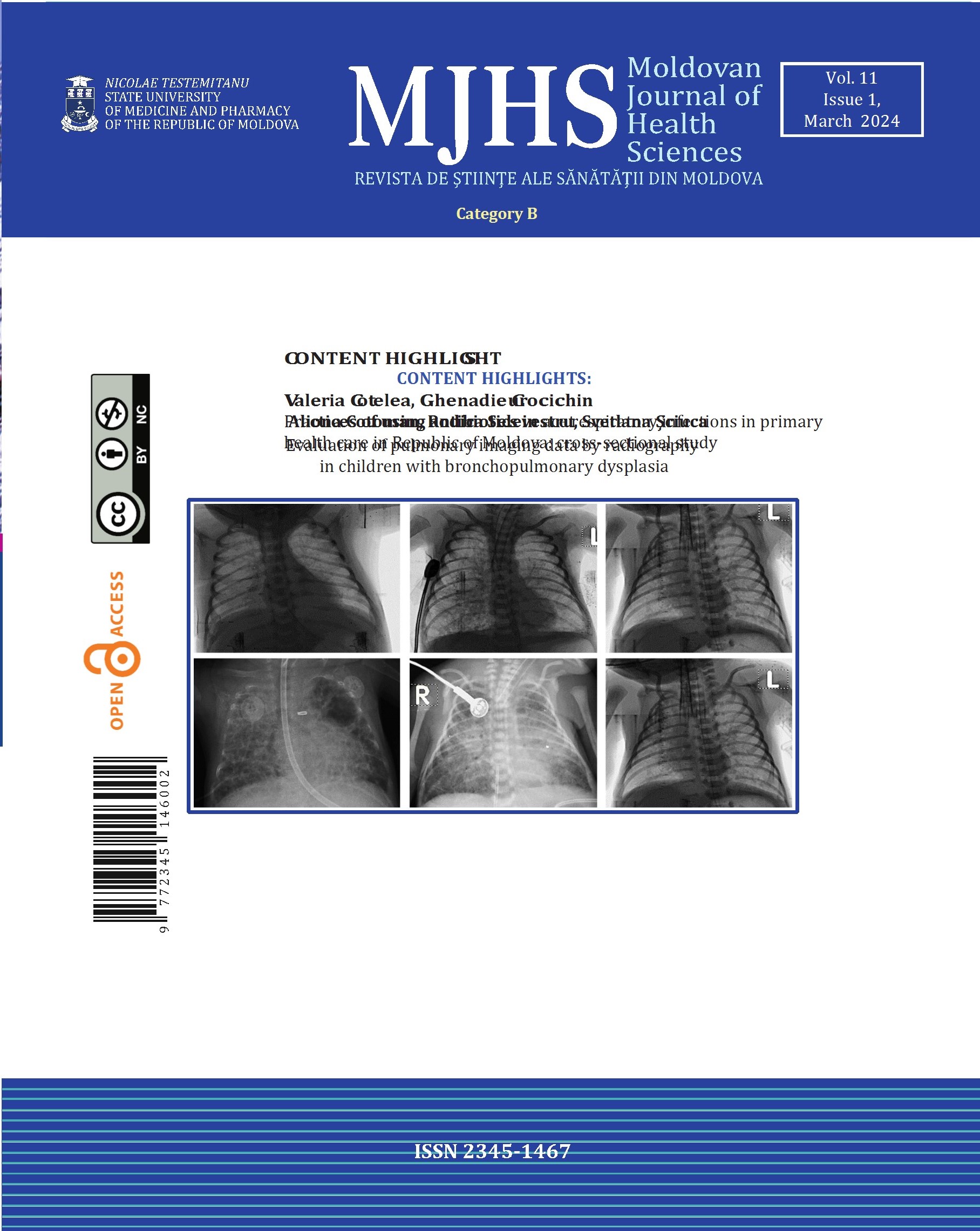
Evaluation of pulmonary imaging data by radiography in children with bronchopulmonary dysplasia

Aliona Cotoman1*, Rodica Selevestru2, Svetlana Şciuca2
https://doi.org/10.52645/MJHS.2024.1.05

Introduction

According to specialized literature, X-rays are also used in the diagnosis and suspicion of chronic broncho-pulmonary disease (bronchopulmonary dysplasia (BPD)), in premature children.

Material and methods

The study included 105 premature babies who were divided into the study group – premature babies with bronchopulmonary dysplasia (BPD) and the control group – premature babies without BPD. Data were statistically analyzed using Microsoft Excel, MedCalc, SPSS and Contingency Table Analysis as a method of evaluating the performance of a diagnostic test.

Results

Radiological changes of discoidal atelectasis type in subjects with BPD were detected in 49.1% in the basic group versus 13.5% in the control group (χ2 = 15.431; p < 0.0001), subsegmental ones 47.2% vs 25.0% (χ2 = 5.586; p = 0.018); pulmonary emphysema areas 62.3% vs 5.8% (χ2 = 37.182; p < 0.0001), opaque fibrosis sectors 50.9% vs 11.5% (χ2 = 18.911; p < 0.0001); signs of pulmonary hypertransparency 47.2% vs 1.9% (χ2 = 28.843; p < 0.0001); microcystic formations 41.5% vs 5.8% (χ2 = 18.482; p < 0.0001). Depending on the severity degrees of BPD, changes such as discoidal atelectasis were noted, in those with mild grade – 50% medium grade – 38.5%, severe grade – 56.3% (χ2 = 16.502; p < 0.001), subsegmental atelectasis 41.7%, vs 38.5% (χ2 = 7.956; p = 0.047), pulmonary emphysema areas 58.3%, vs 38.5%, vs 87.5% (χ2 = 45.138; p < 0.0001); microcystic formations, 33.3%, vs 53.8%, vs 43.8% (χ2 = 20.502; p < 0.0001).

Conclusions

The changes recorded on radiological examination in mild, moderate, and severe cases of BPD in premature babies are of the type discoidal atelectasis, areas of emphysema, opaque sectors of fibrosis, pulmonary hypertransparency, microcystic formations.

View Article

Visual field evaluation following brain injury in school-aged children

Victoria Verejan*, Eugeniu Bendelic
https://doi.org/10.52645/MJHS.2024.1.06

Introduction

Brain injury may affect both afferent and efferent visual pathways. In children it is quite difficult to determine visual disturbances since they are often non cooperative. Visual field examination is an objective evaluation method that can outline visual pathway alteration in the acute period of head trauma.

Materials and methods

Forty-eight patients with persisting visual symptoms after mild traumatic brain injury were examined. A control group of the same size has been evaluated.

Results

Patients in the research group showed an obvious alteration of the fixation capacity of more than 20% in 91.7%-95.8%, while in the control group the fixation capacity was up to 20% in 68.7%-70.8%. The ability to fix false positive points was up to 20% in 43.8%-45.8% patients in the research group and 70.8%-83.3% in the control group. The rate to fix false negative points was within the range of up to 20% for the research group in 93.7%-95.8% and the control group 91.7%-97.9%. The index of localized defects was up to 3dB in 62.5%-70.8% in the research group and predominantly 91.6%-95.8% for the control group. The average elevation index was within the range of < -3dB, 3dB> in 12.5%-20.8% research group and respectively 54.1%-56.2% control group. The graphic interpretation of changes in the visual field revealed a prevalence of the incidence of diffuse retinal depression with relative paracentral scotomas in 64.6%-68.7%.

Conclusions

Based on the results, we can conclude that perimetric examination in the case of brain injured pediatric patients fulfils the requirements of credibility. Perimetric examination could be a landmark in the initial phase of settling post brain injured visual disturbances.

View Article

Diagnosis of maxillary compression syndrome

Sabina Calfa*, Valentina Trifan, Iulian Storojov, Ana-Maria Șeptelici
https://doi.org/10.52645/MJHS.2024.1.07

Introduction

Upper maxillary compression syndrome is characterized by a deficit in transversal development and is recognized within the classification system of the German school. Regardless of the schools’ affiliation according to which this malocclusion is classified, the objective is to determine and select the treatment method with maximum efficiency. Thus, the compression of the upper jaw can also be included as a component in class II subdivision 1 malocclusions, presenting in 2 clinical forms. In addition, the types of palatal suture and their impact on the development of the maxilla in the transverse plane were identified. The purpose of the study was to assess the importance of cone beam computed tomography (CBCT) in providing a comprehensive diagnosis of this malocclusion and formulating an elaborate treatment plan.

Material and methods

After applying the inclusion and exclusion criteria, 165 patients were enrolled in the study. The research included patients with jaw compression syndrome diagnosed orthodontically during the mixed and permanent dentition periods. The patients were divided into 3 groups according to the stages of formation of the medio-palatine suture, correlating with their biological age. The research sample was calculated using ANOVA program: fixed effects, omnibus, one-way Analysis.

Results

Determining the shape and degree of formation of the median palatal suture at the ages studied in the research, favors the selection of modality, type, and speed of expansion. These factors are directly related to the stage of formation of the palatal suture, which may or may not coincide with the patient’s biological age. CBCT is the method of choice for assessing this. Furthermore, the range of movement in millimeters that can be achieved after separating the upper jaw can be determined, regardless of the type of expansion.

Conclusions

Based on the analysis of the data, we can appreciate the variety of expansion methods depending on the degree of formation of the medio-palatine suture. Through a comprehensive paraclinical examination and accurate interpretation, we can establish the definitive diagnosis of this clinical entity and create a treatment plan that minimizes the chance of recurrence.

View Article

Review

Surgical approaches in pituitary neuroendocrine tumors

Dan Croitoru1,2*, Victor Andronachi2, Sergiu Vișnevschi1, Ana-Maria Dumitrașco2
https://doi.org/10.52645/MJHS.2024.1.08

Introduction

Pituitary neuroendocrine tumors account for 3.9-7.4/100,000 of central nervous system tumors in the Western world. They are particularly noteworthy, comprising 10-15% of all cases, with a higher prevalence in the 75-79 age group. In the Republic of Moldova, these tumors account for 34% of cases in postmortem examinations while remain an actual theme of discussion in the ENDO WHO congress and are regarded as a factor, which may influence the quality of life (QOL).

Material and methods

We have critically revised 66 literary sources, which were selected using the PubMed library after introducing the keywords “pituitary adenoma surgical approach”.

Results

The main surgical approaches were the transsphenoidal (transnasal, sublabial and endonasal) and transcranial (subfrontal unilateral/bilateral, fronto-lateral, fronto-temporal and median basilar) while the additional surgical approaches were designed for complicated and unusual pituitary neuroendocrine tumors and included combined versions, multiple surgeries or extended approaches. Numerous factors were influential for the selection of a surgical approach concerning the pituitary neuroendocrine tumors. They are not sensible for a type of pituitary neuroendocrine tumor according to the WHO classification while the size of a tumor may dictate its surgical approach.

Conclusion

Each surgical intervention requires a personalized approach and the critical thinking of the surgical team but most of them can be systematically considered before confronting the tumor in an intraoperative environment because most of the preoperative investigations are proven unreliable. There is no established superior surgical approach for each surgical intervention.

View Article

Oral lichen planus – an oral potentially malignant disorder (OPMD) of the oral cavity

Irina Ivasiuc1*, Eugen Melnic2, Daniela-Elena Costea3,4, Diana Uncuța1
https://doi.org/10.52645/MJHS.2024.1.09

Introduction

Oral lichen planus is a chronic inflammatory disease of unknown etiology, characterized by recurrent lesions, presenting as reticular lesions, sometimes accompanied by atrophic, erosive, and/or ulcerative areas. Despite being one of the most common conditions affecting the oral mucosa, oral lichen planus remains an ailment with undefined etiology and unclear pathogenesis, imprecise management, and uncertain premalignant potential.

Materials and methods

A narrative literature review study was conducted. A bibliographic search was carried out in databases such as PubMed, Hinari, SpringerLink, the National Center of Biotechnology Information, and Medline. Articles published from 1990 to 2023 were selected using various combinations of keywords: “oral lichen planus,” and “epidemiology,” “etiology,” “pathogenesis,” “symptoms,” “management,” “histopathology,” and “malignant transformation.” After processing the data from these databases, 475 full articles were found. The final bibliography comprised 50 relevant sources, considered representative of the materials published on the topic of this synthesis article.

Results

Oral lichen planus is an inflammatory condition associated with T-cell-mediated immune dysfunction. Triggers include autoimmune responses to local antigens, microorganisms, and stress. The disease results from a complex interplay of host factors, lifestyle, and environmental factors leading to T-cell-mediated immune dysregulation. Diagnosis of oral lichen planus is based on clinical features (multiple, bilateral, symmetrically distributed lesions, occurring most commonly on the buccal mucosa, dorsal tongue surface, and gingiva), histopathological findings (predominantly lymphocytic band-like infiltrate in the lamina propria, presence of apoptotic cells in the basal cell layer, absence of epithelial dysplasia), and immune-related changes (deposition of fibrinogen along the basement membrane zone, presence of granular fluorescent deposits containing IgA, IgG, and IgM in colloid bodies).

Conclusions

Oral lichen planus is a chronic inflammatory condition mediated by T-cells in response to various extrinsic antigens, modified autoantigens, or superantigens, with periods of remission and relapse and the potential for malignant transformation. The etiology and pathogenesis of this condition are complex, diagnosis relies on clinical features, histopathological findings, and immunological data, patient treatment is symptomatic, and the potential for malignant transformation varies. Nevertheless, prospective studies with large sample sizes, adequate treatment duration, and long-term follow-up are needed.

View Article

Case study

Obstetrical anesthesia for a patient with multiple sclerosis: case report and literature review

Svetlana Plămădeală1,2, Dmitri Coloman2, Roman Ciubara1,2, Natalia Belîi1,2,3*
https://doi.org/10.52645/MJHS.2024.1.10

Introduction

Multiple sclerosis is an autoimmune disease characterized by chronic inflammation with progressive demyelination and axonal dysfunction. The disease affects about 1 million young adults, 2/3 of which are women of childbearing age, with all patients developing irreversible neurological dysfunction. There is observed a stagnation of the disease during pregnancy, with the return of exacerbations in the postpartum period. Contemporary specialty textbooks neither confirm nor deny the safety of neuraxial anesthesia in patients with central nervous system diseases.

Clinical case

We present the clinical case of a 25-year-old nullipara pregnant (36 weeks and 6 days gestation age), known with relapsing-remitting multiple sclerosis and epilepsy. The woman has relapsing multiple sclerosis symptoms during the last 6 days, reason why is urgently consulted by the anesthesiologist for cesarean delivery.

Management and outcome

The article describes the technique of epidural anesthesia for the obstetrical patient with multiple sclerosis and the course of the perianesthetic evolution, including 1-year follow-up after cesarean section.

Discussions

With the aim of avoiding potential influences on the evolution and progression of the disease, clinical judgment and the choice of anesthetic technique (general vs. neuraxial) depends on several factors: vaginal delivery or caesarean section, the presence of contextual clinical modifiers (native or drug-induced coagulopathy, infection), the urgency of the intervention, and the patient’s cooperation. In case of parturients with multiple sclerosis, all the risks should be rigorously evaluated: on one hand - the additional risk of general anesthesia (risk of aspiration, potential loss of airway control, critical desaturations) and on the other hand - the risk of hypothetical local anesthetic toxicity in the case of neuraxial techniques.

Conclusion

Neuraxial epidural anesthesia is a safe technique in obstetric patients with multiple sclerosis.

View Article

Single visit indirect pulp capping with Biodentine: clinical case report

Diana Trifan*, Diana Uncuța
https://doi.org/10.52645/MJHS.2024.1.11

Introduction

Dental caries is the most widespread dental disease worldwide; it affects population of different ages. If not treated promptly or addressed improperly, dental caries progresses in a deep cavity, with potential signs of pulp inflammation. The minimal invasive principles of treatment of deep carious lesions provide guidelines for the preservation of the dental pulp, techniques known as vital pulp therapies. Among these techniques, indirect pulp capping is a method that has shown high clinical rates of success over time, if performed properly. The applied bioactive dental material plays an important role. The aim of this clinical case report is to describe a clinical case of single-visit indirect pulp capping with Biodentine, along with the algorithm of diagnosis of the pulp health status and treatment used in deep caries lesion in a permanent tooth.

Materials and methods

Description of a clinical case of a permanent upper molar with a deep carious lesion, treated by single-visit indirect pulp capping with Biodentine. Clinical and radiological methods of investigations were used; the patient was assessed after 3 and 6 months after the applied treatment.

Results

The main complaint of sensitivity to sweet stimuli presented by the patient attenuated shortly after receiving the treatment. After 3 and 6 months, the tooth is asymptomatic; the clinical and radiological findings show no evidence of pulp inflammation.

Conclusions

Biodentine showed successful results when used as a bioactive dental material for indirect pulp capping.

View Article

Anniversary