Introduction
After the first record of muscle contraction in 1890 by Marey, electromyography has known a huge development being constantly upgraded and having its applications in different medical pathologies like temporomandibular disorders, dystonia, lesions of cranial nerves, sports medicine etc. [1-3]. Surface electromyography (sEMG) is often used due to the lack of tissue damage, ease of use and it provides often similar data to the needle EMG [4, 5]. Despite its extensive use, recording is influenced by many factors such as electrode type and positioning, fat tissue thickness, sex, age etc. [6, 7]. Different researches have shown that elder patients have less muscle activity than younger ones, which can be explained by the possible decrease of motor units [8]. Dentists often use investigations like x-ray, MRI, functional analysis with standard parameters to assess the skeletal or functional symmetry in patients. In this perspective, asymmetrical work of masticatory muscles can lead to different complications both in dental structures, temporomandibular joint (TMJ) or even muscles themselves. In most cases, the electromyographic activity is assessed as mean values of muscle activity in a time span. However, this data is hard to compare between different groups due to numerous variables that influences the data [2, 5]. EMG activity of masticatory muscles in dentistry is mainly influenced by the occlusal changes that dentists perform. There are many articles that analyze the change in EMG activity in numerous dental procedures like implant-supported dentures, complete dentures, orthodontic treatments etc. [9-12]. In order to be able to use this data for comparison, a group of healthy subjects is required that would have also similar age, ethnicity, gender as the study group.
Material and methods
The study was based on analysis of electromyographic activity of masticatory muscles in 33 dentate subjects (21 women and 12 men) aged between 43-67 years old (mean/SD – 54±1.26 years). Patients were included in the study from three dental clinics based on the following criteria:
Lack of TMJ or masticatory muscle pathology.
Lack of fix dental or implant prostheses that exceed more than 2 teeth per arch.
Edentulous spans that don’t exceed 1 tooth on the same side of dental arch.
Lack of heart pacemakers or any electronic device that may interfere with the muscle signal.
Patients that have signed the informed consent.
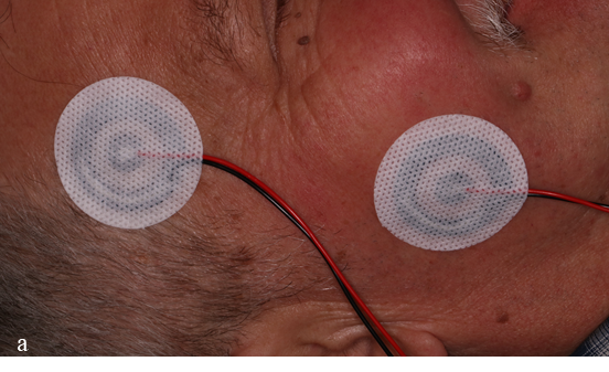
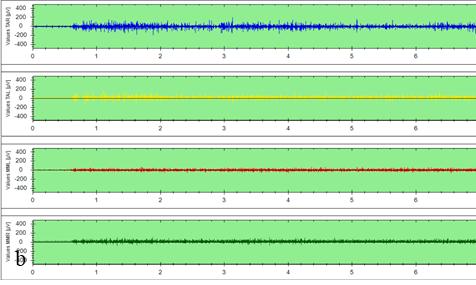
The study was conducted according to the criteria of Helsinki Declaration and the protocol was approved by the Ethic Committee of N. Testemițanu State University of Medicine and Pharmacy on 16th of March 2018, no. 43. Taking into account the age of patients there were no patients completely without dental procedures performed in the past. From overall number of subjects 12 had single unit edentulous spans on one or both arches, 9 had implant supported restorations, 12 had fixed tooth supported restorations, 25 patients had associated tooth attrition with the dentin isles exposed in molars and premolars. Only patients with class I malocclusion were accepted and those with removable prostheses were not accepted in the study. After routine dental examination, patients were sited upright in the chair with the Frankfort line parallel to the ground. Muscles were identified through manual palpation, and the electrodes were positioned on the most prominent area of masseter and anterior temporal muscles (Figure 1a). Placement of bipolar electrodes may influence the amplitude of EMG records [13]. In this research, we have used the 4 channel electromyograph (ForEMG, Quatrotii, Italy) with concentric electrodes which are not such susceptible to positioning. The acquired data are available both as raw and as mean values in a time span (Figure 1 b, c). Patients were instructed to stay relaxed for first 3 seconds of registration then, to clench on their teeth for another 3 seconds. The values were related as percentage of initial values that were obtained during 3s clenching on cotton rolls as described previously by Ferrario [5]. Having the same time span, only the average values were compared. Besides the 4 basic values (TAL, TAR, MML, MMR) another 6 parameters were evaluated (Figure 1c): Percentage overlapping for temporalis (PocTA) and masseter (PocMM), Percentage overlapping for both masseter and temporalis (BAR), torque coefficient (Tors), percentage overlapping of right and left side muscles (Asym), muscular work related to vertical dimension of occlusion (IMPACT). The acquired data were introduced into RStudio software for descriptive statistical analysis. Shapiro-wilk test was applied for normality distribution, mean and median values were calculated.
|
|
|
Fig. 1. Evaluation of masticatory muscle EMG activity. a – placement of concentric electrodes, b – raw data of EMG activity, c – average values in a time span and percentage overlapping coefficients. |
Results
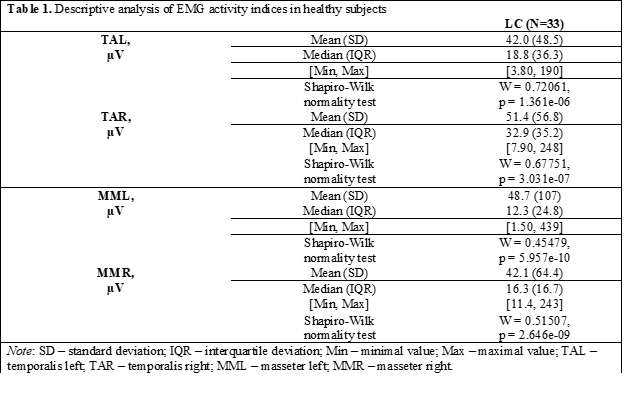
Statistical analysis of electromyographic indices has shown a wide distribution of values that can be explained by the small number of subjects included in the study. However, a wide range of parameters has been also reported in other studies [14]. Statistical data have been shown in Table 1. The average value for temporalis left (TAL) was 42 µV, median 18.8 with minimum of 3.8 and maximum of 190 µV. For temporalis right (TAR) the mean was 51.4 µV, median 32.9, minimal value 7.9 and maximum of 248 µV. Masseter left had a mean of 48.7 µV, median 12.3, minimal value of 1.5 and maximum 439 µV. Masseter right 42.1 µV, median 16.3, minimum value 11.4 and maximum 243 µV.
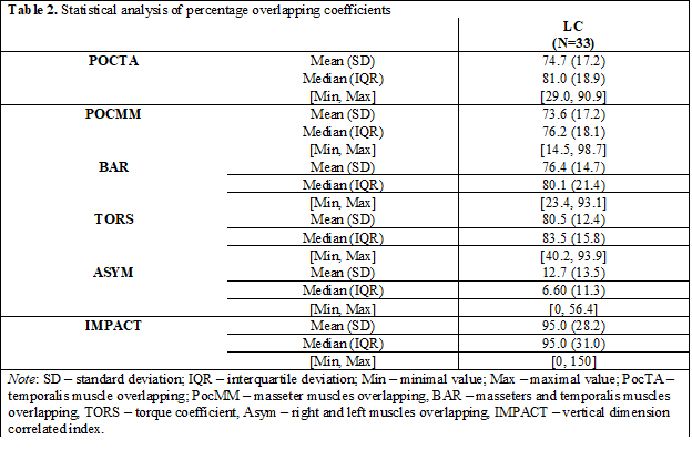
The obtained value can be also analyzed depending on their relation toward the normal range provided by the manufacturer for assessment of muscle symmetrical work. For this, another 6 parameters previously emphasized are available. They are formed depending on the interaction of first 4 main parameters of muscle activity. The result of statistical analysis is given in table 2.
The percentage overlapping coefficients have a range in which the value indicates a symmetrical function. For PocTa and PocMM it is between 83 and 100%, BAR and Tors coefficients have a normal range between 90 and 100% and Asym from -10 to 10% range.
Even though, the subjects in this study had minimal dental procedures, they did not fit perfectly in the normal range provided by the manufacturer. The value with highest deviation from the normal range was IMPACT. This might be due to wider range that this coefficient has (from 85 to 115%) or instability of vertical dimension of occlusion that is changed after full mouth rehabilitation procedures. The overall mean deviation percentage was 20.5% from normal parameters of the device.
Discussions
The obtained data were a part of broader study conducted in a PhD thesis that require healthy patients of middle age to be assessed and compared to the ones that had full mouth implant-supported rehabilitation. The electromyographic signal can vary during the same registration due to different factors that influences the muscle contraction. In order to provide reproducibility of acquired signals there are different protocols that aim to position the electrodes depending on different landmarks [13, 15]. In this case, the concentric electrodes allow their positioning without taking into account the interelectrode distance that can in some cases lead to „crosstalk” signals from other muscles. Analysis of percentage overlapping coefficients allows a better understanding of muscle interaction during dental rehabilitation procedures with the modification of occlusal surfaces [5].
The acquired data has shown that even in cases were dental procedures were minimum; there is a deviation from the normal range in the percentage overlapping coefficients of 20.5%.
The wide range of value distribution inside the sample group shows that surface electromyography is hardly comparable due to high individuality of evaluated parameters. It cannot be said that these parameters are high or low in accordance with the data from the literature because there are various devices available in the market with different electrode types that can provide non-comparable data and different reference values. The registration method is technique sensitive and depends also on the positioning of the electrodes, tissue thickness, age, sex, anatomical peculiarities etc.
The middle-aged patients are hard to be found with most of the teeth sound. Most of the patients already have dental treatment of different type and extent that can disturb the acquired data. Additional to that, the low awareness of Moldovan people towards the oral health leads to early tooth loss, which minimize the number of healthy subjects for establishing normal reference values.
Conclusions
Surface electromyography has been widely used in dentistry to validate the dental treatment and try to create the most suitable restorations that would integrate into stomatognathic system. However, there is a big number of variables that can influence the acquired data among different population. Thus, in order to say if a treatment is valid or interfering with the muscular system, a reference group of the same age, sex and ethnicity is required.
Competing interests
None declared
Authors’ contributions
AM – Conception and design of study; MM – Acquisition of data and manuscript drafting; NC – Analysis and interpretation of data; OS – Revising the manuscript critically for important intellectual content.
Authors’ ORCID IDs
Mihail Mostovei - https://orcid.org/0000-0002-8112-4798
Oleg Solomon https://orcid.org/0000-0002-7341-1711
References
- Nishi S. E., Basri R., Alam M. K. Uses of electromyography in dentistry: An overview with meta-analysis. European Journal of Dentistry, 2016; 10(03): 419–425.
- Klasser G. D., Okeson J. P. The clinical usefulness of surface electromyography in the diagnosis and treatment of temporomandibular disorders. The Journal of the American Dental Association, 2006; 137(6): 763–771.
- Raez, M. B., Hussain, M. S., Mohd-Yasin, F. Techniques of EMG signal analysis: detection, processing, classification and applications. Biological procedures online, 2006; 8, 11–35.
- Belser U. C., Hannam A. G. The contribution of the deep fibers of the masseter muscle to selected tooth-clenching and chewing tasks. The Journal of Prosthetic Dentistry, 1986; 56(5): 629–635.
- Ferrario V. F., Sforza C., Colombo A., Ciusa, V. An electromyographic investigation of masticatory muscles symmetry in normo-occlusion subjects. Journal of Oral Rehabilitation, 2000; 27(1): 33–40.
- Suvinen T.I., Kemppainen P. Review of clinical EMG studies related to muscle and occlusal factors in healthy and TMD subjects. Journal of Oral Rehabilitation. 2007; 34(9):631-44.
- Suvinen TI, Malmberg J, Forster C, Kemppainen P. Postural and dynamic masseter and anterior temporalis muscle repeatability in serial assessments. Journal of Oral Rehabilitation, 2009; 36(11):814-20.
- Jensen R., Fuglsang-Frederiksen A. Quantitative surface EMG of pericranial muscles. Relation to age and sex in a general population. Electroencephalography and Clinical Neurophysiology/Evoked Potentials Section, 1994; 93(3): 175–183.
- Nishi S. E., Rahman N. A., Basri R., Alam M. K., Noor N., Zainal S. A., Husein A. Surface Electromyography (sEMG) Activity of Masticatory Muscle (Masseter and Temporalis) with Three Different Types of Orthodontic Bracket. BioMed research international, 2021; 6642254.
- Hamada T., Kotani H., Kawazoe Y., Yamada S. Effect of occlusal splints on the EMG activity of masseter and temporal muscles in bruxism with clinical symptoms. Journal of Oral Rehabilitation, 1982; 9: 119-23.
- Szyszka-Sommerfeld L., Machoy M., Lipski M., Woźniak K., The Diagnostic Value of Electromyography in Identifying Patients With Pain-Related Temporomandibular Disorders. Frontiers in Neurology, 2019; 10, 180p.
- Sonego M., Goiato M., Santos D. Electromyography evaluation of masseter and temporalis, bite force, and quality of life in elderly patients during the adaptation of mandibular implant-supported overdentures. Clinical Oral Implants Research, 2017; 28(10):e169-e174.
- Sabaneeff, A., Caldas L. D., et al. Proposal of surface electromyography signal acquisition protocols for masseter and temporalis muscles. Research on Biomedical Engineering, 2017; 33(4): 324–330.
- Gracht I., Derks A., Haselhuhn K., Wolfart S. EMG correlations of edentulous patients with implant overdentures and fixed dental prostheses compared to conventional complete dentures and dentates: A systematic review and meta-analysis. Clinical Oral Implants Research, 2016; 28(7): 765-773.
- Hermens HJ., Freriks B., Disselhorst-Klug C., Rau G. Development of recommendations for SEMG sensors and sensor placement procedures. Journal of Electromyography and Kinesiology, 2000; 10(5): 361-74.
