MJHS
Navigation
Journal Navigation
Article Navigation
opened article
Volume 10, Issue 3
September 2023
Cover Image

Article contents

opened journal
Volume 10, Issue 3
September 2023
Cover Image

Abstract

Introduction

Acute appendicitis is among the three most frequent surgical diseases. The lifetime likelihood of developing acute appendicitis is around 7%. The incidence of acute appendicitis reduces with age after adolescence. Several studies evaluated the relevance of the current scores to the general population, mostly children, but a limited number of studies have studied the elderly population. This study aims to assess the clinical effectiveness of the new diagnostic score for the elderly population in comparison to both the Alvarado score and the non-standardized score.

Materials and methods

In order to evaluate the effectiveness of the diagnostic score of acute appendicitis, we examined 78 patients who were admitted to emergency unit of the Saint Archangel Michael Municipal Clinical Hospital during 2018-2021 with the presumptive diagnosis of acute appendicitis. Of all patients admitted, Acute Appendicitis was confirmed on pathological examination in 22 (28.2%) patients. The average age of patients was 73.5±13.5 years (minimum - 60 years, maximum - 87 years). The ratio of males to females was 1:1.6.

Results

Comparative evaluation of the new diagnostic score of acute appendicitis and the non-standardized clinical-echographic examination for acute appendicitis diagnosis showed better performance indicators of the new diagnostic score of acute appendicitis compared to the non-standardized clinical method for acute appendicitis diagnosis. The high sensitivity of the new diagnostic score of acute appendicitis was statistically demonstrated (ƛ2 = 4.32; p < 0.05), a lower rate of missed acute appendicitis cases in the new diagnostic score of acute appendicitis (ƛ2 = 4.32; p < 0.05), the „grey area” is lower in the new diagnostic score of acute appendicitis (ƛ2 = 5.28; p < 0.05), than in the non-standardized diagnosis. It was shown to have a lower rate of acute appendicitis cases in the „grey area” of the total number of acute appendicitis cases (ƛ2 = 4.9; p < 0.05). Benchmarking indicators such as specificity and diagnostic accuracy showed no statistically significant differences. At the same time, a definite increase in specificity and accuracy was observed for the new diagnostic score of acute appendicitis compared to non-standardized clinical diagnosis.

Conclusions

Diagnosing acute appendicitis in elderly patients remains challenging due to the numerous potential diagnoses with similar clinical manifestations that are observed in this patient population. It was necessary to utilize clinical risk-scoring systems that could aid in the prompt identification of patients with acute appendicitis. This study concludes that the New diagnostic score has higher clinical efficiency in diagnosing acute appendicitis in elderly patients. It has a sensitivity of up to 93.15%, compared to the unstandardized clinical method and the Alvarado diagnostic score, and is independent of „risk factors” such as obesity and atypical vermiform appendix localization.

Key Messages

What is not yet known about the issue addressed in the submitted manuscript

The epidemiology and outcomes of acute appendicitis in elderly patients are very different from the

younger population. Elderly patients with acute appendicitis have higher mortality, higher perforation rate, lower diagnostic accuracy, longer delay from onset and manifestation of symptoms, higher rate of postoperative complications, and a higher risk of colon and appendix cancer. Therefore, it is necessary to develop a new diagnostic and remedial approach to treating acute appendicitis in elderly patients.

The research hypothesis

We aim to develop a new diagnostic score for elderly patients and apply it in parallel with the non-standardized diagnosis.

The novelty added by the manuscript to the already published scientific literature

The clinical efficacy of the new score was evaluated in comparison with the Alvarado and a non-standardized diagnostic score.

Introduction 

Acute appendicitis (AA) is among the three most commonly occurring acute surgical diseases. The likelihood of experiencing AA in a lifetime is approximately 7%. The incidence of AA reduces with age after adolescence [1]. Approximately 15% of patients over 60 years of age who present with acute abdominal pain to the Emergency Department receive a final diagnosis of acute appendicitis, which is half as common as in younger patients [2]. Nevertheless, the epidemiology and outcomes of acute appendicitis in elderly patients differ significantly from those of the younger population. First, despite the decrease in incidence, acute appendicitis in elderly patients is marked by significantly higher mortality, which is 8% in the category of patients over 60 years of age, compared to less than 1% among younger patients. A large observational study of 164,579 patients with acute appendicitis, age greater than 60 years was a significant risk factor for mortality by multivariate analysis [3].

All the data suggest that older patients are more likely to have complicated appendicitis with perforation or abscessing compared with other age groups. The rate of complicated appendicitis ranges from 18% to 70% [2, 4-6] (compared to a rate of 3 % and 29% among patients under 60 years). The reason for this major risk of perforation could be the vascular sclerosis that the vermiform appendix develops in elderly patients and the narrowing of the lumen by the phenomenon of fibrosis. In these patients, the muscle layers are infiltrated with fat, so having a fragile structure they have a tendency toward early perforation [7]. These findings together with the delay in diagnosis and treatment could explain a more aggressive evolution of the disease in this population category.

Another finding among the elderly population, who develop acute appendicitis, is the lower rate of correct preoperative diagnosis compared to the younger population [8], with a reported diagnostic accuracy (defined as the percentage of appendices removed with a histological diagnosis of acute appendicitis out of the total number of appendectomies performed) of 64% in patients over 60 years of age versus 78% in other age groups [9]. Moreover, in the vast majority of included studies, the mean time from symptom onset to admission was longer in older patients than in younger patients [9-11].

Focusing on appendectomy, compared with young patients, elderly patients are burdened with higher postoperative mortality, higher postoperative morbidity [12], longer length of hospital stay [13], lower rate of laparoscopic appendectomy, and a higher risk of being subjected to high-throughput investigations [14-15].

In a large Swedish study that included more than 117,000 patients, the mortality rate after appendectomy was strongly influenced by age, with a threefold increase for each decade of age, reaching more than 16% in nonagenarians. Finally, the complication rate in elderly patients with negative appendectomy was significantly higher than in younger patients (25% vs 3%) [16]. 

This raises the question of whether existing clinical scoring systems have sufficient diagnostic accuracy for the diagnosis of acute appendicitis in elderly patients?

The Alvarado score is the most widely studied. Its validity in adult and pediatric patients was summarized in a recent meta-analysis [17] that included 5960 patients in 29 studies. According to Ohle et al., the performance of the score depends on the cutoff value: a clinical cutoff score of < 5 can be applied to „exclude” appendicitis with a sensitivity of 99% (95% CI 97–99%) and a specificity of 43% (36–51%).

According to the Jerusalem guidelines [18], in adult patients the Alvarado score (with a cut-off score < 5) is sensitive enough to exclude acute appendicitis, but is not specific enough in the diagnosis of acute appendicitis.

However, the Alvarado score was developed based on the pattern of presentation of clinical and laboratory variables of a young population (mean age 23.4 - 25.9) [19]. Considering that the complication rate in elderly patients with negative appendectomy is significantly higher than in younger patients (25% vs 3%, p < 0.05) [20], the preoperative diagnosis in these patients must be as accurate as possible.

Although computer tomography (CT) with intravenous (IV) contrast is associated with lower rates of negative appendectomy [21]. Ultrasound  (US) is clearly inferior to CT in sensitivity and negative predictive value for appendicitis, however,  it may be equally useful for excluding appendicitis [22, 23], while CT is especially useful if the appendix is ​​not visualized by US.

The New diagnostic score (DS), which we aimed to compare with the Alvarado score, is a diagnostic score that includes 10 parameters: the positive Kocher symptom- 1 point; positive Blumberg symptom in the right iliac region - 2 points; positive Bartomier-Michelson symptom - 1 point; the presence of nausea and/or vomiting - 1 point; leukocytosis in Complete Blood Count (CBC) 10 x 109/l and more - 1 point; ultrasound determination of Vermiform Appendix (VA) with a diameter greater than 7 mm is estimated at 2 points; VA incompressibility - 1 point; thickening of the peri-appendiceal tissue - 1 point; coprolite in the VA lumen - 1 point; the presence of ultrasound signs of another acute non-appendiceal pathology of the abdominal cavity and/or ultrasound detection of a compressible VA less than 7 mm in diameter - „minus” 3 points.

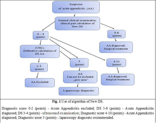
In this scoring system, the total score varies between -3 and 10 points. When obtaining a score below 2 points, the diagnosis of AA is excluded. If adding up the points of the positive clinical and laboratory criteria of AA, a result of 6-7 points will be obtained, and then the diagnosis of AA will be established. In this case, an additional ultrasound will not be necessary, because even the identification of another acute pathology with / or without signs of inflammation of vermiform appendix on UST („minus” 3 points), will not affect the result and the interpretation of the New DS application algorithm, because the final score will be 3 or more points, which definitely indicates that the patient has AA. The patient diagnosed with AA will later undergo urgent surgical treatment.

If the sum of the points of the clinical and laboratory criteria of the New DS will be less than 4 points, an ultrasound of the abdominal cavity will be performed with the additional inclusion of ultrasound signs of AA. If following a general ultrasound examination, the sum of AA points will constitute < 2 points, the diagnosis of AA will be excluded.

When following the general ultrasound evaluation of signs of AA, the number of points will be 3 or more, the diagnosis of AA will be very likely and appendectomy will be indicated.

Material and methods 

There were prospectively analyzed 78 cases (patients), who were admitted to the Emergency Department of Saint Archangel Michael Municipal Clinical Hospital in 2018-2022 with the diagnosis of acute appendicitis (AA). Of all hospitalized patients, AA was confirmed on histological examination in 22 (28.2%) patients.

The average age of patients was 73.5±13.5 years (minimum - 60 years, maximum - 87 years). The ratio of males to females was 1:1.6. Demographic data of patients, including age, sex, duration of hospitalization, and histopathological reports of appendectomy materials were recorded. Analyzing the obtained data, we note that the structure of distribution by sex and age in this group of patients is comparable to that in the group of patients in which the new DS was developed.

The time from the debut of complaints of abdominal pain to hospitalization was: in 9 (11.5%) people - less than 6 hours, from 6 hours to 24 hours - in 28 (35.9%) patients, from 24 hours to 48 hours - in 22 (28.3%) patients, more than 48 hours - in 19 (24.3%) patients.

After data processing, the patients admitted to the study group had the concomitant pathologies noted in table 1.

Table 1. Patient demographic data and characteristics


Associated medical conditions

%

1.

Hypertension

37

47.4

2.

Coronary heart disease

20

25.6

3.

Diabetes

11

14.1

4.

Obesity

5

6.4

5.

Dyscirculatory encephalopathy 

14

17.9

6.

Urolithiasis

7

8.9

7.

Chronic duodenal ulcer

4

5.1

8.

Chronic gynecological pathologies without exacerbation

Uterine myoma

3

3.8

Uterovaginal prolapse 

2

2.6

Pelvic inflammatory disease

2

2.6

Statistical analysis

The SPSS 18 software (SPSS Inc., Chicago, IL, USA) was used for statistical analysis. Numerical data were presented as mean ± standard deviation. The one-sample Kolmogorov–Smirnov test was used to assess the distribution of numerical data. The independent sample t-test was used when the distribution was normal and the Mann-Whitney U test was used for the non-normal distribution. A chi-square test was used to compare between groups. Values ​​with a P value < 0.05 were considered to be statistically significant.

Results

New DS in AA implementation results

In this group of patients, the non-standardized clinical and ultrasonographic diagnosis of AA was used as the main diagnostic method, which was performed by the doctor on call, based on professional knowledge and skills, in the absence of a mandatory research standard and an algorithm for interpreting the obtained data, which is largely subjective. In the Emergency department, the non-standardized diagnosis of AA was made based on clinical, laboratory, and ultrasonographic investigations. The method of diagnosis and the clinical-therapeutic tactic used for all groups of patients were methodologically similar to this diagnostic method (New DS) (Table 2), and its algorithm, which corresponds to all the training principles of the diagnostic algorithm (Fig. 1).

Table 2. The New Diagnostic Score. 

No.

Criterion

Assessment

Score

1.

Kocher symptom

Positive

1

2.

Nausea /vomiting

Present

1

3.

Blumberg symptom in right iliac region

Positive

1

4.

Bartomier-Michelson symptom

Positive

1

5.

Leukocytosis

>10 × 109/l

1

6.

Ultrasound: VA unchanged and /or other pathology

Determined

-3

7.

Ultrasound: increased VA diameter > 7mm

Determined

2

8.

Ultrasound: thickening periappendicular tissue

Determined

1

9.

Ultrasound: VA Incompressibility

Determined

1

10.

Ultrasound: coprolite in VA lumen

Determined

1


Total

Max

Min

10

-3

Note: VA - vermiform appendix; the total score is a sum of criteria points. Minimal Score (-3), maximum (10).


All patients were examined using the same diagnostic equipment. The non-standardized clinical and ultrasonographic method included the use of a general clinical examination, laboratory investigations (CBC, urinalysis), ultrasonography (USG) of the abdominal cavity.

The diagnosis and management of AA patients were specified directly by the on-call surgeon. The general clinical examination was performed in all 78 (100%) patients and consisted of collecting anamnesis, and patient complaints to determine the symptoms of AA with their subsequent interpretation.

Patients in the study group presented the following complaints (Table 3).

Table 3. Diagnostic criteria of patients

No.

Diagnostic criteria

Patients

No.

%

1

Abdominal pain

78

100

2

Nausea

30

38.5

3

Vomiting

41

52.5

4

Kocher's symptoms

32

41.0

5

Gynecological anamnesis 

7

9

6

Intestinal disorders (constipation)

64

82.9

7

Dysuria 

37

47.4

8

Local tenderness (pain) (on palpation in the right iliac region)

78

100

9

Blumberg's symptom in the right iliac region

78

100

10

Bartomier-Michelson's symptom

56

71.8

11

Rovzing's symptom

44

56.4

12

Sitkovsky's symptom

54

69.2

13

Obraztsov's symptom

3

3.8

14

Coupe's symptom

2

2.5

15

Voscresenscky symptom

2

2.5

16

Hyperthermia >37.4oC

65

83.3

17

Tenderness on palpation of the anterior rectal wall

3

3.8

18

Leukocytosis >10×109/l

78

100

19

Deviation of the leukocyte formula >74%

78

100

20

Hematuria, leukocyturia

78

100

21

Free fluid in the abdominal cavity

78

100

22

US signs of unchanged VA or other pathologies of the right iliac region

43

55.1

Note:  US – ultrasound; VA - vermiform appendix.


Results of laboratory examinations of patients

In examined patients, the increase in the number of leukocytes in complete blood count (CBC) >10x109/l was detected in 70 (89.7%) patients. Left shift of (increased neutrophil ratio) more than 74% was found in 58 (74.3%) patients.

Left shift of (increased granulocytes ratio) more than > 6% was found in 47 (60.25%) examined patients. The absence of pathological changes (leukocyturia, hematuria, bacteriuria) in the urinalysis was observed in 44 (56.4%) of the examined patients. Leukocyturia/hematuria was found in 34 (43.5%) cases.

Results of ultrasound

The following ultrasonographic signs were recorded in the examined patients:

  • AV diameter increase > 7 mm was determined in 30 (38.4%) patients.

  • AV incompressibility during compression was observed in 28 (35.8%) patients.

  • Positive „Target” symptom was detected in 39 (50%) of the examined patients.

  • Coprolite in the VA lumen was detected in 5 (6.4%) patients.

  • Thickening of the peri-appendiceal tissue was detected in 16 (20.5%) of the examined patients.

  • Free liquid in the abdominal cavity was detected in 28 (35.8%) patients.

  • Increased blood flow in the VA wall during Doppler examination was observed in 17 (21.7%) patients.

In 23 (29.4%) of the examined patients, ultrasound signs of unchanged AV or other pathologies of the right lower quadrant of the abdomen were detected. The distinctive feature of establishing the diagnosis through a DS is that the surgeon has the possibility of interpreting the results of investigations and symptoms in three categories: positive, negative, and doubtful, which, in our opinion, largely depends on personal qualification and experience. Laboratory diagnosis consisted of CBC and urinalysis, which were performed in all patients included in the clinical trial. In 13 (16.6%) patients, additional biochemical blood analysis was performed (amylase level, urea, creatinine, serum protein level, and bilirubin). Blood glucose analysis was performed in 56 (71.7 %) patients.

Examination of the ultrasound signs of AA can confirm or deny the diagnosis, as well as exclude abdominal surgical pathologies of the gallbladder and pancreas, and some gynecological pathologies.

Overall radiography of the abdomen was performed in 19 (24.3%) patients. Additionally, a gynecologist consulted 12 (15.3%) patients.

 Following the examination, the patients were divided into three groups: the first group of patients, who underwent emergency surgery for AA; the second group of patients - who „accumulated” insufficient data to exclude or to confirm AA, and in our proposed algorithm for the implementation of the New DS was designated by us as a „grey area”, and the third group of patients - in which the diagnosis of AA was excluded.

 In the group of patients, in which AA was excluded, ulcer disease was diagnosed, chronic duodenal ulcer in exacerbation - 2 cases, urolithiasis, right renal colic - 1 case, acute pancreatitis - 2 cases, pelvic inflammatory disease - 2 cases, myxomatous node necrosis - 1, cr. Right ovarian cancer - 1 case, cancer. of cecum - 1, and functional bowel disorders - 3 (3.8%) cases.

 Patients who did not „accumulate” enough data to exclude and confirm AA, 3 (3.8%) were admitted to the hospital, where they were evaluated and monitored dynamically for 72 hours. In all these patients, the diagnosis of AA was excluded.

 Diagnostic laparoscopy was performed in 11 (14.1%) patients, of which 6 (5.1%) patients subsequently underwent laparoscopic appendectomy. From this group (laparoscopy + laparotomy) in 5 (6.4%) patients the diagnosis of AA was confirmed histologically. „Negative” appendectomy due to intraoperative overdiagnosis of AA was performed in one case. Based on the results of diagnostic laparoscopy, AA was excluded in 5 (6.4%) patients. The pathologies diagnosed by diagnostic laparoscopy were destructive acute appendicitis (AA) in 5 (6.4%) patients, simple acute appendicitis in 1 (1.2%) case, terminal ileitis in 1 patient, necrosis of the mammary nodule in 1 patient, ovarian cancer on the right in 2 patients, functional disorders of the intestine in 1 patient.

 Patients in whom the diagnosis of AA was established based on the results obtained from the non-standardized diagnosis, underwent emergency surgical treatment, and laparoscopic appendectomy. At the histopathological examination, the diagnosis of AA was confirmed in 39 (73.5%) of the number of patients initially operated on – 53 (100%) cases. 14 (26.8%) people were found to have undergone „negative” appendectomy. Of 59 (75.6%) patients who underwent appendectomy (initial or after diagnostic laparoscopy), the diagnosis was confirmed in 44 (74.5%) and non-destructive forms of AA were established in 15 (26.4%) patients.

Following the analysis of the „negative” appendectomy protocols, it was demonstrated that in 8 (53.3%) cases, the non-destructive form of VA inflammation was diagnosed by the surgeon intraoperatively, but the appendectomy was still performed due to the surgical approach in the already present right iliac region. In 7 (46.7%) cases, an intraoperative hyper-diagnosis of AA was found, but it was not histologically confirmed.

Overall, AA was histologically confirmed in 45 (57.6%) patients, and in 33 (42.4%) patients, this diagnosis was excluded. Typical VA localization was observed in 31 patients (68.8% of the total number of operated patients). The atypical location of the VA was observed in 14 (31.2%) patients.

New DS implementation results

In parallel with the non-standardized clinical-paraclinical diagnosis of AA, in the group of patients under study, an assessment based on certain criteria of AA symptoms was performed based on the New DS. According to the New DS, the following diagnostic criteria were recorded in the study group: positive Kocher symptom in 9 (11.5%) patients; nausea and/or vomiting in 41 (52.5%) patients; the positive Shchetkin-Blumberg symptom in the right iliac region in 25 (32%) patients; positive Bartomier-Michelson symptom in 17 (21.7%) patients; leukocytosis >10 x 109/l - in 39 (50%) patients.

 The ultrasound data obtained in the patients of this study group showed that the determination of signs of another pathology and/or VA without signs of inflammation was detected in 20 (25.6%) patients; volume increase of VA diameter greater than 7 mm in 21 (26.9%) patients; AV incompressibility was determined in 19 (24.3%) patients; coprolite in the VA lumen - in 4 (5.1%) patients; thickening of the peri-appendiceal tissue - in 16 (20.5%) patients.

Considering that the indications for surgical treatment were established based on New DS, appendectomy was performed in 30 (38.2%) patients from the study group. With the sum of New DS scores >3, surgical intervention was performed in 27 (90%) cases, and histologically AA was confirmed in 21 (70%) patients. In 7 (8.9%) cases from this group of patients, based on the New DS, the diagnosis of AA was excluded, respectively, surgical treatment was avoided. Subsequently, those patients no longer requested specialized medical help.

Patients who accumulated 2 points – 6 (7.6%) cases, were assigned to the „grey area” of the New DS, of which 3 patients underwent diagnostic laparoscopy and subsequent appendectomy through laparotomy in one case. AA was histologically confirmed in 1 patient. The others – 4 (57.2%) patients, avoided appendectomy, AA being excluded. None of the patients with excluded AA required further medical attention.

Out of 41 (52.7%) patients with total New DS results < 2, surgery was performed in 31 (75.6%) patients. Histological AA was confirmed in 5 (12%) patients. The other 10 (24.4%) patients were not operated on, AA being excluded.

When evaluating the effectiveness of the New Diagnostic Score, the following results were obtained: sensitivity - 94.1%, specificity - 73.8%, precision - 79.6%, the size of the „grey area” - 7.6%, the proportion of AA in the „grey area” of the total amount of AA - 4.3%, the proportion of undiagnosed AA cases - 5.9%.

Comparative evaluation of the effectiveness indicators of the New DS. The comparative analysis of the New DS and the non-standardized diagnosis demonstrated the superiority of the respective indicators and the effectiveness of the diagnostic score. The high sensitivity of the New Diagnostic Score was statistically demonstrated (ƛ2 = 4.32; p < 0.05), a lower rate of missed AA cases in New Diagnostic Score (ƛ2 = 4.32; p < 0.05), the „grey area” is smaller in New SD (ƛ2 = 5.28; p < 0.05) than by non-standardized diagnosis. A lower rate of AA cases in the „grey area” of the total AA cases was demonstrated (ƛ2 = 4.9; p < 0.05).

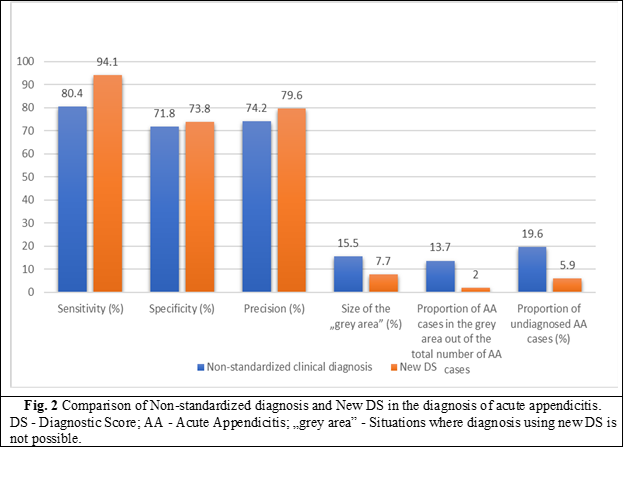
Based on these data, the comparative evaluation indicators such as specificity and diagnostic accuracy did not show significant statistical differences. At the same time, a definite increase in specificity and accuracy is noted in the case of New DS compared to the non-standardized clinical diagnosis.

Risk factors in the diagnosis of AA and evaluation of their impact on the effectiveness of the New DS

 In specialized literature, it is indicated that it is difficult to diagnose AA using the clinical method and unstandardized DS in female patients, at a young age, in patients with atypical VA localization, obesity, and in geriatric patients. This leads to false positive and false negative diagnoses of AA [24 - 27]. We consider these circumstances as risk factors for the clinical diagnosis of AA. Considering the fact that New DS is based on clinical data, we conclude that this criterion may affect its performance indicators.

We considered necessary to study the effectiveness of the New DS in the presence of the indicated risk factors. We evaluated the performance indicators of New DS in the subpopulation with risk factors - obesity, atypical location of VA.

Table 4.  Effectiveness of the New DS analysis according to risk factors

No.

Indicator of performance

General

Atypical location

of VA


р*

Obesity

BMI >25 kg/m2

р*

1

Sensitivity (%)

93.15

92.2

>0.05

93.8

>0.05

2

Specificity (%)

73.06

72.3

>0.05

68.3

>0.05

3

Precision (%)

78.8

77.8

>0.05

79.5

>0.05

4

The size of „the grey area” (%)

7.5

8.3

>0.05

5.5

>0.05

5

The proportion of ADA from the „grey area” out of the total number of AA (%)

2

0

>0.05

3.1

>0.05

6

The ratio of undiagnosed cases of AA (%)

5.84

0

>0.05

3.1

>0.05

Note: VA - vermiform appendix; BMI - body mass index; p - coefficient; ADA -acute destructive appendicitis; AA - acute appendicitis; „grey area” - Situations where diagnosis using new DS is not possible.

Thus, the use of the New Diagnostic Score for the selected subpopulations did not demonstrate statistically significant differences in performance indicators compared to the universal population. This fact indicates the possibility of the universal application of the New DS developed by us on the population of elderly and senile patients, excluding people with central nervous system injuries, as well as obese patients. The sensitivity of New DS in typical localization of VA was 94.1% and in the atypical one - 92.2%. Thus, the atypical location of the AV does not affect the sensitivity of the New DS.

Comparative evaluation of the clinical efficacy of New DS and DS Alvarado. 

According to the Jerusalem guidelines for the diagnosis and management of acute appendicitis in the general population, which recommend the use of scoring systems for the exclusion of AA in elderly patients compared to the low-probability score - DS AA Alvarado, we performed an analysis of the effectiveness of the New DS.

Few studies have evaluated the applicability of existing appendicitis diagnostic scores in the elderly population [28, 29]. A retrospective study of 96 patients over 65 years of age demonstrated that the use of the Alvarado scoring system, with a cut-off of 5, maintains reliability in elderly patients. In fact, the vast majority of patients with morpho-pathologically confirmed appendicitis (86.6%) had an Alvarado score ranging from 5 to 8 and 40% a score of 5 or 6. According to these data, Alvarado scores ranging from 5 to 10 should correspond to an increased risk of appendicitis in the elderly. Another retrospective study performed on 41 patients aged over 65 years presented an area under the curve (AUC) of the Alvarado score for this population of 96.9% with 100% negative and positive predictive values ​​of the two cut-off points of 3 and 6 [30]. In the absence of high-quality evidence dedicated to the elderly, the multitude of experts could not make a strong recommendation; The Alvarado score is suggested for excluding but not diagnosing appendicitis in elderly patients, with a conditional recommendation based on low-quality evidence.

Another Diagnostic Score of acute appendicitis Tzanakis did not include the analysis performed, since, according to the structural-comparative analysis of it and its application algorithm, a very important diagnostic concept is missing in its structure, namely the presence of the „grey area”, due to which, according to the data of the specialized literature, an unacceptably high number of non-destructive forms of AA was admitted (54% of operated patients), which is currently a very low indicator of the clinical efficiency of AA diagnosis.

Also, a comparative evaluation of the clinical effectiveness of the original clinical score with the RIPASA, Christian, Lintula scores, which are not focused on the diagnosis of destructive forms of AA, being developed on the basis of retrospective studies, without using statistical methods to calculate the diagnostic efficiency (MStA), was not performed.

In accordance with the recommendations of the National Clinical Protocol for the diagnosis and treatment of acute Appendicitis adopted and approved by the Moldovan Nicolae Anestiadi Association of Surgeons, all examined patients, according to DS AA Alvarado, were assigned as follows:

  • 0-4 points (AA is unlikely) – 31 (39.7%) patients;

  • 5-6 points (AA is possible and the patient needs observation) – 20 (25.6%) patients;

  • 7-8 points (AA is probable) – 22 (28.2%) patients;

  • 9-10 points (AA confirmed and the patient needs urgent surgical treatment) – 5 (6.4%) patients.

At the same time, we recommend that patients with score results of 7-8 and 9-10 points to be combined into one group, because the formulation of the algorithm for patients of these groups is ambiguous, AA being diagnosed in both cases, constituting 27 (34.6%) patients.

In 27 (34.7%) patients with a score between 7-10 points, surgery was performed in 18 (66.7%) cases. In 9 (33.3%) patients the diagnosis of AA was excluded without surgical intervention. Histologically AA was confirmed in 12 (44.4%) patients.

In 20 (25.6%) patients, with a score of 5-6, 17 (85%) patients underwent surgery, of which in 3 (15%) patients, the diagnosis of AA was excluded without surgery. Histologically AA was confirmed in 6 (30.0%) patients.

In 31 (39.7%) patients, with a score of 0-4, 26 (83.8%) patients underwent surgery; in 5 (16.2%) patients, the diagnosis of AA was excluded without surgery. Histologically AA was confirmed in 9 (29.0%) patients.

For a comparative evaluation of New DS with DS Alvarado with the help of the PASW Statistics 18 program, ROC analysis was performed with the construction of the corresponding curves (Fig. 3).

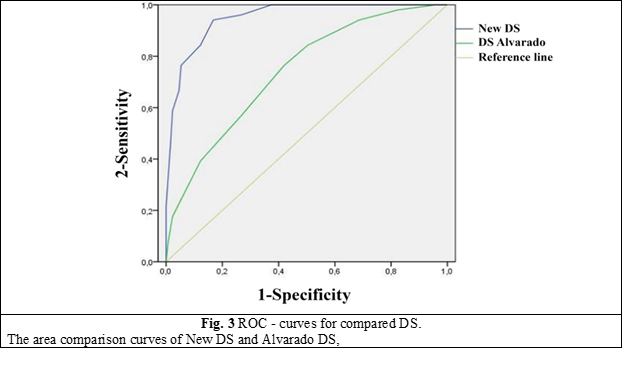
 The area under the curve for New DS was found to be statistically significantly higher compared to DS Alvarado and amounted to 0.95, which corresponds to the excellent quality indicator of the statistical model.

Table 5. ROC metrics (area under the curve) of New DS and Alvarado DS

Diagnostic scores

The area under the curve

95% - confidence interval

New DS 

0.952

0.924

0.981

Alvarado DS

0.739

0.662

0.816

Note:The area comparison curves of New DS and Alvarado DS


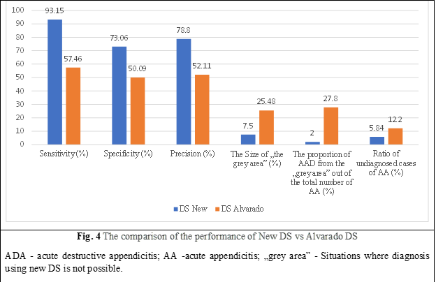
As a result of the comparative evaluation of New DS and Alvarado DS, it was observed that New DS has significantly higher sensitivity, specificity, and accuracy, and the number of undiagnosed AA cases compared to Alvarado DS is lower. If Alvarado's AA DS had been used in undiagnosed AA cases, there would have been 1.6% perforated, gangrenous, and complicated AA.

The size of the „grey area” and the weight of the „grey area” of AA, out of the total number of AA in New DS, was significantly smaller than in DS Alvarado (p < 0.001). The comparative evaluation of the main performance indicators New DS and DS Alvarado is shown in Fig. 4.

Thus, New DS showed greater clinical effectiveness in diagnosing AA in the elderly compared to the non-standardized clinical method and DS Alvarado, a lack of dependence on „risk factors” for diagnosing AA, such as obesity and atypical location of VA.

Discussion 

This study evaluated the acceptability of the Alvarado scores and the developed New DS in determining the diagnosis of AA in elderly patients.

Early diagnosis of AA is quite laborious in elderly people, having a high mortality and morbidity rate. In a study conducted in Finland, the data of 164,579 patients who underwent appendectomy surgery were examined over a 20-year period, and mortality increased 39 times in patients over 60 years. Similarly, the same study determined that negative appendectomy increased four-fold and mortality increased 10-fold. In the literature, the rate of negative appendectomy in geriatric patients ranges from 17% to 31% [3-4]. In the current cohort, the negative appendectomy rate was 28.3%, which is in accordance with the specialized literature.

Due to increasing life expectancy, diseases previously associated with the younger population, including AA, have an increasing incidence among elderly patients [6]. Although the lifetime risk of AA is 7% for the general population, this rate may increase to 10% among the elderly population [2]. As in most diseases, the clinical diagnostic process of AA is more difficult in the geriatric population than in the young. This is due, in part, to altered pain sensations due to impaired nerve conduction as a result of aging and the atypical picture of classical AA [6]. Since a delayed diagnosis will increase the mortality and morbidity of AA, international guidelines and evidence-based medicine guidelins recommend the use of clinical scoring systems in the initial evaluation process of patients [15].

The Alvarado score [17-19] being a 10-point scale based on indications, symptoms, and laboratory data, is one of the most widely used and evaluated scoring systems for the assessment of AA. A score of 5 or 6 points on the Alvarado scale is considered compatible with the diagnosis of AA; a score of 7 or 8 suggests a plausible diagnosis of AA; and a score of 9 or 10 indicates a very likely diagnosis of AA. This diagnostic score was designed to assist clinicians in clinical decision-making by objectively determining which patients should be monitored and evaluated and which should be operated on. The limited research that assessed the relevance of the Alvarado score in the elderly population, retrospectively analyzing 96 patients over 60 years of age, using the Alvarado score system with a cut-off value of 5, demonstrated high efficacy in the elderly [17]. In another study, the Alvarado and Lintula scores were compared in elderly patients undergoing appendectomy, and the former was found to be a more useful predictive tool, with an AUC value of 96.9% [18]. Another research, however, demonstrated that the Alvarado score is ineffective in elderly people [5].

There are, however, some limitations to our study. First, the results obtained by us cannot be generalized to the general population, since they were obtained from a single center. Second, because this study was retrospective, the results may have been influenced by inadequate or erroneous data from hospital records. Another disadvantage is the small group of patients.

Conclusions 

The use of the diagnostic score in the elderly will raise the quality of care, reduce the amount of time it takes to diagnose a similar case and as a result - lead to a reduction in complications and mortality in acute appendicitis. The study of the efficacy of new AA DS by comparative evaluation with the traditional non-standardized clinical diagnosis of AA and Alvarado AA DS demonstrated higher clinical efficiency in diagnosing AA with sensitivity up to 93.15% compared to the non-standardized clinical method and Alvarado AA DS and also does not depend on „risk factors” for AA diagnoses such as obesity and atypical location of AV, due to which we recommend wide application in medical practice.

Competing interests 

None declared.

Patient consent 

Obtained

Ethics approval

This study was approved by the Research Ethics Committee of Nicolae Testemițanu State University of Medicine and Pharmacy (Minutes No. 25, from 21.11.2016).

Author’s ORCID ID

Alexandr Gaitur – https://orcid.org/0009-0005-0512-7515

References

  1. Ceresoli M, Zucchi A, Allievi N, Harbi A, Pisano M, Montori G, Heyer A, Nita GE, Ansaloni L, Coccolini F. Acute appendicitis: epidemiology, treatment and outcomes- analysis of 16544 consecutive cases. World J Gastrointest Surg. 2016;8(10):693-699. doi: 10.4240/wjgs. v8.i10.693. 

  2. Kraemer M, Franke C, Ohmann C, Yang Q; Acute Abdominal Pain Study Group. Acute appendicitis in late adulthood: incidence, presentation, and outcome. Results of a prospective multicenter acute abdominal pain study and a review of the literature. Langenbecks Arch Surg. 2000 Nov;385(7):470-481. doi: 10.1007/s004230000165.

  3. Kotaluoto S, Ukkonen M, Pauniaho SL, Helminen M, Sand J, Rantanen T. Mortality related to appendectomy; a population based analysis over two decades in Finland. World J Surg. 2017;41(1):64-69. doi: 10.1007/s00268-016-3688-6. 

  4. Franz MG, Norman J, Fabri PJ. Increased morbidity of appendicitis with advancing age. Am Surg. 1995;61(1):40-44. 

  5. Shchatsko A, Brown R, Reid T, Adams S, Alger A, Charles A. The utility of the Alvarado score in the diagnosis of acute appendicitis in the elderly. Am Surg. 2017;83(7):793-798. 

  6. Lau WY, Fan ST, Yiu TF, Chu KW, Lee JM. Acute appendicitis in the elderly. Surg Gynecol Obstet. 1985;161(2):157-160. 

  7. Pokharel N, Sapkota P, Kc B, Rimal S, Thapa S, Shakya R. Acute appendicitis in elderly patients: a challenge for surgeons. Nepal Med Coll J. 2011;13(4):285-288. 

  8. Horattas MC, Guyton DP, Wu D. A reappraisal of appendicitis in the elderly. Am J Surg. 1990;160(3):291-293. doi: 10.1016/S0002-9610(06)80026-7. 

  9. Körner H, Söndenaa K, Söreide JA, Andersen E, Nysted A, Lende TH, Kjellevold KH. Incidence of acute nonperforated and perforated appendicitis: age-specific and sex-specific analysis. World J Surg. 1997;21(3):313-317. doi: 10.1007/s002689900235. 

  10. Zbierska K, Kenig J, Lasek A, Rubinkiewicz M, Wałęga P. Differences in the clinical course of acute appendicitis in the elderly in comparison to younger population. Pol Przegl Chir. 2016;88(3):142-146. doi: 10.1515/pjs-2016-0042. 

  11. Lunca S, Bouras G, Romedea NS. Acute appendicitis in the elderly patient: diagnostic problems, prognostic factors and outcomes. Rom J Gastroenterol. 2004;13(4):299-303.

  12. Bhullar JS, Chaudhary S, Cozacov Y, Lopez P, Mittal VK. Acute appendicitis in the elderly: diagnosis and management still a challenge. Am Surg. 2014 Nov;80(11):E295-E297. 


  13. Segev L, Keidar A, Schrier I, Rayman S, Wasserberg N, Sadot E. Acute appendicitis in the elderly in the twenty-first century. J Gastrointest Surg. 2015;19(4):730-735. doi: 10.1007/s11605-014-2716-9.

  14. Sülberg D, Chromik AM, Kersting S, Meurer K, Tannapfel A, Uhl W, Mittelkötter U. Altersappendizitis [Appendicitis in the elderly]. Chirurg. 2009;80(7):608-614. doi: 10.1007/s00104-009-1754-4. German. 

  15. Fugazzola P, Ceresoli M, Agnoletti V, Agresta F, Amato B, Carcoforo P,  The SIFIPAC/WSES/SICG/SIMEU guidelines for diagnosis and treatment of acute appendicitis in the elderly (2019 edition). World J Emerg Surg. 2020 Mar 10;15(1):19. doi: 10.1186/s13017-020-00298-0. 

  16. Blomqvist PG, Andersson RE, Granath F, Lambe MP, Ekbom AR. Mortality after appendectomy in Sweden, 1987-1996. Ann Surg. 2001;233(4):455-460. doi: 10.1097/00000658-200104000-00001. 

  17. Ohle R, O'Reilly F, O'Brien KK, Fahey T, Dimitrov BD. The Alvarado score for predicting acute appendicitis: a systematic review. BMC Med. 2011;9:139. doi: 10.1186/1741-7015-9-139. 

  18. Di Saverio S, Birindelli A, Kelly MD, Catena F, Weber DG, Sartelli M, et al. WSES Jerusalem guidelines for diagnosis and treatment of acute appendicitis. World J Emerg Surg. 2016 Jul 18;11:34. doi: 10.1186/s13017-016-0090-5. 

  19. Alvarado A. A practical score for the early diagnosis of acute appendicitis. Ann Emerg Med. 1986;15(5):557-564. doi: 10.1016/S0196-0644(86)80993-3. 

  20. Harbrecht BG, Franklin GA, Miller FB, Smith JW, Richardson JD. Acute appendicitis - not just for the young. Am J Surg. 2011;202(3):286-290. doi: 10.1016/j.amjsurg.2010.08.017. 

  21. Krajewski S, Brown J, Phang PT, Raval M, Brown CJ. Impact of computed tomography of the abdomen on clinical outcomes in patients with acute right lower quadrant pain: a meta-analysis. Can J Surg. 2011;54(1):43-53. doi: 10.1503/cjs.023509. 

  22. Terasawa T, Blackmore CC, Bent S, Kohlwes RJ. Systematic review: computed tomography and ultrasonography to detect acute appendicitis in adults and adolescents. Ann Intern Med. 2004;141(7):537-546. doi: 10.7326/0003-4819-141-7-200410050-00011. 

  23. Doria AS, Moineddin R, Kellenberger CJ, Epelman M, Beyene J, Schuh S, Babyn PS, Dick PT. US or CT for diagnosis of appendicitis in children and adults? A meta-analysis. Radiology. 2006;241(1):83-94. doi: 10.1148/radiol.2411050913.

  24. Marudanayagam R, Williams GT, Rees BI. Review of the pathological results of 2660 appendicectomy specimens. J Gastroenterol. 2006;41(8):745-749. doi: 10.1007/s00535-006-1855-5.

  25. Pooler BD, Lawrence EM, Pickhardt PJ. MDCT for suspected appendicitis in the elderly: diagnostic performance and patient outcome. Emerg Radiol. 2011;19(1):27-33. doi: 10.1007/s10140-011-1002-3.

  26. Barreto SG, Travers E, Thomas T, Mackillop C, Tiong L, Lorimer M, Williams R. Acute perforated appendicitis: an analysis of risk factors to guide surgical decision making. Indian J Med Sci. 2010;64(2):58-65. doi: 10.4103/0019-5359.94401. 

  27. Gürleyik G, Gürleyik E. Age-related clinical features in older patients with acute appendicitis. Eur J Emerg Med. 2003;10(3):200-203. doi: 10.1097/00063110-200309000-00008. 

  28. Konan A, Hayran M, Kılıç YA, Karakoç D, Kaynaroğlu V. Scoring systems in the diagnosis of acute appendicitis in the elderly. Ulus Travma Acil Cerrahi Derg. 2011;17(5):396-400. doi: 10.5505/tjtes.2011.03780. 

  29. Agafonoff S, Hawke I, Khadra M, Munnings V, Notaras L, Wadhwa S, Burton R. The influence of age and gender on normal appendicectomy rates. Aust N Z J Surg. 1987;57(11):843-846. doi: 10.1111/j.1445-2197.1987.tb01277.x.

  30. Rub R, Margel D, Soffer D, Kluger Y. Appendicitis in the elderly: what has changed? Isr Med Assoc J. 2000;2(3):220-223. 

More articles

Research Harmfulness of prooxidants in bronchopulmonary dysplasia in preterm children
Mariana Ceahlău1*, Rodica Selevestru1,2, Olga Tagadiuc3, Svetlana Șciuca1,2
https://doi.org/10.52645/MJHS.2023.3.01
Oxidative stress can be defined as the imbalance of the redox state of a certain system including living one (organelle, cell, organ/tissue), which excessively produces reactive oxygen and/or reactive nitrogen species (ROS/RNS) that exceed the capacity of the antioxidant defense system, which have the ability to slow down or even prevent the oxidative damage of macromolecules. Oxidative stress is a pathogenic mechanism of a large variety of diseases, including pulmonary one.
Research Predicting sympathovagal balance using parameters of breathing patterns in abdominal breathing
Abdominal breathing is utilized as a non-pharmacological treatment method for various stress-related conditions and autonomic dysfunctions. The objective of the study was to determine the predictors in the modulation of sympathovagal balance, as indicated by the ratio of low frequency to high frequency power of heart rate variability, by utilizing the respiratory pattern parameters recorded during the abdominal breathing model.
Research The impact of imuheptin and imupurin on cytokine profile and antioxidant status in rat model of inflammation
Ina Guțu1*, Nicolae Bacinschi1, Valentin Gudumac2
https://doi.org/10.52645/MJHS.2023.3.03
Insects, throughout evolution, have developed a huge arsenal of active compounds, which they use to defend themselves against enemies and diseases, at the same time in recent years insects have shown great interest as a source of food rich in biologically active substances. Research in recent decades has shown that insects produce a variety of proteins and peptides with antibacterial, antifungal, antiviral, immunomodulatory, anti-inflammatory, antioxidant, antitumor, hepatoprotective, antithrombotic, antihypertensive and detoxifying activity during or after contact with the microbial agent or unfavourable factor.
Research Early results of endovascular treatment using percutaneous vacuum-assisted thromboaspiration in acute lower limb ischemia
Alexandru Predenciuc1*, Vasile Culiuc1,2, Dumitru Casian1,2
https://doi.org/10.52645/MJHS.2023.3.05
Open balloon thrombectomy and embolectomy remain the preferred initial option in the management of acute lower limb ischemia (ALI), but various endovascular techniques have become accessible and are growing in popularity. The aim of the study was to assess our early experience with percutaneous vacuum-assisted thromboaspiration using the Penumbra/Indigo® system for non-traumatic ALI.
Review Venous and arterial endothelium: markers of dysfunction and pathophysiological significance
Victor Ojog1*, Svetlana Lozovanu1
https://doi.org/10.52645/MJHS.2023.3.06
Endothelial dysfunction is a result of complex pathogenic interface involving inflammation, oxidative stress, disorders of endothelization and hemostasis etc., in both arteries and veins, leading to a lot of cardiovascular diseases. Identifying markers with high predictive value has an important diagnostic and prognostic significance.
Review Current affairs in the use of medical ozone. Biological effects. Mechanisms of action
Natalia Cernei1*, Serghei Șandru1, Serghei Cobîlețchi1, Ion Grabovschi2, Ivan Cîvîrjîc1, Ruslan Baltaga1,3
https://doi.org/10.52645/MJHS.2023.3.08
Oxygen-ozone therapy stands as a medically endorsed practice confirmed by numerous international clinical studies. Various authors have illustrated the beneficial clinical outcomes of ozone therapy in terms of its capacity to regulate redox balance, cellular inflammatory responses, and adaptation to ischemia/reperfusion processes. Ozone therapy extends to encompass a range of viral infections, inflammatory disorders, and degenerative ailments, used as both monotherapy and as an adjunct to unified conventional therapies.Introduction Ozone (O3), a gas discovered in the mid-19th century and composed of three oxygen atoms, represents a highly reactive allotropic form of oxygen. It exhibits high solubility in plasma, extracellular fluids, and water (approximately 10 times more soluble in water than conventional oxygen). At room temperature, it is unstable, causing rapid decomposition into ordinary diatomic oxygen. Notably, its half-life measures 25 minutes at 30°C, 40 minutes at 20°C, and 140 minutes at 0°C [1-10]. Medical ozone is a blend of oxygen and ozone derived from medical-grade oxygen through the utilization of a medical ozone generator. This medical ozone contains a concentration of 1-5% ozone and 90-95% pure medical oxygen, or 10-80 μg/mL (0.21-1.68 μmol/ml) of ozone per milliliter of blood. Ozone therapy stands as a current and significant avenue of research in contemporary medicine [1, 3-5, 7, 9, 10-15]. Oxygen-ozone therapy is a medically validated practice supported by numerous international clinical studies. Nowadays, many clinical trials have shown its beneficial effects on the modulation of the oxidoreduction balance, cellular inflammation state, and adaptation to ischemia/reperfusion processes. Ozonotherapy is an effective, safe, feasible, and easy-to-perform technique, which finds applications in various inflammatory, infectious, degenerative diseases, as well as in rehabilitation following acute cardiac and cerebral ischemic events. It demonstrates good efficacy both as an independent treatment and, notably, as an adjunct to conventional therapies [3-5, 7, 11, 16-22]. By incorporating this medical practice, patients can attain significant clinical benefits. When combined with standard therapies, it often leads to reduced medication dosages, complication rates, treatment duration, medication toxicity, and medical expenses. It also addresses the issue of bacterial resistance to antibiotics [2, 4, 18, 19, 21, 23]. In the context of the aforementioned, the purpose of this article is to present a synthesis of the most recent findings regarding ozone's mechanisms of action. Materials and methods To achieve the outlined purpose, an initial search of specialized scientific publications was conducted. These were identified through the Google Search engine, namely, PubMed, Hinari, SpringerLink, the National Center of Biotechnology Information, and Medline. The article selection criteria encompassed contemporary data regarding the mechanisms of action of ozone therapy, utilizing the following keywords: “ozone”, “ozone therapy”, “ozone mechanisms of action”, “biological effects of ozone”, “antioxidant effect”, “anti-inflammatory effect”, and “immunomodulatory effect.” These keywords were employed in various combinations to optimize search efficiency. For the advanced selection of bibliographic sources, the following filters were used: full-text articles, articles in English, articles published between 1990 and 2022. After a preliminary analysis of the titles, original articles, editorials, articles of narrative synthesis, taxonomy, and meta-analysis were selected, which contained up-to-date information and contemporary concepts regarding the mechanisms of ozone therapy. Furthermore, a search was conducted within the reference lists of the identified sources to highlight additional relevant publications that were not found during the initial database searches. The information from the publications included in the bibliography was gathered, organized, evaluated, and synthesized, showcasing the key aspects of the contemporary understanding of ozone's mechanisms of action, namely, its antioxidant capacity, vascular and hematological modulation, immune system activation, as well as its anti-inflammatory, bactericidal, virucidal, and fungicidal effects. To minimize the potential systematic errors (bias) in the study, a meticulous search was conducted within databases to identify a maximum number of relevant publications for the study's purpose. Only studies that satisfy validity criteria were evaluated, rigorous exclusion criteria for articles under consideration were applied, and a comprehensive review was conducted of both positive outcome studies and those that did not highlight the treatment's benefits. If necessary, additional sources of information were consulted to clarify some concepts. Duplicate publications and articles that did not meet the purpose of the article and were not available for full viewing were excluded from the list of publications generated by the search engine. Results Following the data processing, as identified by the Google Search engine and from databases such as PubMed, Hinari, SpringerLink, National Center of Biotechnology Information, and Medline, in accordance with the search criteria, a total 475 articles on the topic of ozone therapy were found. After a primary analysis of the titles, 59 articles were eventually deemed relevant for the given synthesis. Upon repeated review of these sources, a final selection of 52 relevant publications was ultimately made in alignment with the intended purpose. The final bibliography of this work comprises 52 articles that have been considered representative of the materials published on the subject of this synthesis article. Publications, the content of which did not reflect the relevant topic, despite being selected by the search program, as well as articles that were not accessible for open viewing through the HINARI (Health Internet Work Access to Research Initiative) database or available in the scientific medical library of the Nicolae Testemițanu State University of Medicine and Pharmacy, were subsequently removed from the list. Although ozone is the most potent natural oxidant, capable of oxidizing a wide range of organic and inorganic substances, and has the potential for cytotoxicity, researchers believe that under controlled conditions, it possesses numerous therapeutic effects. Moreover, the reactivity to ozone can be effectively mitigated by the blood and cellular antioxidant system [1, 2, 15, 24-28]. Initially used as an empirical approach, oxygen-ozone therapy has now evolved to a stage where the majority of ozone's biological mechanisms of action have been extensively studied and elucidated, these findings being found within the fields of biochemistry, physiology, and pharmacology [18, 22, 25, 27-29]. Ozone is not a pharmaceutical medicine but rather a regulatory molecule capable of generating bioactive mediators. The effects of ozone have been demonstrated to be consistent, safe, and associated with minimal preventable side effects [2, 30]. Chronic oxidative stress, chronic inflammatory processes, and immune overactivation are present and highly detrimental in a wide variety of diseases. The effectiveness of ozone therapy is determined by moderate oxidative stress, resulting from the interaction of ozone with the biological components of the body, triggering an endogenous cascade of biochemical reactions [31]. Ozone can function as an oxidant either directly, when it dissolves in plasma and other biological fluids, immediately reacting with polyunsaturated fatty acids, antioxidants, cysteine-rich proteins, and carbohydrates; or indirectly, by generating reactive oxygen species (ROS) and lipid oxidation products (LOP) [25, 28, 32-37]. At the onset of ozone therapy, an endogenous cascade is triggered, releasing bioactive substances in response to transient and moderately induced oxidative stress by ozone ('oxidative eustress'). Ozone can easily induce this oxidative stress due to its plasma solubility. Reacting with polyunsaturated fatty acids and water, ozone forms ROS in human fluids and tissues. The main molecule among ROS is hydrogen peroxide (H2O2) – a non-radical oxidant. Concurrently, ozone also gives rise to LOP – the lipoperoxide radical, hydroperoxides, malondialdehyde, isoprostanes, ozoneides, alkenes, and predominantly, 4-hydroxynonenal. ROS and LOP are the effector molecules responsible for modulating several biological and therapeutic effects in the body following ozone therapy [3, 8, 13, 27, 34, 37-39]. Having reacted with a number of biomolecules, ozone disappears, and hydrogen peroxide, the main molecule of ROS, and other mediators rapidly diffuse into cells, activating various metabolic pathways with numerous biological and therapeutic effects [3, 5, 27, 28, 35, 40]. Therefore, ROS and LOP are “biological messengers of ozone” and are responsible for the biological and therapeutic effects of ozone. ROSs are short-acting early messengers and are responsible for immediate biological effects, while LOPs are important late and long-term messengers [3, 5, 10, 13, 14, 17, 28, 36]. The formation of ROS in plasma occurs extremely quickly (less than a minute), accompanied by a moderate and transient decrease in the antioxidant capacity of the blood (from 5% to 25%). However, this antioxidant capacity returns to normal within 15-20 minutes [3, 9, 17, 28, 31, 40, 41]. Discussion Although not fully comprehended, the present article will delve into the mechanisms underlying the antioxidant, anti-inflammatory, immunomodulatory, antimicrobial, antiviral, and analgesic effects of ozone. The antioxidative capacity is considered one of the key impacts of ozone therapy. Moderate oxidative stress induced by ozone within the therapeutic range (10-80 μg/mL), most commonly 30-45 μg/mL (the 'physiological' dose of ozone), physiologically effective and recommended levels for systemic application, elicits controlled low doses of ROS acting primarily as signaling molecules, thereby stimulating the formation of LOP. ROS triggers the activation of nuclear erythroid factor 2 (Nrf2), well known as a pivotal regulator of manifold cytoprotective responses, responsible for upregulating antioxidant enzyme activity. In response to transient, moderate oxidative stress, the levels of antioxidant enzymes such as superoxide dismutase, glutathione peroxidase, glutathione S-transferase, catalase, and heme oxygenase-1 increase. Thus, moderate, transient, and repetitive oxidative stress causes an intense modulation of antioxidants in the body. A multitude of cells across various organs upregulate the synthesis of antioxidants, which are capable of significantly countering excessive ROS, thereby alleviating chronic oxidative stress, which is present and extremely harmful in a variety of diseases Consequently, ozone, either through oxidative preconditioning or adaptation to chronic oxidative stress, safeguards tissue integrity against ROS-induced damage, fostering a balance between antioxidant and pro-oxidant factors while preserving cellular redox balance [27, 30, 41-45]. Nrf2 and nuclear factor kappa Β (NF-κB) represent the primary signaling pathways through which ozone exerts its effects. Nrf2 activation regulates cell defense and maintains cellular homeostasis [36]. Furthermore, ozone therapy fosters adaptation to oxidative stress by gently triggering the immune system, releasing growth factors, and/or activating metabolic pathways that contribute to maintaining redox balance [38]. By activating Nrf2, LOP induces oxidative stress proteins, including heme-oxygenase-1 (HO-1), another inhibitor of the NF-κB pathway and one of the most crucial antioxidant defense enzymes. Through inhibiting the high expression level of hypoxia-inducible factor-1α (HIF-1α), it contributes to reducing the production of proinflammatory cytokines, directly activating anti-inflammatory cytokines, enhancing antioxidant protection, and consequently, safeguarding cellular integrity [8, 28, 30, 44-46]. Thus, ozone mimics acute oxidative stress which, when properly balanced, is not harmful, but can trigger several beneficial biochemical mechanisms. It can reactivate the intra- and extracellular antioxidant system, thereby reversing chronic oxidative stress in various inflammatory, degenerative processes, etc. During ozone treatment, cells throughout the body receive gradual and subtle impulses of LOP, significant long-term messengers that play a crucial role in up-regulating antioxidant enzymes in multiple cell types while rebalancing the oxidant/antioxidant system [46, 47]. Vascular and Hematological Modulation. Ozone serves as a catalyst for transmembrane oxygen flow. The increase in cellular oxygen levels resulting from ozone therapy enhances the efficiency of the mitochondrial respiratory chain. Moreover, ozone amplifies the production of prostacyclin, a widely acknowledged vasodilator [2, 6, 8, 26]. The effects of ozone on oxygen metabolism can be explained by promoting (1) changes in the rheological properties of blood (reversal of erythrocyte aggregation, increased flexibility and elasticity of red blood cells, favoring the transport and delivery of tissue oxygen), leading to enhanced blood flow in microcirculation; (2) increasing the speed of glycolysis in erythrocytes; and (3) the release of substances (adenosine triphosphate, nitric oxide, and prostaglandins) that may contribute to reducing peripheral vascular resistance and increasing oxygen supply to tissues [18, 25, 35, 37, 40, 48-50]. Hydrogen peroxide (H2O2) diffuses from the plasma into the cellular cytoplasm and serves as the triggering stimulus. Depending on the cell type, various biochemical pathways can be simultaneously activated in red blood cells, white blood cells, and platelets, leading to a multitude of biological effects [10, 28, 32]. The Impact of Ozone on Erythrocytes. Erythrocytes are the focus of ROS. During erythropoiesis, submicromolar concentrations of LOP positively regulate the synthesis of antioxidant enzymes. Consequently, ozone therapy increases the glycolytic rate by enhancing intracellular adenosine triphosphate production. This approach intensifies erythrocyte generation, yielding metabolically enhanced erythrocytes (super-endowed erythrocytes) capable of more effectively transporting and delivering oxygen to tissues, including ischemic tissues, thereby correcting hypoxia in vascular diseases [7, 12, 25, 27, 28, 39, 48]. Coupled with increased nitric oxide synthase activity, there is a significant increase in nitric oxide, an essential element in maintaining optimal levels of vasodilation and blood perfusion [1, 6, 8, 40]. Ozone therapy, through careful regulation of ozone dosage, stimulates the production of antioxidant enzymes within the system (catalase, glutathione peroxidase, and superoxide dismutase) while mitigating excessive formation of ROS, thereby reducing chronic oxidative stress [1, 6, 12, 14, 39, 43, 49]. The impact of ozone on leukocytes. Ozone acts as a mild cytokine and serves as a cytokine inducer by lymphocytes and monocytes, thereby enhancing the immune system's activity. This stimulation fosters intercellular matrix synthesis and contributes to the healing process [1, 12, 32, 35, 37, 39]. The Impact of ozone on platelets. Hydrogen Peroxide (H2O2) and other ROS generated through blood ozonation initiate a cascade of enzymatic reactions. These reactions gradually elevate intracellular Ca levels and trigger the release of prostaglandins (F2a and E2), leading to irreversible platelet aggregation. Increased levels of growth factors released from platelets, mobilization of endogenous stem cells, and stimulation of neoangiogenesis promote tissue regeneration, as well as healing of injuries and wounds [27, 49, 51]. Thus, the impact of ozone on oxygen metabolism is explained by how it alters the blood's rheological properties (reversing red blood cell clumping, enhancing the flexibility and elasticity of red blood cells, promoting the transport and delivery of oxygen to tissues). This, in turn, facilitates blood flow in the microcirculation, increases glycolysis in red blood cells, and triggers the production of substances (such as adenosine triphosphate, nitric oxide, and prostaglandins) that help reduce peripheral vascular resistance. Activation of the immune system. Ozone promotes an increase in the production of interferon-γ (IFN-γ) and some cytokines, with interleukin-2 (IL-2) being the primary one, subsequently triggering a whole cascade of immunological reactions [1, 2]. It has been shown that ROS, including H2O2 and LOP generated by ozone therapy, can easily diffuse into plasma cells and activate NF-κB, inducing the production of immunoactive cytokines in normal cells (IL-2, tumor necrosis factor alpha - TNF-a, IL-6 and IFN-γ), thereby enhancing the immune response [9, 13, 14, 26, 32, 40, 44]. Ozone indirectly activates the innate (non-specific) immune system by enhancing phagocytosis and promoting the synthesis of cytokines and interleukins in neutrophils and leukocytes. It also triggers the components of both cellular and humoral immunity [8, 26, 33, 39]. Within mononuclear cells, ozone stimulates immune responses by modulating the NF-kB transcription factor, thereby reactivating the suppressed immune system [27, 28]. Furthermore, ROSs trigger the activation of the immune system, which acts through monocytes and lymphocytes, promoting the production of a variety of cytokines (IL-1, IL-2, IL-6, IFN-β, IFN-γ, TNFα) [6, 49]. Thus, ozone induces mild immune system activation by stimulating neutrophils and initiating the synthesis of certain cytokines that trigger a whole cascade of immunological responses. Bactericidal, virucidal and fungicidal action of ozone. Ozone used in vitro acts directly on the membrane of bacterial cells (direct oxidative effect), disrupting and damaging the integrity of bacterial cell membranes, oxidizing phospholipids and lipoproteins, thereby impeding their enzymatic function. Additionally, ozone damages the viral capsids, disturbing their structure and interfering with the virus-cell interaction, leading to disruption in the reproductive cycle. When it comes to fungi, ozone inhibits cell growth by perturbing intracellular homeostasis, resulting from the compromised barrier properties of the plasma membrane [2, 6, 12, 16, 26, 44, 48, 50]. Although ozone is one of the most potent disinfectants, used in various ways, it cannot deactivate any pathogens (bacteria, viruses, and fungi) in vivo. This is because pathogens are well protected, especially within cells, by the cell's powerful antioxidant system. Consequently, ozone acts as a gentle enhancer of the immune system by activating neutrophils and stimulating the synthesis of certain cytokines [1, 10, 19, 22, 28, 39, 46]. The anti-inflammatory effect is revealed in ozone's ability to influence the inflammatory cascade by oxidizing biologically active substances (arachidonic acid and its derivatives - prostaglandins), which participate in the development and sustenance of the inflammatory process. Additionally, ozone significantly reduces the levels of pro-inflammatory cytokines (IL-1β, IL-6, IL-8, and TNF-α) without any signs of toxicity or recorded side effects [8, 26, 30, 31]. These cytokines induce the prostaglandin E2 pathway, which causes pain or increases the sensitivity of nerve roots to other algogenic substances (such as bradykinin) [31]. Severe oxidative stress, triggered by high concentrations of ozone, along with proinflammatory cytokines (IL-1β, IL-6, IL-8, TNF-α), activate NF-κB, a key regulator of the inflammatory response and muscle atrophy. This contributes to an increased inflammatory response and tissue damage, including the release of other inflammatory factors that enhance the migration of eosinophils and neutrophils [9, 13, 17, 47, 49]. On the contrary, mild oxidative stress induced by precise and small doses of ozone activates Nrf2. The latter indirectly inhibits the pro-inflammatory mechanism driven by the NF-kB pathway. As a result, there is a reduction in NF-κB activity along with a modification in the expression of inflammatory cytokines associated with NF-kB activity. This triggers an anti-inflammatory effect, leading to a decrease in IL-1, IL-2, IL-6, IL-7, and TNFα, as well as an increase in interleukins such as IL-4, IL-10, IL-13, and the transforming growth factor beta – TGF-β [11, 13, 19, 38, 43, 49, 50]. Nrf2 also plays an important role in intracellular inflammatory signaling pathways. Triggering the Nrf2-antioxidant signal can dampen NF-kB activity, leading to the downregulation of the inflammatory response by suppressing essential inflammatory mediators and cytokines (IL-6, IL-8, and TNF-a) [31, 38, 42, 50]. Moreover, a small amount of H2O2 stimulates the NF-kB pathway, which is typically balanced out by the Nrf2's blocking action, resulting in an immunomodulatory effect [11]. The analgesic effect of ozone is ensured by the oxidation of the byproducts of albuminolysis, known as algopeptides, which act on the nerve endings in the damaged tissue and determine the intensity of the pain response. Additionally, the analgesic effect is attributed to the restoration of the antioxidant system and, subsequently, the reduction of harmful molecular byproducts from lipid peroxidation [26]. Recent preclinical studies have elucidated the role of ROS in hyperalgesia by activating N-methyl-D-aspartate receptors [11]. Following ozone therapy, there has been a demonstrated increase in antioxidant molecules (serotonin and endogenous opioids), which induce pain relief by stimulating antinociceptive pathways [31, 39]. Data from scientific research acknowledge that the mechanisms of action of ozone are due to: (1) a decrease in the production of inflammatory mediators; (2) oxidation (inactivation) of metabolic mediators of pain; (3) improvement of local blood microcirculation leading to improved oxygen delivery to tissues; (4) elimination of toxins and resolution of physiological disorders that generate pain [42, 52]. Therefore, ozone exhibits pleiotropic properties, extending beyond its exclusive role as an antioxidant, anti-inflammatory, or immunomodulatory one. It also encompasses the capacity to employ ROS as a signaling molecule rather than merely as intracellular toxic substances. In existing experimental models and clinical studies, the anti-inflammatory, antioxidant, regenerative and immunomodulatory effects of ozone therapy have been associated with several molecular mechanisms, the main ones being the NF-kB/Nrf2 balance and IL-6 and IL-1β expression. NF-kB and Nrf2 are the most studied and important transcription factors and regulatory proteins that control the expression of a wide range of genes, encoding proteins involved in a multitude of vital biological functions, including those associated with redox status, immunity, and inflammatory responses. Additionally, indirectly through these pathways, LOP initiates the HIF-1α, HO-1, and NO/iNOS pathways. The main pharmacological effects of medical ozone through ozone-produced peroxides are as follows: (1) increased oxygen release by erythrocytes due to activated metabolism; (2) immunomodulation due to leukocyte activation; and (3) regulation of cellular antioxidants via Nrf2 signaling. Ozone therapy can elicit the following biological reactions: (a) improved blood circulation and oxygen delivery to ischemic tissue; (b) optimization of overall metabolism by improving oxygen delivery; (c) regulation of cellular antioxidant enzymes and induction of HO-1; (d) triggering a mild immune system activation and intensified release of growth factors; (e) providing a state of well-being in most patients, probably due to stimulation of the neuroendocrine system. Conclusions 1. Ozone induces both mild and moderate oxidative stress. When appropriately balanced, this stress poses no harm; instead, it can initiate several beneficial biochemical mechanisms. These mechanisms, in turn, reactivate the intracellular and extracellular antioxidant systems, effectively countering long-term oxidative stress in various inflammatory and degenerative processes, etc. Cells throughout the body receive small and gradual bursts of lipid oxidation products, important late and long-term messengers that are responsible for activating antioxidant enzymes in many cell types to rebalance the oxidant/antioxidant system. 2. The impact of ozone on oxygen metabolism is explained by changes in the blood's rheological properties. This involves reversing red blood cell aggregation, enhancing the flexibility and elasticity of hemoglobin, and promoting the efficient transport and delivery of oxygen to tissues. This process also facilitates blood flow within microcirculation, speeds up glycolysis within red blood cells, and triggers the release of substances like adenosine triphosphate, nitric oxide, and prostaglandins, which help to reduce peripheral vascular resistance. 3. Ozone triggers a slight activation of the immune system by up-regulating and activating neutrophils and promoting the synthesis of cytokines (IL-2, TNF-a, IL-6, and IFN-γ), setting off a chain reaction of immune responses. 4. Ozone therapy induces the following biological responses: enhanced blood circulation and oxygen delivery to ischemic tissue, regulation of cellular antioxidant enzymes, mild immune system activation, and intensified release of growth factors. 5. Ozone is an inherently toxic gas that should never be inhaled, cannot be stored, and must be handled with caution. Generally, no toxic effects were reported, and only the respiratory tract was found to be highly sensitive to inhaled ozone since the respiratory mucosal cells contain a minimal amount of antioxidants and are extremely susceptible to oxidation. 6. Although ozone ranks among the most potent disinfectants, being employed in various ways, it cannot neutralize any pathogens (bacteria, viruses, and fungi) in vivo, since pathogens are effectively shielded by the strong blood and cellular antioxidant system. Furthermore, elevated ozone concentrations induce severe oxidative stress, prompting increased inflammatory responses and tissue damage. Competing interests None declared. Authors’ contribution NC and RB conceived the study, participated in the study design and assisted in drafting the manuscript. SȘ and IC performed the analysis and data interpretation. IG drafted the manuscript. SC conceived the significant revision of the manuscript and provided significant intellectual involvement. The authors have read and approved the final version of the manuscript. Authors’ ORCID IDs Natalia Cernei – https://orcid.org/0000-0002-2031-5881 Serghei Șandru – https://orcid.org/0000-0002-2973-9154 Ion Grabovschi – https://orcid.org/0000-0002-7716-9926 Ivan Cîvîrjîc – https://orcid.org/0000-0002-1360-5485 Ruslan Baltaga – https://orcid.org/0000-0003-0659-4877 References 1. Cakir R. General aspects of ozone therapy [Internet]. In: Atroshi F, editor. Pharmacology and nutritional intervention in the treatment of disease. London: IntechOpen, 2014 [cited 2023 Apr 12]. Available from: http://dx.doi.org/10.5772/57470 2. Elvis A.M., Ekta J.S. Ozone therapy: a clinical review. J. Nat. Sci. Biol. Med., 2011 Jan; 2 (1): 66-70. doi: 10.4103/0976-9668.82319. 3. Bocci V. Ozone: a new medical drug. 2nd ed. New York: Springer, 2011; 315p. https://doi.org/10.1007/978-90-481-9234-2. 4. Allorto N. Oxygen-ozone therapy: an extra weapon for the general practitioners and their patients. Ozone Ther., 2019 July; 4 (2): 8424. doi: 10.4081/ozone.2019.8424. 5. Baeza-Noci J., Cabo-Soler J.R., Moraleda-Gómez M. et al. Revisión WFOT sobre Ozonoterapia Basada en Evidencias. Comité Científico Asesor de la WFOT – 2015 [WFOT Review on Evidence-Based Ozone Therapy. WFOT Scientific Advisory Committee – 2015]. Brescia: World Federation of Ozone Therapy, 2015; 117p. Spanish. https://semdor.es/wp-content/uploads/2021/10/WFOT-OZONE-2015-ESP.pdf. 6. Scassellati C., Ciani M., Galoforo A., Zanardini R., Bonvicini C., Geroldi C. Molecular mechanisms in cognitive frailty: potential therapeutic targets for oxygen-ozone treatment. Mech. Ageing Dev., 2020 Mar; 186: 111210. doi: 10.1016/j.mad.2020.111210. 7. Yousefi B., Banihashemian S., Feyzabadi Z., Hasanpour S., Kokhaei P., Abdolshahi A. et al. Potential therapeutic effect of oxygen-ozone in controlling of COVID-19 disease. Med. Gas Res., 2022 Apr-Jun; 12 (2): 33-40. doi: 10.4103/2045-9912.325989. 8. Smith N., Wilson A., Gandhi J., Vatsia S., Khan S. Ozone therapy: an overview of pharmacodynamics, current research, and clinical utility. Med. Gas Res., 2017 Oct 17; 7 (3): 212-9. doi: 10.4103/2045-9912.215752. 9. Sagai M., Bocci V. Mechanisms of action involved in ozone therapy: is healing induced via a mild oxidative stress? Med. Gas Res., 2011 Dec 20; 1:29. doi: 10.1186/2045-9912-1-29. 10. Bocci V., Borrelli E., Travagli V., Zanardi I. The ozone paradox: ozone is a strong oxidant as well as a medical drug. Med. Res. Rev., 2009 Jul; 29 (4): 646-82. doi: 10.1002/med.20150. 11. Hidalgo-Tallón F., Torres-Morera L., Baeza-Noci J., Carrillo-Izquierdo M., Pinto-Bonilla R. Updated review on ozone therapy in pain medicine. Front. Physiol., 2022 Feb 23; 13: 840623. doi: 10.3389/fphys.2022.840623. 12. Manjunath R., Singla D., Singh A. Ozone revisited. J. Adv. Oral Res., 2015 May-Aug; 6 (2): 5-9. https://doi.org/10.1177/222941122015020. 13. Cenci A., Macchia I., La Sorsa V., Sbarigia C., Di Donna V., Pietraforte D. Mechanisms of action of ozone therapy in emerging viral diseases: immunomodulatory effects and therapeutic advantages with reference to SARS-CoV-2. Front Microbiol., 2022 Apr 21; 13: 871645. doi: 10.3389/fmicb.2022.871645. 14. du Plessis L.H., van der Westhuizen F.H., Kotzé H.F. The protective effect of plasma antioxidants during ozone autohemotherapy. Afr. J. Biotechnol., 2008 Aug; 7 (14): 2472-7. doi: 10.4314/ajb.v7i14.59039. 15. Rowen R.J. Ozone and oxidation therapies as a solution to the emerging crisis in infectious disease management: a review of current knowledge and experience. Med. Gas Res., 2019 Oct-Dec; 9 (4): 232-7. doi: 10.4103/2045-9912.273962. 16. Tizaoui C. Ozone: a potential oxidant for COVID-19 virus (SARSCoV-2). Ozone Sci. Eng., 2020 Jul; 42 (5): 378-85. https://doi.org/10.1080/01919512.2020.1795614. 17. Zanardi I., Borrelli E., Valacchi G., Travagli V., Bocci V. Ozone: a multifaceted molecule with unexpected therapeutic activity. Curr. Med. Chem., 2016; 23 (4): 304-14. doi: 10.2174/0929867323666151221150420. 18. Radzimierska M., Śmigielski K. Ozone and products of ozonation in medical use. Biomed J. Sci. Tech. Res., 2019 Apr; 17 (1): 002959. doi: 10.26717/BJSTR.2019.17.002959. 19. de Sire A., Agostini F., Lippi L., Mangone M., Marchese S., Cisari C. et al. Oxygen-Ozone therapy in the rehabilitation field: state of the art on mechanisms of action, safety and effectiveness in patients with musculoskeletal disorders. Biomolecules, 2021 Feb 26; 11 (3): 356. doi: 10.3390/biom11030356. 20. Wen Q., Liu D., Wang X., Zhang Y., Fang S., Qiu X. et al. A systematic review of ozone therapy for treating chronically refractory wounds and ulcers. Int. Wound J., 2022 May; 19 (4): 853-70. doi: 10.1111/iwj.13687. 21. Mendes C., Cunha M., Ferreira A., Santos E. Ozone therapy as a coadjuvant in wound healing and pain reduction. Millenium, 2021 Sep; 2 (9): 131-8. https://doi.org/10.29352/mill029e.25237. 22. Smith A., Oertle J., Warren D., Pratoet D. Ozone therapy: a critical physiological and diverse clinical evaluation with regard to immune modulation, anti-infectious properties, anti-cancer potential, and impact on anti-oxidant enzymes. Open J. Mol. Integr. Physiol., 2015 Aug; 5 (3): 37-48. doi: 10.4236/ojmip.2015.53004. 23. León Fernández O.S., Oru G.T., Hansler R.V., Cabreja G.L., Espinosa I.S., Polo Vega J.P. et al. Medical Ozone: the pharmacological mechanisms accounting for its effectiveness against COVID-19/SARS-COV-2. J. Med. Clin. Res. Rev., 2021 Mar; 5 (3): 1-10. doi: 10.33425/2639-944X.1199. 24. Bocci V. Biological and clinical effects of ozone. Has ozone therapy a future in medicine? Br. J. Biomed. Sci., 1999 Apr; 56 (4): 270-9. 25. Di Mauro R., Cantarella G., Bernardini R., Di Rosa M., Barbagallo I., Distefano A. et al. The biochemical and pharmacological properties of Ozone: the smell of protection in acute and chronic diseases. Int. J. Mol. Sci., 2019 Feb 1; 20 (3): 634. doi: 10.3390/ijms20030634. 26. Maslennikov O.V., Kontorshchikova C.N., Gribkova I.A. Ozone therapy in practice. Health manual [Internet]. Nizhny Novgorod (Russia); 2008 [cited 2023 Feb 23]. 42 p. Available from: https://absoluteozone.com/wp-content/uploads/2022/05/Ozone-Therapy-in-Practice.pdf. 27. Tricarico G., Travagli V. The relationship between Ozone and human blood in the course of a well-controlled, mild, and transitory oxidative eustress. Antioxidants (Basel), 2021 Dec 4; 10 (12): 1946. doi: 10.3390/antiox10121946. 28. Borrelli E., Bocci V. Basic biological and therapeutic effects of ozone therapy in human medicine [Internet]. In: Munter R, editor. Ozone science and technology. Oxford, UK: Encyclopedia of Life Support Systems, 2010 [cited 2022 Apr 18]. Available from: https://www.eolss.net/sample-chapters/c07/E6-192-15-00.pdf 29. Inal M., Dokumacioglu A., Özcelik E., Ucar O. The effects of ozone therapy and coenzyme Q₁₀ combination on oxidative stress markers in healthy subjects. Ir. J. Med. Sci., 2011 Sep; 180 (3): 703-7. doi: 10.1007/s11845-011-0675-7. 30. Chirumbolo S., Valdenassi L., Simonetti V., Bertossi D., Ricevuti G., Franzini M. et al. Insights on the mechanisms of action of ozone in the medical therapy against COVID-19. Int. Immunopharmacol., 2021 Jul; 96: 107777. doi: 10.1016/j.intimp.2021.107777. 31. de Sire A., Marotta N., Ferrillo M., Agostini F., Sconza C., Lippi L. et al. Oxygen-ozone therapy for reducing pro-inflammatory cytokines serum levels in musculoskeletal and temporomandibular disorders: a comprehensive review. Int. J. Mol. Sci., 2022 Feb 25; 23 (5): 2528. doi: 10.3390/ijms23052528. 32. Bocci V., Zanardi I., Travagli V. Has oxygen-ozonetherapy a future in medicine? J. Exp. Integr. Med., 2011 Jan; 1: 5-11. doi: 10.5455/jeim.161210.ir.002. 33. Néri J., Lomba E., Karam A., Reis S., Marchionni A., Medrado A. Ozone therapy influence in the tissue repair process: a literature review. J. Oral Diagn., 2017 Jul; 2: e20170032. doi: 10.5935/2525-5711.20170032. 34. Bocci V., Valacchi G., Corradeschi F., Fanetti G. Studies on the biological effects of ozone: 8. Effects on the total antioxidant status and on interleukin-8 production. Mediators Inflamm., 1998 Jul; 7 (5): 313-7. doi: 10.1080/09629359890820. 35. Bocci V. Oxygen-ozone therapy: a critical evaluation. Dordrect: Kluwer Academic Publishers, 2002. 440 p. http://doi.org/10.1007/978-94-015-9952-8. 36. Viebahn-Haensler R., León Fernández O. Ozone in Medicine. The low-dose ozone concept and its basic biochemical mechanisms of action in chronic inflammatory diseases. Int. J. Mol. Sci., 2021 Jul 23; 22 (15): 7890. doi: 10.3390/ijms22157890. 37. Bocci V. Ozone as Janus: this controversial gas can be either toxic or medically useful. Mediators Inflamm., 2004 Feb; 13 (1): 3-11. doi: 10.1080/0962935062000197083. 38. Scassellati C., Galoforo A., Bonvicini C., Esposito C., Ricevuti G. Ozone: a natural bioactive molecule with antioxidant property as potential new strategy in aging and in neurodegenerative disorders. Ageing Res. Rev., 2020 Nov; 63: 101138. doi: 10.1016/j.arr.2020.101138. 39. Bocci V., Zanardi I., Travagli V. Ozone acting on human blood yields a hormetic dose-response relationship. J. Transl. Med., 2011 May 17; 9: 66. doi: 10.1186/1479-5876-9-66. 40. International Scientific Committee of Ozonetherapy (ISCO3). Madrid Declaration on Ozone Therapy [Internet]. 2nd ed. Madrid: ISCO3; 2012 [cited 2023 Feb 23]. 50 p. Available from: https://bornclinic.com/wp-content/uploads/2020/12/OZONE-THERAPY.pdf. 41. Thorp J., Thorp K., Thorp E., Viglione D. Ozone preconditioning in viral disease. Virol. Curr. Res. [Internet], 2022; 6 (S1): 001 [cited 2023 Feb 23]. Available from: https://www.hilarispublisher.com/open-access/ozone-preconditioning-in-viral-disease.pdf. 42. Chirumbolo S., Varesi A., Franzini M., Valdenassi L., Pandolfi S., Tirelli U. et al. The mito-hormetic mechanisms of ozone in the clearance of SARS-CoV2 and in the COVID-19 therapy. Biomedicines, 2022 Sep 12; 10 (9): 2258. doi: 10.3390/biomedicines10092258. 43. Pivotto A.P., Banhuk F.W., Staffen I.V., Daga M.A., Ayala T.S., Menolli R.A. Clinical uses and molecular aspects of ozone therapy: a review. OnLine J. Biol. Sci., 2020 Feb; 20 (1): 37-49. https://doi.org/10.3844/ojbsci.2020.37.49. 44. Obeid B.M. Ozone autohemotherapy: possible mechanisms of anti-viral action and anti oxidative. J. Infect. Dis. Epidemiol., 2020 Apr; 6: 117. doi: 10.23937/2474-3658/1510117. 45. Galiè M., Covi V., Tabaracci G., Malatesta M. The role of Nrf2 in the antioxidant cellular response to medical ozone exposure. Int. J. Mol. Sci., 2019 Aug 17; 20 (16): 4009. doi: 10.3390/ijms20164009. 46. Martínez-Sánchez G., Schwartz A., Donna V. Potential cytoprotective activity of ozone therapy in SARS-CoV-2/COVID-19. Antioxidants (Basel), 2020 May 6; 9 (5): 389. doi: 10.3390/antiox9050389. 47. Ranaldi G., Villani E., Franza L. Rationale for ozone-therapy as an adjuvant therapy in COVID-19: a narrative review. Med. Gas Res., 2020 Jul-Sep; 10 (3): 134-8. doi: 10.4103/2045-9912.289462. 48. Bodrug N., Barba D., Istrati V., Botezatu A. Eficacitatea terapiei cu ozon în medicină [Effectiveness of ozone therapy in medicine]. Chișinău: Medicina, 2012. 116 p. Romanian. 49. Dias E.N., Andrade K.F., Silveira R.S., Machado R.R.P. [The acting of ozoniotherapy in wounds, neuropathies, infections and inflammations: a systematic review]. Braz. J. Dev., 2021; 7 (5): 48604-29. Portuguese. https://doi.org/10.34117/bjdv.v7i5.29786. 50. Borges F., Meyer P., Jahara R., de Morais Carreiro E., Antonuzzo P., Picariello F. et al. Fundamentals of the use of ozone therapy in the treatment of aesthetic disorders: a review. J. Biosci. Med., 2021 Dec; 9: 40-70. doi: 10.4236/jbm.2021.912005. 51. Fitzpatrick E., Holland O., Vanderlelie J. Ozone therapy for the treatment of chronic wounds: a systematic review. Int. Wound J., 2018 Aug; 15 (4): 633-44. doi: 10.1111/iwj.12907. 52. Franzini M., Valdenassi L., Pandolfi S., Tirelli U., Ricevuti G., Simonetti V. et al. The biological activity of medical ozone in the hormetic range and the role of full expertise professionals. Front Public Health, 2022 Sep 16; 10: 979076. doi: 10.3389/fpubh.2022.979076.
Review The role of the lateral pterygoid muscle in temporomandibular disorders
Vitalie Pântea1*, Felicia Tabără1, Mariana Ceban1, Veronica Burduja1, Lilian Nistor2, Olga Ursu3
https://doi.org/10.52645/MJHS.2023.3.09
The clinical concept that would argue that the activity of the lateral pterygoid muscle, being disturbed, would play an important role as an etiological factor in temporomandibular joint dysfunctions is still widely accepted, being also a decisive factor in the correct choice of the treatment plan. However, because of the fact that very few research and clear evidence were conducted and presented to support completely that concept, it continues to remain a very controversial one.
Case study Laser ureteroscopic endopyelotomy efficacy in pyeloureteral junction stenosis
Vladimir Caraion1*, Eduard Pleșca2, Andrei Mezu2, Corneliu Maximciuc2
https://doi.org/10.52645/MJHS.2023.3.10
Pyeloureteral junction stenosis (PUJS) is a condition that affects urinary drainage at level of the renal pelvis and upper ureter. It is found in approximately 1 in 500 newborns, with a higher prevalence in males (2:1 ratio). PUJS is the main cause of congenital hydronephrosis and can also be caused by other specific pathologies. Endoscopic management is the primary treatment for PUJS, particularly in cases of aperistaltic and <2cm intrinsic ureteral stenosis without aberrant vessels.
Case study Treatment of deep carious lesions with mineral trioxide aggregate: clinical case report
Diana Trifan*, Diana Uncuța
https://doi.org/10.52645/MJHS.2023.3.11
Deep carious lesions are a dental disease widely spread among population of all ages. From clinical point of view, they have little symptoms and go unnoticed by the patients a long time, until they provoke dental pulp inflammations. If diagnosed and treated properly, the tooth can be treated conservatively with certain techniques of pulp vitality preservation. An important role in this process plays the innate capacity of regeneration of the pulp-dentine complex and the enhanced stimulating properties of new biomaterials used in dentistry. The aim of this clinical case report is to describe the clinical manifestations and the diagnostic algorithm used in deep caries and to establish a clinical guideline of treatment of deep carious lesion with a calcium silicate hydraulic cement.