MJHS
Navigation
Journal Navigation
Article Navigation
opened article
Volume 10, Issue 3
September 2023
Cover Image

Article contents

opened journal
Volume 10, Issue 3
September 2023
Cover Image

Abstract

Introduction

Oxidative stress can be defined as the imbalance of the redox state of a certain system including living one (organelle, cell, organ/tissue), which excessively produces reactive oxygen and/or reactive nitrogen species (ROS/RNS) that exceed the capacity of the antioxidant defense system, which have the ability to slow down or even prevent the oxidative damage of macromolecules. Oxidative stress is a pathogenic mechanism of a large variety of diseases, including pulmonary one.

Material and methods

81 preterm born children included in the study were divided into the main group – preterm children with bronchopulmonary dysplasia (BPD), and the control group – preterm children without BPD. The comparison groups were prospectively evaluated clinical, instrumental and laboratory (TPA, prooxidant-antioxidant balance, nitric oxide metabolistes and MDA). Data were statistically analyzed using Microsoft Excel, MedCalc and SPSS and Contingency Table Analysis as a way to evaluate the performance of a diagnostic test.

Results

In preterm children with BPD were found to be decreased by 29% (p < 0.001) the prooxidant-antioxidant balance (PAB) and the nitric oxide metabolistes (NO) level by 12% (p < 0.001) compared to children in the control group. The assessment of tissue oxidative damage markers revealed a significant 62% (p < 0.001) increase in malonic dialdehyde (MDA) content and a 4.86-fold (p < 0.001) increase in total prooxidant activity (TPA) in children with bronchopulmonary dysplasia compared to children in the control group. Our study confirms that TPA, PAB, MDA and NO values are reliable markers of hypoxic tissue damage at children with bronchopulmonary dysplasia and can be recommended for assessing the intensity of oxidative stress.

Conclusions

Pulmonary bronchodysplasia is characterized by the imbalance of prooxidant-antioxidant processes with the exacerbation of prooxidant ones that trigger the oxidative/nitrosative stress and the deterioration of vital chemical compounds.

Key Messages

What is not yet known on the issue addressed in the submitted manuscript

Changes in markers of oxidative stress, in particular in preterm children with bronchopulmonary dysplasia have not yet been studied.

The research hypothesis

Preterm infants with pulmonary bronchodysplasia may develop oxidative stress, which may affect their health.

The novelty added by manuscript to the already published scientific literature

The assessment of biochemical markers established a significant increase in oxidative stress in preterm children with bronchopulmonary dysplasia. This was revealed by the prooxidant-antioxidant disbalance and a decrease of the nitric oxide metabolites content, associated with lipid damage as evidenced by the increase in malondialdehyde (MDA) levels.

Introduction

There is increasing evidence linking exposure to increased oxygen concentration and oxidative stress (OS) to the development of chronic bronchopulmonary disease, making the lung of preterm infants more susceptible to various diseases such as respiratory distress syndrome, bronchopulmonary dysplasia (BPD), and persistent pulmonary hypertension. Existing research in the field shows that although BPD is a disease with a multifactorial pathogenesis, the major risk factors are exposure to hyperoxia and the action of reactive oxygen species (ROS) [1, 2]. 

From conception to birth, mammals (including humans) develop under conditions of physiological hypoxia, with fetal arterial and venous pO2 values rarely exceeding ~4 kPa (30 mmHg) which constitutes ~4% O2. Thus, the entire process of organogenesis takes place in hypoxia, despite the fact that fetal hemoglobin has a significantly higher affinity for oxygen compared to that of an adult. The birth process is accompanied by an increase of oxidative stress, and this continues in the first months of life due to exposure to increased concentrations of oxygen provided by breathing. The role of oxidative stress triggered by hyperoxia in development is still unclear, but ROS are known to be involved in signal transduction, they also play an important role in immune function, are important regulators of circulation, activate cellular growth factors, remove dysfunctional proteins by oxidation and are essential for the functioning of cellular organelles [3].

It has been demonstrated that before and at birth in infants there is an increase in signs of oxidative stress, which indirectly reveals the increase in antioxidant capacity with the aim of adapting to the amplification of oxidative processes induced by the spontaneous inhalation of oxygen. At the same time, these adaptive reactions are missing or significantly underdeveloped in premature babies, especially those very premature and those with very low birth weight. Deficiencies of antioxidant protection mechanisms in these children are exacerbated by pulmonary insufficiency, lack of alveolar surfactant, underdevelopment of antioxidant enzymes. Subsequently, premature infants are much more likely to be hyperoxic and will develop oxidative stress following even short-term exposure to high levels of oxygen [4].

As a result of this phenomenon, cellular homeostasis is affected. The balance between the production and excessive accumulation of reactive oxygen species (ROS) in cells and the detoxification capacity due to the lack of endogenous antioxidants is inclined towards the amplification of oxidative processes, which causes tissue damage. Exogenous factors that stimulate ROS production have been shown to have both beneficial and deleterious cellular effects, thus either participating in cellular signaling or causing macromolecular damage [1, 2].

In case of the preterm babies, prolonged exposure to elevated oxygen concentrations can affect and alter the normal development of the lung tissue, triggering developmental disorders such as BPD. Some relevant studies regarding human BPD reported that increased ROS production is associated with impaired lung development [5].

Free radicals have unpaired electrons and are extremely reactive, but at the same time unstable, having low activation energy and short lifetime. It should be noted that they can act as both oxidants and antioxidants (reducers), a phenomenon explained by their ability to donate or accept an electron from other molecules [6].

The generation of ROS occurs in a series of redox reactions, which are the basis of many processes that take place in cells. Under physiological conditions, ROS are produced by the body as part of normal metabolic processes such as the electron transporting chain, Fenton and Haber-Weiss reactions etc. A series of pathological conditions and diseases, including those associated with hypoxia and ischemia/reperfusion, can disrupt the balance between the generation of ROS and the antioxidant system capacity to neutralize them, which causes oxidative stress. The amplification of the levels of intracellular ROS, causes damage of different macromolecules which will alter their function and the cell state [7, 8].

Oxidative stress is often associated with nitrosative stress, due to the interaction of nitric oxide with superoxide radical anion with the formation of peroxynitrite (ONOO‾), which, being a highly reactive radical, can cause enzyme inhibition, lipid peroxidation, protein and DNA damage, etc. The multitude of harmful processes initiated by NO metabolites can end with the induction of apoptosis and significant tissue damage [9-11].

Lipid hydroperoxides (LOOH), unsaturated aldehydes (MDA, 4-hydroxy-2-nonenal, 2-propenal or acrolein) and isoprostanes are relatively stable primary products of the lipid peroxidation process [6]. The well-known product of lipid damage produced by oxidative stress, malonic dialdehyde (MDA), is a recognized biomarker of oxidative stress, cell membrane damage, but also tissue and cell oxidative damage [12]. MDA is formed, as a result of the peroxidation process of polyunsaturated FA, either in the presence of a large number of free oxygen radicals from sialic acid and deoxyribose, or from the phospholipid structure of cell membranes [13, 14]. 

MDA is considered the most mutagenic product, in contrast to 4-hydroxy-2-nonenal, which is considered the most toxic. There are three mechanisms by which the damaging effect of lipid oxidation products is explained: the damage to the integrity of the cell membrane, the ability to add an additional ROS gene, or the degradation into reactive compounds, which have the potential of damaging DNA, proteins and lipids [6, 7]. The effects of lipid peroxidation in cells are loss of cell membrane properties, inactivation of many membrane receptors, and increased influx of calcium ions. These events alter permeability, membrane electrical potential and intercellular communication [15].

A number of commonly used methods for the assessment of oxidative/nitrosative stress (methods for measuring lipid and protein oxidation end products) are described. Nevertheless, there is an issue that is still addressed – the ability of oxidative/nitrosative stress, measured in plasma, to reflect the tissue processes, along with the need for a simple laboratory method to characterize an oxidative stress „profile” related to growth and maturation in physiological conditions and different diseases [15].

In children with DBP who endure chronic hypoxia due to respiratory impairment, we assume an alteration of these mechanisms. And our study was initiated as a challenge to have answers to these complex questions.

The aim of the research was to evaluate markers of oxidative/nitrosative stress measured in the serum of preterm children with bronchopulmonary dysplasia and to analyze their performance as a diagnostic test.

Material and methods

The research was carried out as part of the doctoral project „Prooxidant and antioxidant status in bronchopulmonary dysplasia in premature children”.

To describe the results of the assessment of oxidative stress markers in children with BPD, an analytical analysis based on a cohort study was performed. In this regard, 81 follow-up records of patients admitted to the Institute of Mother and Child (Chisinau, Republic of Moldova) with positive history of preterm births, postnatal oxygen therapy in respiratory distress were documented and analyzed. The patients were examined according to the same protocol, which included the complex examination and contained the information from the outpatient medical record (F112/e), the inpatient medical record (F003/e). Comparison groups were evaluated prospectively, through clinical, laboratory, instrumental examination. The children were divided into main group (children born preterm with BPD) and control group (children born preterm without BPD). Data analysis was performed according to the methodology described in „Basics of Epidemiology and Research Methods” [16].

The biochemical investigations were carried out according to methods adapted by the collaborators of the Biochemistry Laboratory of Nicolae Testemițanu State University of Medicine and Pharmacy for the Synergy H1 (Hydrid Reader) microplate spectrofluorometer (BioTek Instruments, USA) and the Power Wave HT spectrophotometer (BioTek Instruments, USA).

For the analysis of markers of interest, venous blood samples (5 mL) were collected, which were centrifuged for 10 minutes at 3000 revolutions/minute. The serum was separated and transferred to Eppendorf tubes and stored at -45°C separately until biochemical testing. All samples were coded.

The prooxidant-antioxidant balance (PAB) was performed by the method described by Toloue Pouya V. et al. [17], modified by Pantea V. et al. [18]. The method is based on the capacity of the free radicals, peroxides and antioxidants, contained in the blood sample, to interact with the TMB (3,3′,5,5′-tetramethylbenzidine) or TMB cation, that will determine changes of the solution color. PAB values were calculated according to the calibration curve data and expressed in arbitrary units.

Determination of oxidative stress marker – malonic dialdehyde and total prooxidant activity, was performed according to the procedure described by Galaktionova LP. et al. [19], and modified by Gudumac V. et al. [20]. The method is based on the spectrophotometric identification of the colored trimethine complex, resulting from the interaction of thiobarbituric acid with DAM. The concentration of DAM in the sample is directly proportional to the intensity of the staining. The final result was expressed in μM/L.

Determination of nitrosative stress marker – nitric oxide metabolites, was performed according to the procedure described by Меtеlskaya VА. et al. [21], modified by Gudumac V. et al. [22]. The principle of the method consists in the deproteinization of the biological material, the reduction of nitrates into nitrites, the processing of the supernatant with the Griss reagent, and the subsequent measurement of the optical density of the reaction product. The calculation of the nitrite concentration was carried out with the help of the calibration curve, built on the basis of successive dilutions of the standard solution of sodium nitrite and was expressed in μmol/L.

The data were statistically processed by operating electronic computerized assessment techniques of the degree of relationship between the evaluated parameters of the patients in the study groups, using Microsoft Excel, MedCalc (DeLong et al., 1988) and SPSS and Contingency Table Analysis as a way to summarize the performance of a diagnostic test [23-25].

Results

An oxidant is any compound that can accept electrons, including oxygen. On the other hand, a substance that donates electrons, is a reducing agent. The redox reactions are essential to the many processes that take place in cells. For specific biological systems, the terms prooxidant and antioxidant are equivalent in chemistry to the terms oxidant and reductant [15]. Many radicals are unstable and highly reactive. Behaving as oxidants or reductants, they have the ability to give an electron or accept an electron from other molecules, and homeostasis between them is important for optimal functioning of the system [14].

The evaluation of the redox status in the blood of the preterm children with and without BPD revealed the prevalence of oxidative processes in the children of the main group compared to the children of the control group (table 1).
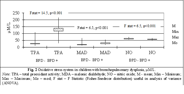
The values ​​of total prooxidant activity (TPA) in children with BPD (41 children) was equal to 138.2±6.1 µM/L, value significantly increased compared to TPA in children without BPD (40 children) equal to 28.4±2.3 µM/L, Fstat = 14.5, p < 0.0001 (table 1, fig. 2).

A 4.86-fold increase (p < 0.001) of the total prooxidant activity was identified in preterm children with BPD compared to those without lung damage, a phenomenon that reveals the intensification of the production and accumulation of prooxidants of different nature, which can amplify the oxidation processes up to the level of oxidative/nitrosative stress (table 1, fig. 2).

Table 1. Values of markers of oxidative stress in children with bronchopulmonary dysplasia

Marker

Control group

(n = 40)

Main group

(n = 41)

The veracity of differences between groups

TPA (μM/L)

28.4±14.3

100%

138.2±38.9

486%

p < 0.001

PAB (U)

140.3±15.2

100%

99.6±15.8

71%

p < 0.001

MDA (μM/L)

20.4±8.2

100%

33.0±8.9

162%

p < 0.001

NO metabolites (μM/L)

64.9±3.8

100%

57.1±4.8

88%

p < 0.001

Note: TPA – total prooxidant activity; PAB – prooxidant/antioxidant balance; MDA – malonic dialdehyde; NO – nitric oxide. Variables are presented as Mean±SD.

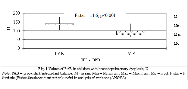
PAB in children with BPD (41 children) is equal to 99.6±2.5 µM/L with minimum value of 65.8 µM/L, median – 101.8 µM/L, maximum – 140.3 µM/L, mode – 76.4 µM/L, compared to PAB concentration in children without BPD (40 children) which have a significant difference between groups equal to 140.3±2.4 µM/L (minimum value of 105.9 µM/L, median – 140.7 µM/L, maximum – 176.8 µM/L, mode – 131.7 µM/L), Fstat = 11.6, p < 0.00001 (table 1, fig. 2).

A major, statistically significant decrease of PAB by 29% (p < 0.001) was identified in preterm children from the group with bronchopulmonary dysplasia compared to children from the control group, which attests to the decrease in the total level of antioxidants with the inclination of the balance towards the formation and accumulation of prooxidants and the definite establishment of the prooxidant status (fig. 1).

Malonic dialdehyde (MDA), in children with BPD (41 children) was  33.1±1.39 µM/L with minimum value of ​​18.3 µM/L, median – 31.8 µM/L, maximum – 55.4 µM/L, mode – 25.4 µM/L, compared to the MDA level in children without DBP (40 children) which was ​​20.4±1.3 µM/L (minimum value ​​of 12.9 µM/L, median – 19.1 µM/L, maximum – 66.8 µM/L, mode – 17.3 µM/L), and presents a significant difference between batches (Fstat = 6.5, p < 0.0001) (table 1, fig. 2).

The installation of the prooxidant status in preterm children from the group with bronchopulmonary dysplasia, initiated the atypical oxidation of lipids via the peroxidative pathway, which was manifested by a significant increase of 62% (p < 0.001) in the content of MDA, the final product of the peroxidation of unsaturated fatty acids, mainly from cell membranes phospholipids (fig. 2).

Nitric oxide (NO) in children with BPD (41 children) is equal to 57.13±0.75 µM/L with minimum value of 49.6 µM/L, median – 57.4 µM/L, maximum – 66.7 µM/L, mode – 59.7 µM/L, compared to the concentration of nitric oxide in children without BPD (40 children) which has a significant difference between groups equal to 64.9±0.6 µM/L (minimum values of 55.8 µM/L, median – 65.1 µM/L, maximum – 74.4 µM/L, mode – 59.7 µM/L), Fstat = 7.9, p < 0.00001 (table 1, fig. 2). 

A statistically significant decrease in the level of NO metabolites by 12% (p < 0.001) was revealed in premature infants from the group with bronchopulmonary dysplasia compared with children in the control group, which may indicate the use of NO in reactions that cause reactive forms of nitrogen (peroxynitrite, protonated peroxynitrite), thereby contributing to the induction of nitrosative stress and the deepening of the redox imbalance (fig. 2).

We can conclude that preterm children with BPD are characterized by the amplification of ROS/NRS production reactions, the establishment of a prooxidant status and the triggering of OS/NS, which ultimately causes damage to biomolecules and cellular macromolecular structures (membranes).

Next are presented the derivations of multiple measures using the four outcomes of the 2×2 contingency table for the prooxidant system to assess utility as diagnostic tests in children with BPD. 

The analysis using ROC curves (Receiver Operating Characteristics) was chosen as a statistical model. These are two-dimensional curves of the values ​​of a diagnostic test that ends with the evaluation of this examination applied to each patient or their comparison [23]. 

The assessment of the usefulness of determining the TPA, for highlighting children at risk of developing oxidative stress, was carried out by means of the ROC curve, which is an excellent way to compare diagnostic tests (fig. 3).

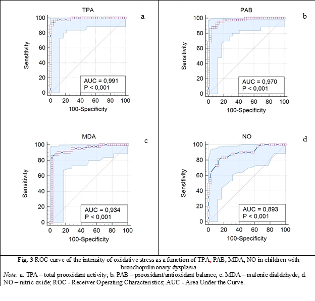
          When the variable under study cannot distinguish between the two groups, i.e., where there is no difference between the two distributions, the area will be equal to 0.5 (the ROC curve will coincide with the diagonal). When there is a perfect separation of the values of the two groups, i.e., there is no overlapping of the distributions, the area under the ROC curve equals 1 (the ROC curve will reach the upper left corner of the plot) (fig. 3). A reflection of the identification of prooxidant processes depending on TPA can be the area located below the level of the ROC curve equal to 0.99 (AUC is equal to 0.991: 95%CI 0.98-1, p = 0.000) (table 2). So, our study confirms the importance of evaluating prooxidant process according to TPA concentration, which has been shown to be a useful test.

Area under the curve of the ROC curve for PAB values was obtained equal to 0.97: 95% CI 0.905-0.999, p = 0.000 (table 2).

Table 2. Area under the curve (AUC) for the values of markers of oxidative stress in children with bronchopulmonary dysplasia.


AUC

Standard Error

p

95% Confidence Interval

Lower Bound

Upper Bound

TPA (μM/L)

0.991

0.008

0.000

0.976

1.000

PAB (U)

0.97

0.016

0.000

0.905

0.995

MDA (μM/L)

0.934

0.032

0.000

0.871

0.997

NO (μM/L)

0.893

0.035

0.000

0.804

0.951

Note: TPA – total prooxidant activity; PAB – prooxidant/antioxidant balance; MDA – malonic dialdehyde; NO – nitric oxide; AUC - Area Under the Curve; p – signification. 

          In the case of the malondialdehyde test, the minimum sensitivity of the test was of 2.4%: 95%CI, 0.01-0.12 for MDA concentration less than 20 µM/L (characteristic only of a child with DBP). The specificity was also minimal with the highest value reaching only 4.9%: 95%CI, 0.05-0.6, which confirms levels higher than 20 µM/L of MDA in only 20 children from the control group (without DBP), χ2 = 24.6, p < 0.0001 (fig. 3). 

Area under the ROC curve equal to 0.934: 95%CI 0.87-0.99, p = 0.000 values for MDA content (µM/L) in children with BPD (fig. 3, table 2). 

And the last demonstration concerns the area under the curve of NO – nitric oxide which was obtained equal to 0.893: 95%CI 0.804-0.951, p = 0.000 (table 2).

Discussions

According to literature data, multiple harmful gestational factors, which can affect the growth and development of the product of conception during the entire intrauterine period, are reported.

During postnatal development, the preterm born child is even more influenced by the convergence of the multitude of endogenous and exogenous factors that have damaged the health status. A dominant focus of the modern experimental studies demonstrates the harmfulness of oxidative stress, as the ultimate goal through the generation of free radicals (FR) and as a result the occurrence of cellular, tissue and organic damage [26, 27].

Our study, based on a prospective evaluation of two groups of children: the main group - preterm infants with BPD and the control group - preterm infants who did not develop BPD, examined clinically, paraclinical and instrumentally, contributes to the study of changes in oxidative stress markers in preterm infants who have developed bronchopulmonary dysplasia, the analysis of these data using various contingency tables and their generalization by performing a diagnostic test.

In multiple series of papers, and a huge variety of laboratory methods and statistical models have been developed and used to measure oxidative stress intensity and its consequences. Researchers such as Ferguson K, Gunko V O, Abiaka C, Machado L in their studies, determine biomarkers using various methods [28-30]. At the same time, all these methods are considered to be quite difficult, since oxidative stress biomarkers are very reactive and have very short half-lives.

Thus, the conducted study is within the limits of the research level, which is of scientific value in terms of the assessed markers of oxidative stress. These markers evaluated in premature children with bronchopulmonary dysplasia compared to those without this lung damage revealed the presence of significant oxidative stress in those enrolled in the main group. Total prooxidant activity increased by 386% (p < 0.001), the balance between prooxidants and antioxidants shifted towards the former (PAB - 29%, p < 0.001) and at the same time the NO content significantly decreased (-12%, p < 0.001). Our research also found lipid damage with the accumulation of the end product of lipid peroxidation – DAM (+62%, p < 0.001).

However, the role of oxidative stress in neonatal lung injury is much more complex and not fully studied. Despite the multiple existing data and different research environments under development, clinical practice is limited in the ability to detect very early preterm newborns who have susceptibility to develop a lung pathology, therefore the markers used currently cannot fully predict the lung damage that may follow in these children. But the detection and monitoring of lung lesions related to oxidative stress, namely through the use of non-invasive methods of detecting different oxidation products, remains to have a predictive and very useful role in the clinical setting.

While a growing body of evidence supports the role of oxidative stress, it appears that the complexity of this multifactorial condition cannot be captured by a single marker. Instead, researchers should move on to develop and validate specific panels of biomarkers that can more reliably predict certain pathological states that evolve with impaired lung function. Early diagnosis and treatment of oxidative stress-related lung diseases may be essential to prevent adverse effects that may spread beyond the neonatal period. Indeed, few of the biomarkers developed to date have been qualified for neonatal lung disease and their analysis has been limited by research settings.

Our study confirms that TPA, PAB, MDA and NO values are reliable markers of hypoxic tissue damage in children with bronchopulmonary dysplasia and can be recommended for assessing the intensity of oxidative stress. Last but not least, the currently available evidence, including our results highlights the need for further studies on a larger scale and with longer follow-up periods to obtain more precise results and allow serial detection of oxidative stress biomarkers.

Conclusions

Oxidative stress is a major contributor to lung injury in preterm children with bronchopulmonary dysplasia, fact confirmed by significantly higher values of total prooxidant activity (4.86 times, p < 0.001) and MDA (by 62%, p < 0.001) along with concomitant decrease of the prooxidnat/antioxidant and NO metabolites levels in the blood of these children. The phenomenon reveals an increase in the production and accumulation of prooxidants of various nature, which enhances oxidation processes and causes damage to biomolecules and cellular macromolecular structures, membranes in particular.

Thus, our study confirms the importance of evaluating pro-oxidant processes according to the concentrations of TPA, PAB, MDA, NO, which have been demonstrated as useful tests.

Competing interests

None declared. 

Authors’ contribution

Authors contributed equally to the literature searching, conceptual highlighting of the material as well as writing of the manuscript. The authors read and approved the final version of the manuscript.

Authors’ ORCID IDs

Mariana Ceahlau – https://orcid.org/0009-0009-3322-344X 

Rodica Selevestru – https://orcid.org/0000-0002-8923-3075 

Olga Tagadiuc – https://orcid.org/0000-0002-5503-8052 

Svetlana Șciuca – https://orcid.org/0000-0003-1091-9419

References

  1. Cannavò L, Perrone S, Viola V, Marseglia L, Di Rosa G, Gitto E. Oxidative stress and respiratory diseases in preterm newborns. Int J Mol Sci. 2021;22(22):12504. doi: 10.3390/ijms222212504. 

  2. Lee JW, Davis JM. Future applications of antioxidants in preterm infants. Curr Opin Pediatrics. 2011;23:161-166. doi: 10.1097/MOP.0b013e3283423e51.

  3. Maltepe E, Saugstad O. Oxygen in health and disease: regulation of oxygen homeostasis - clinical implications. Pediatr Res. 2009;65(3):261-268. https://doi.org/10.1203/PDR.0b013e31818fc83f.

  4. Kaarteenaho-Wiik R, Kinnula VL. Distribution of antioxidant enzymes in developing human lung, respiratory distress syndrome, and bronchopulmonary dysplasia. J Histochem Cytochem. 2004;52(9):1231-1240. doi: 10.1369/jhc.4A6291.2004.

  5. Wang J, Dong W. Oxidative stress and bronchopulmonary dysplasia. Gene. 2018;678:177-183. doi: 10.1016/j.gene.2018.08.031.

  6. Halliwell B, Gutteridge JMC. Free radicals in biology and medicine. Oxford: Clarendon Press, Oxford University Press; 2000. p. 160-5.

  7. Chandrasekaran A, Idelchik MDPS, Melendez JA. Redox control of senescence and age-related disease. Redox Biol. 2017;11:91-102. doi: 10.1016/j.redox.2016.11.005.

  8. Sies H, Jones DP. Reactive oxygen species (ROS) as pleiotropic physiological signaling agents. Nat Rev Mol Cell Biol. 2020 Jul;21(7):363-383. doi: 10.1038/s41580-020-0230-3.

  9. Liguori I, Russo G, Curcio F, et al. Oxidative stress, aging, and diseases. Clin Interv Aging. 2018;2018(13):757-772. doi: 10.2147/CIA.S158513.

  10. Pavlovschi E. Markerii biochimici ai retinopatiei hypertensive [Biochemical markers of hypertensive retinopathy] [dissertation]. Chișinău: Doctoral School in Medical Sciences; 2022. 118 p. Romanian.

  11. Lazăr C, Vozian M, Pantea V, Şveţ I, Mishina A, Tagadiuc O. The effect of controlled reperfusion on experimental ovarian torsion. Russ Open Med J. 2019;8(4):1-5. doi: 10.15275/rusomj.2019.0404.

  12. Riggins JN, Marnett LJ. Mutagenicity of the malondialdehyde oligomerization products 2-(3'-oxo-1'-propenyl)-malondialdehyde and 2,4-dihydroxymethylene-3-(2,2-dimethoxyethyl) glutaraldehyde in Salmonella. Mutat Res. 2001 Oct 18;497(1-2):153-7. doi: 10.1016/s1383-5718(01)00253-4.

  13. Moris D, Spartalis M, Tzatzaki E, Spartalis E, Karachaliou GS, Triantafyllis AS, Karaolanis GI, Tsilimigras DI, Theocharis S. The role of reactive oxygen species in myocardial redox signaling and regulation. Ann Transl Med. 2017 Aug;5(16):324. doi: 10.21037/atm.2017.06.17.

  14. Clark RH, Gerstmann DR, Jobe AH, Moffitt ST, Slutsky AS, Yoder BA. Lung injury in neonates: causes, strategies for prevention, and long-term consequences. J Pediatr. 2001 Oct;139(4):478-86. doi: 10.1067/mpd.2001.118201.

  15. Granot E, Kohen R. Oxidative stress in childhood - in health and disease states. Clin Nutr. 2004 Feb;23(1):3-11. doi: 10.1016/s0261-5614(03)00097-9.

  16. Spinei L, Stefăneţ S, Moraru C. Notiuni de baza de epidemiologie şi metode de cercetare [Basic knowledge of epidemiology and research methods]. Chisinau: Bons Offices; 2006. 224 p. Romanian.

  17. Toloue Pouya V, Hashemy SI, Shoeibi A, Nosrati Tirkani A, Tavallaie S, et al. Serum prooxidant-antioxidant balance, advanced oxidized protein products (AOPP) and protein carbonyl in patients with stroke. Razavi Int J Med. 2016;4(2):e38203.

  18. Pantea V, Șveț I, Tagadiuc O, Gudumac V, Andronache L, inventors. Procedeu de determinare a balanței pro-oxidante – antioxidante [Procedure for determining the pro-oxidant - antioxidant balance]. Republic of Moldova patent no. 5669. 2018 Oct 24. Romanian.

  19. Galaktionova LP, Molchanov AV, El’chaninova SA, Varshavskii BIa. Sostoianie perekisnogo okisleniia u bol’nykh s iazvennoi bolezn’iu zheludka i dvenadtsatiperstnoi kishki [The state of peroxidation in patients with peptic ulcer of the stomach and duodenum]. Klin Lab Diagn.1998;(6):10-14. Russian.

  20. Gudumac V, Tagadiuc O, Andronache L, Ștîrba O, Pantea V, inventors. Procedeu de dozare a dialdehidei malonice în materialul biologic [Malonic dialdehyde dosage procedure in biological material]. Republic of Moldova patent no. 5157. 2012 Dec 14. Romanian.

  21. Metel’skaia VA, Gumanova NG. Skrining-metod opredeleniia urovnia metabolitov oksida azota v syvorotke krovi cheloveka [Screening-method for nitric oxide metabolites determination in human serum]. Klin Lab Diagn. 2005;(6):15-18. Russian.

  22. Gudumac V, Tagadiuc O, Rîvneac V, Sardari V, Pantea V, et al. Investigații biochimice. Vol. 2: Micrometode [Biochemical investigations. Vol. 2: Micromethods]. Chisinau: “Elena-VI”; 2010. 97 p. ISBN 978-9975-106-06-1. Romanian.

  23. Băicuş C. Medicina bazată pe dovezi: cum înţelegem studiile [Evidence-based medicine: how we understand studies]. Bucharest: Editura Medicală; 2007. 144 p. Romanian.

  24. Everitt BS. The Cambridge dictionary of statistics. 2nd ed. Cambridge, UK: Cambridge University Press; 2002. 410 p. ISBN 052181099X.

  25. Powers, DMW. Evaluation: from precision, recall and F-score to ROC, informedness, markedness & correlation. J Mach Learning Technol. 2011;2(1):37-63.

  26. Ferrante G, Carota G, Li Volti G, Giuffrè M. Biomarkers of oxidative stress for neonatal lung disease. Front Pediatr. 2021 Feb 18;9:618867. doi: 10.3389/fped.2021.618867. 

  27. Perrone S, Santacroce A, Picardi A, Buonocore G. Fetal programming and early identification of newborns at high risk of free radical-mediated diseases. World J Clin Pediatr. 2016;5(2):172-81. doi: 10.5409/wjcp.v5.i2.172.

  28. Arogbokun O, Rosen E, Keil AP, Milne GL, Barrett E, Nguyen R, Bush NR, Swan SH, Sathyanarayana S, Ferguson KK. Maternal oxidative stress biomarkers in pregnancy and child growth from birth to age 6. J Clin Endocrinol Metab. 2021 Apr 23;106(5):1427-1436. doi: 10.1210/clinem/dgab018. 

  29. Abiaka C, Machado L. Nitric oxide and antioxidant enzymes in venous and cord blood of late preterm and term omani mothers. Sultan Qaboos Univ Med J. 2012 Aug;12(3):300-5. doi: 10.12816/0003143. 

  30. Gunko VO, Pogorelova TN, Linde VA. Proteomic profiling of the blood serum for prediction of premature delivery. Bull Exp Biol Med. 2016;161(6):829-832. https://doi.org/10.1007/s10517-016-3522-z.

More articles

Research Predicting sympathovagal balance using parameters of breathing patterns in abdominal breathing
Abdominal breathing is utilized as a non-pharmacological treatment method for various stress-related conditions and autonomic dysfunctions. The objective of the study was to determine the predictors in the modulation of sympathovagal balance, as indicated by the ratio of low frequency to high frequency power of heart rate variability, by utilizing the respiratory pattern parameters recorded during the abdominal breathing model.
Research The impact of imuheptin and imupurin on cytokine profile and antioxidant status in rat model of inflammation
Ina Guțu1*, Nicolae Bacinschi1, Valentin Gudumac2
https://doi.org/10.52645/MJHS.2023.3.03
Insects, throughout evolution, have developed a huge arsenal of active compounds, which they use to defend themselves against enemies and diseases, at the same time in recent years insects have shown great interest as a source of food rich in biologically active substances. Research in recent decades has shown that insects produce a variety of proteins and peptides with antibacterial, antifungal, antiviral, immunomodulatory, anti-inflammatory, antioxidant, antitumor, hepatoprotective, antithrombotic, antihypertensive and detoxifying activity during or after contact with the microbial agent or unfavourable factor.
Research Clinical effectiveness study of the new diagnostic score of acute appendicitis in the elderly
Acute appendicitis is among the three most frequent surgical diseases. The lifetime likelihood of developing acute appendicitis is around 7%. The incidence of acute appendicitis reduces with age after adolescence. Several studies evaluated the relevance of the current scores to the general population, mostly children, but a limited number of studies have studied the elderly population. This study aims to assess the clinical effectiveness of the new diagnostic score for the elderly population in comparison to both the Alvarado score and the non-standardized score.
Research Early results of endovascular treatment using percutaneous vacuum-assisted thromboaspiration in acute lower limb ischemia
Alexandru Predenciuc1*, Vasile Culiuc1,2, Dumitru Casian1,2
https://doi.org/10.52645/MJHS.2023.3.05
Open balloon thrombectomy and embolectomy remain the preferred initial option in the management of acute lower limb ischemia (ALI), but various endovascular techniques have become accessible and are growing in popularity. The aim of the study was to assess our early experience with percutaneous vacuum-assisted thromboaspiration using the Penumbra/Indigo® system for non-traumatic ALI.
Review Venous and arterial endothelium: markers of dysfunction and pathophysiological significance
Victor Ojog1*, Svetlana Lozovanu1
https://doi.org/10.52645/MJHS.2023.3.06
Endothelial dysfunction is a result of complex pathogenic interface involving inflammation, oxidative stress, disorders of endothelization and hemostasis etc., in both arteries and veins, leading to a lot of cardiovascular diseases. Identifying markers with high predictive value has an important diagnostic and prognostic significance.
Review Current affairs in the use of medical ozone. Biological effects. Mechanisms of action
Natalia Cernei1*, Serghei Șandru1, Serghei Cobîlețchi1, Ion Grabovschi2, Ivan Cîvîrjîc1, Ruslan Baltaga1,3
https://doi.org/10.52645/MJHS.2023.3.08
Oxygen-ozone therapy stands as a medically endorsed practice confirmed by numerous international clinical studies. Various authors have illustrated the beneficial clinical outcomes of ozone therapy in terms of its capacity to regulate redox balance, cellular inflammatory responses, and adaptation to ischemia/reperfusion processes. Ozone therapy extends to encompass a range of viral infections, inflammatory disorders, and degenerative ailments, used as both monotherapy and as an adjunct to unified conventional therapies.Introduction Ozone (O3), a gas discovered in the mid-19th century and composed of three oxygen atoms, represents a highly reactive allotropic form of oxygen. It exhibits high solubility in plasma, extracellular fluids, and water (approximately 10 times more soluble in water than conventional oxygen). At room temperature, it is unstable, causing rapid decomposition into ordinary diatomic oxygen. Notably, its half-life measures 25 minutes at 30°C, 40 minutes at 20°C, and 140 minutes at 0°C [1-10]. Medical ozone is a blend of oxygen and ozone derived from medical-grade oxygen through the utilization of a medical ozone generator. This medical ozone contains a concentration of 1-5% ozone and 90-95% pure medical oxygen, or 10-80 μg/mL (0.21-1.68 μmol/ml) of ozone per milliliter of blood. Ozone therapy stands as a current and significant avenue of research in contemporary medicine [1, 3-5, 7, 9, 10-15]. Oxygen-ozone therapy is a medically validated practice supported by numerous international clinical studies. Nowadays, many clinical trials have shown its beneficial effects on the modulation of the oxidoreduction balance, cellular inflammation state, and adaptation to ischemia/reperfusion processes. Ozonotherapy is an effective, safe, feasible, and easy-to-perform technique, which finds applications in various inflammatory, infectious, degenerative diseases, as well as in rehabilitation following acute cardiac and cerebral ischemic events. It demonstrates good efficacy both as an independent treatment and, notably, as an adjunct to conventional therapies [3-5, 7, 11, 16-22]. By incorporating this medical practice, patients can attain significant clinical benefits. When combined with standard therapies, it often leads to reduced medication dosages, complication rates, treatment duration, medication toxicity, and medical expenses. It also addresses the issue of bacterial resistance to antibiotics [2, 4, 18, 19, 21, 23]. In the context of the aforementioned, the purpose of this article is to present a synthesis of the most recent findings regarding ozone's mechanisms of action. Materials and methods To achieve the outlined purpose, an initial search of specialized scientific publications was conducted. These were identified through the Google Search engine, namely, PubMed, Hinari, SpringerLink, the National Center of Biotechnology Information, and Medline. The article selection criteria encompassed contemporary data regarding the mechanisms of action of ozone therapy, utilizing the following keywords: “ozone”, “ozone therapy”, “ozone mechanisms of action”, “biological effects of ozone”, “antioxidant effect”, “anti-inflammatory effect”, and “immunomodulatory effect.” These keywords were employed in various combinations to optimize search efficiency. For the advanced selection of bibliographic sources, the following filters were used: full-text articles, articles in English, articles published between 1990 and 2022. After a preliminary analysis of the titles, original articles, editorials, articles of narrative synthesis, taxonomy, and meta-analysis were selected, which contained up-to-date information and contemporary concepts regarding the mechanisms of ozone therapy. Furthermore, a search was conducted within the reference lists of the identified sources to highlight additional relevant publications that were not found during the initial database searches. The information from the publications included in the bibliography was gathered, organized, evaluated, and synthesized, showcasing the key aspects of the contemporary understanding of ozone's mechanisms of action, namely, its antioxidant capacity, vascular and hematological modulation, immune system activation, as well as its anti-inflammatory, bactericidal, virucidal, and fungicidal effects. To minimize the potential systematic errors (bias) in the study, a meticulous search was conducted within databases to identify a maximum number of relevant publications for the study's purpose. Only studies that satisfy validity criteria were evaluated, rigorous exclusion criteria for articles under consideration were applied, and a comprehensive review was conducted of both positive outcome studies and those that did not highlight the treatment's benefits. If necessary, additional sources of information were consulted to clarify some concepts. Duplicate publications and articles that did not meet the purpose of the article and were not available for full viewing were excluded from the list of publications generated by the search engine. Results Following the data processing, as identified by the Google Search engine and from databases such as PubMed, Hinari, SpringerLink, National Center of Biotechnology Information, and Medline, in accordance with the search criteria, a total 475 articles on the topic of ozone therapy were found. After a primary analysis of the titles, 59 articles were eventually deemed relevant for the given synthesis. Upon repeated review of these sources, a final selection of 52 relevant publications was ultimately made in alignment with the intended purpose. The final bibliography of this work comprises 52 articles that have been considered representative of the materials published on the subject of this synthesis article. Publications, the content of which did not reflect the relevant topic, despite being selected by the search program, as well as articles that were not accessible for open viewing through the HINARI (Health Internet Work Access to Research Initiative) database or available in the scientific medical library of the Nicolae Testemițanu State University of Medicine and Pharmacy, were subsequently removed from the list. Although ozone is the most potent natural oxidant, capable of oxidizing a wide range of organic and inorganic substances, and has the potential for cytotoxicity, researchers believe that under controlled conditions, it possesses numerous therapeutic effects. Moreover, the reactivity to ozone can be effectively mitigated by the blood and cellular antioxidant system [1, 2, 15, 24-28]. Initially used as an empirical approach, oxygen-ozone therapy has now evolved to a stage where the majority of ozone's biological mechanisms of action have been extensively studied and elucidated, these findings being found within the fields of biochemistry, physiology, and pharmacology [18, 22, 25, 27-29]. Ozone is not a pharmaceutical medicine but rather a regulatory molecule capable of generating bioactive mediators. The effects of ozone have been demonstrated to be consistent, safe, and associated with minimal preventable side effects [2, 30]. Chronic oxidative stress, chronic inflammatory processes, and immune overactivation are present and highly detrimental in a wide variety of diseases. The effectiveness of ozone therapy is determined by moderate oxidative stress, resulting from the interaction of ozone with the biological components of the body, triggering an endogenous cascade of biochemical reactions [31]. Ozone can function as an oxidant either directly, when it dissolves in plasma and other biological fluids, immediately reacting with polyunsaturated fatty acids, antioxidants, cysteine-rich proteins, and carbohydrates; or indirectly, by generating reactive oxygen species (ROS) and lipid oxidation products (LOP) [25, 28, 32-37]. At the onset of ozone therapy, an endogenous cascade is triggered, releasing bioactive substances in response to transient and moderately induced oxidative stress by ozone ('oxidative eustress'). Ozone can easily induce this oxidative stress due to its plasma solubility. Reacting with polyunsaturated fatty acids and water, ozone forms ROS in human fluids and tissues. The main molecule among ROS is hydrogen peroxide (H2O2) – a non-radical oxidant. Concurrently, ozone also gives rise to LOP – the lipoperoxide radical, hydroperoxides, malondialdehyde, isoprostanes, ozoneides, alkenes, and predominantly, 4-hydroxynonenal. ROS and LOP are the effector molecules responsible for modulating several biological and therapeutic effects in the body following ozone therapy [3, 8, 13, 27, 34, 37-39]. Having reacted with a number of biomolecules, ozone disappears, and hydrogen peroxide, the main molecule of ROS, and other mediators rapidly diffuse into cells, activating various metabolic pathways with numerous biological and therapeutic effects [3, 5, 27, 28, 35, 40]. Therefore, ROS and LOP are “biological messengers of ozone” and are responsible for the biological and therapeutic effects of ozone. ROSs are short-acting early messengers and are responsible for immediate biological effects, while LOPs are important late and long-term messengers [3, 5, 10, 13, 14, 17, 28, 36]. The formation of ROS in plasma occurs extremely quickly (less than a minute), accompanied by a moderate and transient decrease in the antioxidant capacity of the blood (from 5% to 25%). However, this antioxidant capacity returns to normal within 15-20 minutes [3, 9, 17, 28, 31, 40, 41]. Discussion Although not fully comprehended, the present article will delve into the mechanisms underlying the antioxidant, anti-inflammatory, immunomodulatory, antimicrobial, antiviral, and analgesic effects of ozone. The antioxidative capacity is considered one of the key impacts of ozone therapy. Moderate oxidative stress induced by ozone within the therapeutic range (10-80 μg/mL), most commonly 30-45 μg/mL (the 'physiological' dose of ozone), physiologically effective and recommended levels for systemic application, elicits controlled low doses of ROS acting primarily as signaling molecules, thereby stimulating the formation of LOP. ROS triggers the activation of nuclear erythroid factor 2 (Nrf2), well known as a pivotal regulator of manifold cytoprotective responses, responsible for upregulating antioxidant enzyme activity. In response to transient, moderate oxidative stress, the levels of antioxidant enzymes such as superoxide dismutase, glutathione peroxidase, glutathione S-transferase, catalase, and heme oxygenase-1 increase. Thus, moderate, transient, and repetitive oxidative stress causes an intense modulation of antioxidants in the body. A multitude of cells across various organs upregulate the synthesis of antioxidants, which are capable of significantly countering excessive ROS, thereby alleviating chronic oxidative stress, which is present and extremely harmful in a variety of diseases Consequently, ozone, either through oxidative preconditioning or adaptation to chronic oxidative stress, safeguards tissue integrity against ROS-induced damage, fostering a balance between antioxidant and pro-oxidant factors while preserving cellular redox balance [27, 30, 41-45]. Nrf2 and nuclear factor kappa Β (NF-κB) represent the primary signaling pathways through which ozone exerts its effects. Nrf2 activation regulates cell defense and maintains cellular homeostasis [36]. Furthermore, ozone therapy fosters adaptation to oxidative stress by gently triggering the immune system, releasing growth factors, and/or activating metabolic pathways that contribute to maintaining redox balance [38]. By activating Nrf2, LOP induces oxidative stress proteins, including heme-oxygenase-1 (HO-1), another inhibitor of the NF-κB pathway and one of the most crucial antioxidant defense enzymes. Through inhibiting the high expression level of hypoxia-inducible factor-1α (HIF-1α), it contributes to reducing the production of proinflammatory cytokines, directly activating anti-inflammatory cytokines, enhancing antioxidant protection, and consequently, safeguarding cellular integrity [8, 28, 30, 44-46]. Thus, ozone mimics acute oxidative stress which, when properly balanced, is not harmful, but can trigger several beneficial biochemical mechanisms. It can reactivate the intra- and extracellular antioxidant system, thereby reversing chronic oxidative stress in various inflammatory, degenerative processes, etc. During ozone treatment, cells throughout the body receive gradual and subtle impulses of LOP, significant long-term messengers that play a crucial role in up-regulating antioxidant enzymes in multiple cell types while rebalancing the oxidant/antioxidant system [46, 47]. Vascular and Hematological Modulation. Ozone serves as a catalyst for transmembrane oxygen flow. The increase in cellular oxygen levels resulting from ozone therapy enhances the efficiency of the mitochondrial respiratory chain. Moreover, ozone amplifies the production of prostacyclin, a widely acknowledged vasodilator [2, 6, 8, 26]. The effects of ozone on oxygen metabolism can be explained by promoting (1) changes in the rheological properties of blood (reversal of erythrocyte aggregation, increased flexibility and elasticity of red blood cells, favoring the transport and delivery of tissue oxygen), leading to enhanced blood flow in microcirculation; (2) increasing the speed of glycolysis in erythrocytes; and (3) the release of substances (adenosine triphosphate, nitric oxide, and prostaglandins) that may contribute to reducing peripheral vascular resistance and increasing oxygen supply to tissues [18, 25, 35, 37, 40, 48-50]. Hydrogen peroxide (H2O2) diffuses from the plasma into the cellular cytoplasm and serves as the triggering stimulus. Depending on the cell type, various biochemical pathways can be simultaneously activated in red blood cells, white blood cells, and platelets, leading to a multitude of biological effects [10, 28, 32]. The Impact of Ozone on Erythrocytes. Erythrocytes are the focus of ROS. During erythropoiesis, submicromolar concentrations of LOP positively regulate the synthesis of antioxidant enzymes. Consequently, ozone therapy increases the glycolytic rate by enhancing intracellular adenosine triphosphate production. This approach intensifies erythrocyte generation, yielding metabolically enhanced erythrocytes (super-endowed erythrocytes) capable of more effectively transporting and delivering oxygen to tissues, including ischemic tissues, thereby correcting hypoxia in vascular diseases [7, 12, 25, 27, 28, 39, 48]. Coupled with increased nitric oxide synthase activity, there is a significant increase in nitric oxide, an essential element in maintaining optimal levels of vasodilation and blood perfusion [1, 6, 8, 40]. Ozone therapy, through careful regulation of ozone dosage, stimulates the production of antioxidant enzymes within the system (catalase, glutathione peroxidase, and superoxide dismutase) while mitigating excessive formation of ROS, thereby reducing chronic oxidative stress [1, 6, 12, 14, 39, 43, 49]. The impact of ozone on leukocytes. Ozone acts as a mild cytokine and serves as a cytokine inducer by lymphocytes and monocytes, thereby enhancing the immune system's activity. This stimulation fosters intercellular matrix synthesis and contributes to the healing process [1, 12, 32, 35, 37, 39]. The Impact of ozone on platelets. Hydrogen Peroxide (H2O2) and other ROS generated through blood ozonation initiate a cascade of enzymatic reactions. These reactions gradually elevate intracellular Ca levels and trigger the release of prostaglandins (F2a and E2), leading to irreversible platelet aggregation. Increased levels of growth factors released from platelets, mobilization of endogenous stem cells, and stimulation of neoangiogenesis promote tissue regeneration, as well as healing of injuries and wounds [27, 49, 51]. Thus, the impact of ozone on oxygen metabolism is explained by how it alters the blood's rheological properties (reversing red blood cell clumping, enhancing the flexibility and elasticity of red blood cells, promoting the transport and delivery of oxygen to tissues). This, in turn, facilitates blood flow in the microcirculation, increases glycolysis in red blood cells, and triggers the production of substances (such as adenosine triphosphate, nitric oxide, and prostaglandins) that help reduce peripheral vascular resistance. Activation of the immune system. Ozone promotes an increase in the production of interferon-γ (IFN-γ) and some cytokines, with interleukin-2 (IL-2) being the primary one, subsequently triggering a whole cascade of immunological reactions [1, 2]. It has been shown that ROS, including H2O2 and LOP generated by ozone therapy, can easily diffuse into plasma cells and activate NF-κB, inducing the production of immunoactive cytokines in normal cells (IL-2, tumor necrosis factor alpha - TNF-a, IL-6 and IFN-γ), thereby enhancing the immune response [9, 13, 14, 26, 32, 40, 44]. Ozone indirectly activates the innate (non-specific) immune system by enhancing phagocytosis and promoting the synthesis of cytokines and interleukins in neutrophils and leukocytes. It also triggers the components of both cellular and humoral immunity [8, 26, 33, 39]. Within mononuclear cells, ozone stimulates immune responses by modulating the NF-kB transcription factor, thereby reactivating the suppressed immune system [27, 28]. Furthermore, ROSs trigger the activation of the immune system, which acts through monocytes and lymphocytes, promoting the production of a variety of cytokines (IL-1, IL-2, IL-6, IFN-β, IFN-γ, TNFα) [6, 49]. Thus, ozone induces mild immune system activation by stimulating neutrophils and initiating the synthesis of certain cytokines that trigger a whole cascade of immunological responses. Bactericidal, virucidal and fungicidal action of ozone. Ozone used in vitro acts directly on the membrane of bacterial cells (direct oxidative effect), disrupting and damaging the integrity of bacterial cell membranes, oxidizing phospholipids and lipoproteins, thereby impeding their enzymatic function. Additionally, ozone damages the viral capsids, disturbing their structure and interfering with the virus-cell interaction, leading to disruption in the reproductive cycle. When it comes to fungi, ozone inhibits cell growth by perturbing intracellular homeostasis, resulting from the compromised barrier properties of the plasma membrane [2, 6, 12, 16, 26, 44, 48, 50]. Although ozone is one of the most potent disinfectants, used in various ways, it cannot deactivate any pathogens (bacteria, viruses, and fungi) in vivo. This is because pathogens are well protected, especially within cells, by the cell's powerful antioxidant system. Consequently, ozone acts as a gentle enhancer of the immune system by activating neutrophils and stimulating the synthesis of certain cytokines [1, 10, 19, 22, 28, 39, 46]. The anti-inflammatory effect is revealed in ozone's ability to influence the inflammatory cascade by oxidizing biologically active substances (arachidonic acid and its derivatives - prostaglandins), which participate in the development and sustenance of the inflammatory process. Additionally, ozone significantly reduces the levels of pro-inflammatory cytokines (IL-1β, IL-6, IL-8, and TNF-α) without any signs of toxicity or recorded side effects [8, 26, 30, 31]. These cytokines induce the prostaglandin E2 pathway, which causes pain or increases the sensitivity of nerve roots to other algogenic substances (such as bradykinin) [31]. Severe oxidative stress, triggered by high concentrations of ozone, along with proinflammatory cytokines (IL-1β, IL-6, IL-8, TNF-α), activate NF-κB, a key regulator of the inflammatory response and muscle atrophy. This contributes to an increased inflammatory response and tissue damage, including the release of other inflammatory factors that enhance the migration of eosinophils and neutrophils [9, 13, 17, 47, 49]. On the contrary, mild oxidative stress induced by precise and small doses of ozone activates Nrf2. The latter indirectly inhibits the pro-inflammatory mechanism driven by the NF-kB pathway. As a result, there is a reduction in NF-κB activity along with a modification in the expression of inflammatory cytokines associated with NF-kB activity. This triggers an anti-inflammatory effect, leading to a decrease in IL-1, IL-2, IL-6, IL-7, and TNFα, as well as an increase in interleukins such as IL-4, IL-10, IL-13, and the transforming growth factor beta – TGF-β [11, 13, 19, 38, 43, 49, 50]. Nrf2 also plays an important role in intracellular inflammatory signaling pathways. Triggering the Nrf2-antioxidant signal can dampen NF-kB activity, leading to the downregulation of the inflammatory response by suppressing essential inflammatory mediators and cytokines (IL-6, IL-8, and TNF-a) [31, 38, 42, 50]. Moreover, a small amount of H2O2 stimulates the NF-kB pathway, which is typically balanced out by the Nrf2's blocking action, resulting in an immunomodulatory effect [11]. The analgesic effect of ozone is ensured by the oxidation of the byproducts of albuminolysis, known as algopeptides, which act on the nerve endings in the damaged tissue and determine the intensity of the pain response. Additionally, the analgesic effect is attributed to the restoration of the antioxidant system and, subsequently, the reduction of harmful molecular byproducts from lipid peroxidation [26]. Recent preclinical studies have elucidated the role of ROS in hyperalgesia by activating N-methyl-D-aspartate receptors [11]. Following ozone therapy, there has been a demonstrated increase in antioxidant molecules (serotonin and endogenous opioids), which induce pain relief by stimulating antinociceptive pathways [31, 39]. Data from scientific research acknowledge that the mechanisms of action of ozone are due to: (1) a decrease in the production of inflammatory mediators; (2) oxidation (inactivation) of metabolic mediators of pain; (3) improvement of local blood microcirculation leading to improved oxygen delivery to tissues; (4) elimination of toxins and resolution of physiological disorders that generate pain [42, 52]. Therefore, ozone exhibits pleiotropic properties, extending beyond its exclusive role as an antioxidant, anti-inflammatory, or immunomodulatory one. It also encompasses the capacity to employ ROS as a signaling molecule rather than merely as intracellular toxic substances. In existing experimental models and clinical studies, the anti-inflammatory, antioxidant, regenerative and immunomodulatory effects of ozone therapy have been associated with several molecular mechanisms, the main ones being the NF-kB/Nrf2 balance and IL-6 and IL-1β expression. NF-kB and Nrf2 are the most studied and important transcription factors and regulatory proteins that control the expression of a wide range of genes, encoding proteins involved in a multitude of vital biological functions, including those associated with redox status, immunity, and inflammatory responses. Additionally, indirectly through these pathways, LOP initiates the HIF-1α, HO-1, and NO/iNOS pathways. The main pharmacological effects of medical ozone through ozone-produced peroxides are as follows: (1) increased oxygen release by erythrocytes due to activated metabolism; (2) immunomodulation due to leukocyte activation; and (3) regulation of cellular antioxidants via Nrf2 signaling. Ozone therapy can elicit the following biological reactions: (a) improved blood circulation and oxygen delivery to ischemic tissue; (b) optimization of overall metabolism by improving oxygen delivery; (c) regulation of cellular antioxidant enzymes and induction of HO-1; (d) triggering a mild immune system activation and intensified release of growth factors; (e) providing a state of well-being in most patients, probably due to stimulation of the neuroendocrine system. Conclusions 1. Ozone induces both mild and moderate oxidative stress. When appropriately balanced, this stress poses no harm; instead, it can initiate several beneficial biochemical mechanisms. These mechanisms, in turn, reactivate the intracellular and extracellular antioxidant systems, effectively countering long-term oxidative stress in various inflammatory and degenerative processes, etc. Cells throughout the body receive small and gradual bursts of lipid oxidation products, important late and long-term messengers that are responsible for activating antioxidant enzymes in many cell types to rebalance the oxidant/antioxidant system. 2. The impact of ozone on oxygen metabolism is explained by changes in the blood's rheological properties. This involves reversing red blood cell aggregation, enhancing the flexibility and elasticity of hemoglobin, and promoting the efficient transport and delivery of oxygen to tissues. This process also facilitates blood flow within microcirculation, speeds up glycolysis within red blood cells, and triggers the release of substances like adenosine triphosphate, nitric oxide, and prostaglandins, which help to reduce peripheral vascular resistance. 3. Ozone triggers a slight activation of the immune system by up-regulating and activating neutrophils and promoting the synthesis of cytokines (IL-2, TNF-a, IL-6, and IFN-γ), setting off a chain reaction of immune responses. 4. Ozone therapy induces the following biological responses: enhanced blood circulation and oxygen delivery to ischemic tissue, regulation of cellular antioxidant enzymes, mild immune system activation, and intensified release of growth factors. 5. Ozone is an inherently toxic gas that should never be inhaled, cannot be stored, and must be handled with caution. Generally, no toxic effects were reported, and only the respiratory tract was found to be highly sensitive to inhaled ozone since the respiratory mucosal cells contain a minimal amount of antioxidants and are extremely susceptible to oxidation. 6. Although ozone ranks among the most potent disinfectants, being employed in various ways, it cannot neutralize any pathogens (bacteria, viruses, and fungi) in vivo, since pathogens are effectively shielded by the strong blood and cellular antioxidant system. Furthermore, elevated ozone concentrations induce severe oxidative stress, prompting increased inflammatory responses and tissue damage. Competing interests None declared. Authors’ contribution NC and RB conceived the study, participated in the study design and assisted in drafting the manuscript. SȘ and IC performed the analysis and data interpretation. IG drafted the manuscript. SC conceived the significant revision of the manuscript and provided significant intellectual involvement. The authors have read and approved the final version of the manuscript. Authors’ ORCID IDs Natalia Cernei – https://orcid.org/0000-0002-2031-5881 Serghei Șandru – https://orcid.org/0000-0002-2973-9154 Ion Grabovschi – https://orcid.org/0000-0002-7716-9926 Ivan Cîvîrjîc – https://orcid.org/0000-0002-1360-5485 Ruslan Baltaga – https://orcid.org/0000-0003-0659-4877 References 1. Cakir R. General aspects of ozone therapy [Internet]. In: Atroshi F, editor. Pharmacology and nutritional intervention in the treatment of disease. London: IntechOpen, 2014 [cited 2023 Apr 12]. Available from: http://dx.doi.org/10.5772/57470 2. Elvis A.M., Ekta J.S. Ozone therapy: a clinical review. J. Nat. Sci. Biol. Med., 2011 Jan; 2 (1): 66-70. doi: 10.4103/0976-9668.82319. 3. Bocci V. Ozone: a new medical drug. 2nd ed. New York: Springer, 2011; 315p. https://doi.org/10.1007/978-90-481-9234-2. 4. Allorto N. Oxygen-ozone therapy: an extra weapon for the general practitioners and their patients. Ozone Ther., 2019 July; 4 (2): 8424. doi: 10.4081/ozone.2019.8424. 5. Baeza-Noci J., Cabo-Soler J.R., Moraleda-Gómez M. et al. Revisión WFOT sobre Ozonoterapia Basada en Evidencias. Comité Científico Asesor de la WFOT – 2015 [WFOT Review on Evidence-Based Ozone Therapy. WFOT Scientific Advisory Committee – 2015]. Brescia: World Federation of Ozone Therapy, 2015; 117p. Spanish. https://semdor.es/wp-content/uploads/2021/10/WFOT-OZONE-2015-ESP.pdf. 6. Scassellati C., Ciani M., Galoforo A., Zanardini R., Bonvicini C., Geroldi C. Molecular mechanisms in cognitive frailty: potential therapeutic targets for oxygen-ozone treatment. Mech. Ageing Dev., 2020 Mar; 186: 111210. doi: 10.1016/j.mad.2020.111210. 7. Yousefi B., Banihashemian S., Feyzabadi Z., Hasanpour S., Kokhaei P., Abdolshahi A. et al. Potential therapeutic effect of oxygen-ozone in controlling of COVID-19 disease. Med. Gas Res., 2022 Apr-Jun; 12 (2): 33-40. doi: 10.4103/2045-9912.325989. 8. Smith N., Wilson A., Gandhi J., Vatsia S., Khan S. Ozone therapy: an overview of pharmacodynamics, current research, and clinical utility. Med. Gas Res., 2017 Oct 17; 7 (3): 212-9. doi: 10.4103/2045-9912.215752. 9. Sagai M., Bocci V. Mechanisms of action involved in ozone therapy: is healing induced via a mild oxidative stress? Med. Gas Res., 2011 Dec 20; 1:29. doi: 10.1186/2045-9912-1-29. 10. Bocci V., Borrelli E., Travagli V., Zanardi I. The ozone paradox: ozone is a strong oxidant as well as a medical drug. Med. Res. Rev., 2009 Jul; 29 (4): 646-82. doi: 10.1002/med.20150. 11. Hidalgo-Tallón F., Torres-Morera L., Baeza-Noci J., Carrillo-Izquierdo M., Pinto-Bonilla R. Updated review on ozone therapy in pain medicine. Front. Physiol., 2022 Feb 23; 13: 840623. doi: 10.3389/fphys.2022.840623. 12. Manjunath R., Singla D., Singh A. Ozone revisited. J. Adv. Oral Res., 2015 May-Aug; 6 (2): 5-9. https://doi.org/10.1177/222941122015020. 13. Cenci A., Macchia I., La Sorsa V., Sbarigia C., Di Donna V., Pietraforte D. Mechanisms of action of ozone therapy in emerging viral diseases: immunomodulatory effects and therapeutic advantages with reference to SARS-CoV-2. Front Microbiol., 2022 Apr 21; 13: 871645. doi: 10.3389/fmicb.2022.871645. 14. du Plessis L.H., van der Westhuizen F.H., Kotzé H.F. The protective effect of plasma antioxidants during ozone autohemotherapy. Afr. J. Biotechnol., 2008 Aug; 7 (14): 2472-7. doi: 10.4314/ajb.v7i14.59039. 15. Rowen R.J. Ozone and oxidation therapies as a solution to the emerging crisis in infectious disease management: a review of current knowledge and experience. Med. Gas Res., 2019 Oct-Dec; 9 (4): 232-7. doi: 10.4103/2045-9912.273962. 16. Tizaoui C. Ozone: a potential oxidant for COVID-19 virus (SARSCoV-2). Ozone Sci. Eng., 2020 Jul; 42 (5): 378-85. https://doi.org/10.1080/01919512.2020.1795614. 17. Zanardi I., Borrelli E., Valacchi G., Travagli V., Bocci V. Ozone: a multifaceted molecule with unexpected therapeutic activity. Curr. Med. Chem., 2016; 23 (4): 304-14. doi: 10.2174/0929867323666151221150420. 18. Radzimierska M., Śmigielski K. Ozone and products of ozonation in medical use. Biomed J. Sci. Tech. Res., 2019 Apr; 17 (1): 002959. doi: 10.26717/BJSTR.2019.17.002959. 19. de Sire A., Agostini F., Lippi L., Mangone M., Marchese S., Cisari C. et al. Oxygen-Ozone therapy in the rehabilitation field: state of the art on mechanisms of action, safety and effectiveness in patients with musculoskeletal disorders. Biomolecules, 2021 Feb 26; 11 (3): 356. doi: 10.3390/biom11030356. 20. Wen Q., Liu D., Wang X., Zhang Y., Fang S., Qiu X. et al. A systematic review of ozone therapy for treating chronically refractory wounds and ulcers. Int. Wound J., 2022 May; 19 (4): 853-70. doi: 10.1111/iwj.13687. 21. Mendes C., Cunha M., Ferreira A., Santos E. Ozone therapy as a coadjuvant in wound healing and pain reduction. Millenium, 2021 Sep; 2 (9): 131-8. https://doi.org/10.29352/mill029e.25237. 22. Smith A., Oertle J., Warren D., Pratoet D. Ozone therapy: a critical physiological and diverse clinical evaluation with regard to immune modulation, anti-infectious properties, anti-cancer potential, and impact on anti-oxidant enzymes. Open J. Mol. Integr. Physiol., 2015 Aug; 5 (3): 37-48. doi: 10.4236/ojmip.2015.53004. 23. León Fernández O.S., Oru G.T., Hansler R.V., Cabreja G.L., Espinosa I.S., Polo Vega J.P. et al. Medical Ozone: the pharmacological mechanisms accounting for its effectiveness against COVID-19/SARS-COV-2. J. Med. Clin. Res. Rev., 2021 Mar; 5 (3): 1-10. doi: 10.33425/2639-944X.1199. 24. Bocci V. Biological and clinical effects of ozone. Has ozone therapy a future in medicine? Br. J. Biomed. Sci., 1999 Apr; 56 (4): 270-9. 25. Di Mauro R., Cantarella G., Bernardini R., Di Rosa M., Barbagallo I., Distefano A. et al. The biochemical and pharmacological properties of Ozone: the smell of protection in acute and chronic diseases. Int. J. Mol. Sci., 2019 Feb 1; 20 (3): 634. doi: 10.3390/ijms20030634. 26. Maslennikov O.V., Kontorshchikova C.N., Gribkova I.A. Ozone therapy in practice. Health manual [Internet]. Nizhny Novgorod (Russia); 2008 [cited 2023 Feb 23]. 42 p. Available from: https://absoluteozone.com/wp-content/uploads/2022/05/Ozone-Therapy-in-Practice.pdf. 27. Tricarico G., Travagli V. The relationship between Ozone and human blood in the course of a well-controlled, mild, and transitory oxidative eustress. Antioxidants (Basel), 2021 Dec 4; 10 (12): 1946. doi: 10.3390/antiox10121946. 28. Borrelli E., Bocci V. Basic biological and therapeutic effects of ozone therapy in human medicine [Internet]. In: Munter R, editor. Ozone science and technology. Oxford, UK: Encyclopedia of Life Support Systems, 2010 [cited 2022 Apr 18]. Available from: https://www.eolss.net/sample-chapters/c07/E6-192-15-00.pdf 29. Inal M., Dokumacioglu A., Özcelik E., Ucar O. The effects of ozone therapy and coenzyme Q₁₀ combination on oxidative stress markers in healthy subjects. Ir. J. Med. Sci., 2011 Sep; 180 (3): 703-7. doi: 10.1007/s11845-011-0675-7. 30. Chirumbolo S., Valdenassi L., Simonetti V., Bertossi D., Ricevuti G., Franzini M. et al. Insights on the mechanisms of action of ozone in the medical therapy against COVID-19. Int. Immunopharmacol., 2021 Jul; 96: 107777. doi: 10.1016/j.intimp.2021.107777. 31. de Sire A., Marotta N., Ferrillo M., Agostini F., Sconza C., Lippi L. et al. Oxygen-ozone therapy for reducing pro-inflammatory cytokines serum levels in musculoskeletal and temporomandibular disorders: a comprehensive review. Int. J. Mol. Sci., 2022 Feb 25; 23 (5): 2528. doi: 10.3390/ijms23052528. 32. Bocci V., Zanardi I., Travagli V. Has oxygen-ozonetherapy a future in medicine? J. Exp. Integr. Med., 2011 Jan; 1: 5-11. doi: 10.5455/jeim.161210.ir.002. 33. Néri J., Lomba E., Karam A., Reis S., Marchionni A., Medrado A. Ozone therapy influence in the tissue repair process: a literature review. J. Oral Diagn., 2017 Jul; 2: e20170032. doi: 10.5935/2525-5711.20170032. 34. Bocci V., Valacchi G., Corradeschi F., Fanetti G. Studies on the biological effects of ozone: 8. Effects on the total antioxidant status and on interleukin-8 production. Mediators Inflamm., 1998 Jul; 7 (5): 313-7. doi: 10.1080/09629359890820. 35. Bocci V. Oxygen-ozone therapy: a critical evaluation. Dordrect: Kluwer Academic Publishers, 2002. 440 p. http://doi.org/10.1007/978-94-015-9952-8. 36. Viebahn-Haensler R., León Fernández O. Ozone in Medicine. The low-dose ozone concept and its basic biochemical mechanisms of action in chronic inflammatory diseases. Int. J. Mol. Sci., 2021 Jul 23; 22 (15): 7890. doi: 10.3390/ijms22157890. 37. Bocci V. Ozone as Janus: this controversial gas can be either toxic or medically useful. Mediators Inflamm., 2004 Feb; 13 (1): 3-11. doi: 10.1080/0962935062000197083. 38. Scassellati C., Galoforo A., Bonvicini C., Esposito C., Ricevuti G. Ozone: a natural bioactive molecule with antioxidant property as potential new strategy in aging and in neurodegenerative disorders. Ageing Res. Rev., 2020 Nov; 63: 101138. doi: 10.1016/j.arr.2020.101138. 39. Bocci V., Zanardi I., Travagli V. Ozone acting on human blood yields a hormetic dose-response relationship. J. Transl. Med., 2011 May 17; 9: 66. doi: 10.1186/1479-5876-9-66. 40. International Scientific Committee of Ozonetherapy (ISCO3). Madrid Declaration on Ozone Therapy [Internet]. 2nd ed. Madrid: ISCO3; 2012 [cited 2023 Feb 23]. 50 p. Available from: https://bornclinic.com/wp-content/uploads/2020/12/OZONE-THERAPY.pdf. 41. Thorp J., Thorp K., Thorp E., Viglione D. Ozone preconditioning in viral disease. Virol. Curr. Res. [Internet], 2022; 6 (S1): 001 [cited 2023 Feb 23]. Available from: https://www.hilarispublisher.com/open-access/ozone-preconditioning-in-viral-disease.pdf. 42. Chirumbolo S., Varesi A., Franzini M., Valdenassi L., Pandolfi S., Tirelli U. et al. The mito-hormetic mechanisms of ozone in the clearance of SARS-CoV2 and in the COVID-19 therapy. Biomedicines, 2022 Sep 12; 10 (9): 2258. doi: 10.3390/biomedicines10092258. 43. Pivotto A.P., Banhuk F.W., Staffen I.V., Daga M.A., Ayala T.S., Menolli R.A. Clinical uses and molecular aspects of ozone therapy: a review. OnLine J. Biol. Sci., 2020 Feb; 20 (1): 37-49. https://doi.org/10.3844/ojbsci.2020.37.49. 44. Obeid B.M. Ozone autohemotherapy: possible mechanisms of anti-viral action and anti oxidative. J. Infect. Dis. Epidemiol., 2020 Apr; 6: 117. doi: 10.23937/2474-3658/1510117. 45. Galiè M., Covi V., Tabaracci G., Malatesta M. The role of Nrf2 in the antioxidant cellular response to medical ozone exposure. Int. J. Mol. Sci., 2019 Aug 17; 20 (16): 4009. doi: 10.3390/ijms20164009. 46. Martínez-Sánchez G., Schwartz A., Donna V. Potential cytoprotective activity of ozone therapy in SARS-CoV-2/COVID-19. Antioxidants (Basel), 2020 May 6; 9 (5): 389. doi: 10.3390/antiox9050389. 47. Ranaldi G., Villani E., Franza L. Rationale for ozone-therapy as an adjuvant therapy in COVID-19: a narrative review. Med. Gas Res., 2020 Jul-Sep; 10 (3): 134-8. doi: 10.4103/2045-9912.289462. 48. Bodrug N., Barba D., Istrati V., Botezatu A. Eficacitatea terapiei cu ozon în medicină [Effectiveness of ozone therapy in medicine]. Chișinău: Medicina, 2012. 116 p. Romanian. 49. Dias E.N., Andrade K.F., Silveira R.S., Machado R.R.P. [The acting of ozoniotherapy in wounds, neuropathies, infections and inflammations: a systematic review]. Braz. J. Dev., 2021; 7 (5): 48604-29. Portuguese. https://doi.org/10.34117/bjdv.v7i5.29786. 50. Borges F., Meyer P., Jahara R., de Morais Carreiro E., Antonuzzo P., Picariello F. et al. Fundamentals of the use of ozone therapy in the treatment of aesthetic disorders: a review. J. Biosci. Med., 2021 Dec; 9: 40-70. doi: 10.4236/jbm.2021.912005. 51. Fitzpatrick E., Holland O., Vanderlelie J. Ozone therapy for the treatment of chronic wounds: a systematic review. Int. Wound J., 2018 Aug; 15 (4): 633-44. doi: 10.1111/iwj.12907. 52. Franzini M., Valdenassi L., Pandolfi S., Tirelli U., Ricevuti G., Simonetti V. et al. The biological activity of medical ozone in the hormetic range and the role of full expertise professionals. Front Public Health, 2022 Sep 16; 10: 979076. doi: 10.3389/fpubh.2022.979076.
Review The role of the lateral pterygoid muscle in temporomandibular disorders
Vitalie Pântea1*, Felicia Tabără1, Mariana Ceban1, Veronica Burduja1, Lilian Nistor2, Olga Ursu3
https://doi.org/10.52645/MJHS.2023.3.09
The clinical concept that would argue that the activity of the lateral pterygoid muscle, being disturbed, would play an important role as an etiological factor in temporomandibular joint dysfunctions is still widely accepted, being also a decisive factor in the correct choice of the treatment plan. However, because of the fact that very few research and clear evidence were conducted and presented to support completely that concept, it continues to remain a very controversial one.
Case study Laser ureteroscopic endopyelotomy efficacy in pyeloureteral junction stenosis
Vladimir Caraion1*, Eduard Pleșca2, Andrei Mezu2, Corneliu Maximciuc2
https://doi.org/10.52645/MJHS.2023.3.10
Pyeloureteral junction stenosis (PUJS) is a condition that affects urinary drainage at level of the renal pelvis and upper ureter. It is found in approximately 1 in 500 newborns, with a higher prevalence in males (2:1 ratio). PUJS is the main cause of congenital hydronephrosis and can also be caused by other specific pathologies. Endoscopic management is the primary treatment for PUJS, particularly in cases of aperistaltic and <2cm intrinsic ureteral stenosis without aberrant vessels.
Case study Treatment of deep carious lesions with mineral trioxide aggregate: clinical case report
Diana Trifan*, Diana Uncuța
https://doi.org/10.52645/MJHS.2023.3.11
Deep carious lesions are a dental disease widely spread among population of all ages. From clinical point of view, they have little symptoms and go unnoticed by the patients a long time, until they provoke dental pulp inflammations. If diagnosed and treated properly, the tooth can be treated conservatively with certain techniques of pulp vitality preservation. An important role in this process plays the innate capacity of regeneration of the pulp-dentine complex and the enhanced stimulating properties of new biomaterials used in dentistry. The aim of this clinical case report is to describe the clinical manifestations and the diagnostic algorithm used in deep caries and to establish a clinical guideline of treatment of deep carious lesion with a calcium silicate hydraulic cement.