MJHS
Navigation
Journal Navigation
Article Navigation
opened article
Volume 10, Issue 3
September 2023
Cover Image

Article contents

opened journal
Volume 10, Issue 3
September 2023
Cover Image

Abstract

Introduction

Abdominal breathing is utilized as a non-pharmacological treatment method for various stress-related conditions and autonomic dysfunctions. The objective of the study was to determine the predictors in the modulation of sympathovagal balance, as indicated by the ratio of low frequency to high frequency power of heart rate variability, by utilizing the respiratory pattern parameters recorded during the abdominal breathing model.

Materials and methods

The study involved a group of 101 healthy subjects, where the breathing pattern was recorded using a respiratory induction plethysmograph. Heart activity was estimated through electrocardiography, followed by heart rate variability analysis during both resting and abdominal breathing. Eight parameters of the breathing pattern were recorded in the subjects during resting breathing and abdominal breathing, presumed to be predictors of the ratio of low frequency to high frequency power of heart rate variability. Separate predictive models were created for this ratio for both the resting and abdominal breathing types.

Results

The multilinear regression analysis revealed that the primary predictor with the highest predictive power for determining the balance between sympathetic and parasympathetic cardiac influence, as indicated by the low frequency spectral power to high frequency spectral power ratio, in individuals practicing abdominal breathing is Tidal Volume (unstandardized coefficient = 5.007). This was followed by the duration of expiration (coefficient = -3.831) and respiratory minute-volume (coefficient = 4.415), both of which were recorded during resting breathing. In the abdominal breathing model, the most effective predictors were found to be time-related parameters, specifically the frequency of breathing during abdominal breathing (coefficient = -5.953), the duration of the inspiratory phase (coefficient = -4.037), and the duration of the expiration phase (coefficient = -4.194).

Conclusions

Abdominal breathing has the potential to normalize sympathovagal balance by adjusting the duration of inspiration or expiration. Further studies should be conducted to investigate the practical application of breathing pattern parameters in restoring the low frequency to high frequency (LF/HF) ratio, particularly in disorders characterized by elevated sympathovagal balance.

Key Messages

What is not yet known about the issue addressed in the submitted manuscript

The respiratory parameters involved in the change of sympathovagal balance when resting breathing changes to abdominal breathing. Is this change benefic or no?

The research hypothesis

Parameters of breathing pattern in resting breathing can be predictors of sympathovagal balance in abdominal breathing.

The novelty added by manuscript the already published scientific literature

Abdominal breathing can normalize sympathovagal balance by modulating the duration of inspiration or expiration.

Introduction

Currently, there is a focus on psychophysiological research in the field of breathing, aiming to understand how various controlled respiratory patterns influence heart rate variability (HRV) [1]. Abdominal (diaphragmatic) breathing, an essential component of protocols that enhance the amplitude of Respiratory Sinus Arrhythmia (RSA), forms the basis of treatment methods for a range of stress-related conditions and autonomic dysfunctions [2, 3].

Respiratory Sinus Arrhythmia (RSA) is characterized by rhythmic fluctuations in heart rate (HR) throughout the respiratory cycle. HR increases during inspiration and decreases during expiration. RSA, as a component of Heart Rate Variability (HRV), is regarded as an indicator of autonomic homeostasis and adaptability [4]. However, despite numerous studies on this subject, much remains unknown regarding the relationship between specific respiratory strategies and RSA [5].

HRV measurements encompass both time and frequency domain variables. Frequency domain HRV metrics include low frequency power (LF), high frequency power (HF), normalized low frequency power (LFn), normalized high frequency power (HFn), and the LF/HF ratio. In healthy adults, the typical resting breathing rate ranges from 9 to 24 breaths per minute [3]. Respiratory sinus arrhythmia (RSA), which is modulated by the parasympathetic nervous system (PNS), occurs within the high-frequency range of 0.15-0.4 Hz [6, 7, 8]. LF serves as a marker of the cardiac sympathetic nervous system (SNS) [8, 9]; however, some studies have not been able to confirm this association [10, 11]. Several studies have suggested that LF is likely influenced by both the SNS and PNS, as well as baroreflex modulation of autonomic flows [11-14].

Previously, the LF/HF ratio was considered an indicator of cardiac autonomic balance, where an increase in the ratio indicated SNS dominance, and a decrease indicated PNS dominance [8]. However, recent studies have demonstrated that the LF/HF ratio may not necessarily reflect SNS or PNS influence [6, 13, 15]. The LF/HF ratio is influenced by various factors, including vagal activity, SNS activity, and respiratory parameters [13, 14], and its interpretation should take into account the individual variations of LF and HF components of heart rate variability [13].

The objective of the study was to identify predictors associated with the modulation of sympathovagal balance, as expressed by the LF/HF index, utilizing respiratory pattern parameters recorded during abdominal breathing.

Materials and methods

The study was conducted on a group of 101 subjects from March 2017 to February 2019 at the Department of Human Physiology and Biophysics, Nicolae Testemiţanu State University of Medicine and Pharmacy. The average age of the individuals included in the study was 33.5 years (ranging from 19 to 60 years old). Subjects with pulmonary and cardiac pathologies were excluded.

All participants signed an informed agreement to be included in this study, which was approved by the Ethical Committee of “Nicolae Testemițanu” State University of Medicine and Pharmacy, with minutes no. 15 dated 11.01.2016.

The recording of breathing patterns was performed using a respiratory induction plethysmograph (RIP) VISURESP (RBI instruments, France) to measure movements of the abdomen and thorax [16]. Additionally, the capnograph CapnoStreamTM 20 (Medtronic, USA) was used to record the partial pressure of CO2 in the expired air at the end of expiration (etCO2). The respiratory parameters measured included tidal volumes (Vt), the duration of the respiratory cycle (Tt), respiratory frequency (FR), inspiratory time (Ti), expiratory time (Te), average inspiratory flow (Vt/Ti), respiratory minute volume (MVR), and etCO2. The recording of ECG signals was performed using the computer system Biopac MP-100. The data processing was conducted using the software Kubios HRV Standard (version 3.2.0, 2019), with manual removal of artifacts. The spectral analysis of the RR interval variation involved calculating the power of the components: LF (low frequency power, in ms²) in the 0.04-0.15 Hz range, and HF (high frequency power, in ms²) in the > 0.15 Hz range.

The experimental protocol included recording the respiratory signals and ECG in a supine position. During the recording, the subjects were asked to breathe quietly, not talk, and avoid additional movements.

  1. Resting period (RR) - for 5 minutes in physical, mental, and emotional rest periods (the first minute was excluded from calculations to exclude artifacts obtained from the application and accommodation movements of subjects in the device's jacket).
  2. Abdominal respiration (AR) - the subjects used abdominal (diaphragmatic) breathing. To perform this type of breathing, the movement of the rib cage was restricted using a chest corset.

The statistical analysis included descriptive statistics, multivariate statistics (ANOVA), and regression analysis. The analysis was performed using IBM SPSS Statistics version 22.0 software (Statistical Package for the Social Sciences 22.0, IBM Corp., Armonk, NY, USA).

Results

Our study utilized seven parameters of the breathing pattern as presumed predictors of the LF/HF ratio. These parameters were recorded during resting breathing and abdominal breathing in the subjects. We developed predictive models for each type of breathing, incorporating these parameters.

Resting respiration. The descriptive analysis of the research group, subjected to statistical analysis (Table 1), revealed the following findings:

  • Tidal volume: The tidal volume ranged from 0.27 l to 0.66 l, with an average of 0.466 l. The standard deviation was 0.1012;
  • Inspiratory time at rest: The inspiratory time varied between 1.15 s and 2.41 s, with an average of approximately 1.64 s. The standard deviation was 0.3555;
  • Duration of free expiration: The duration of free expiration ranged from 1.14 s to 4.64 s. The average duration was 4.64 s, with a standard deviation of 0.8714;
  • Total duration of respiratory cycle: The total duration of the respiratory cycle ranged from 2.32 s to 7.05 s. The mean duration was 4.06 s, with a standard deviation of 1.17 s;
  • Vt/Ti ratio: The Vt/Ti ratio varied between 0.20 l/s and 0.39 l/s. The mean value was 0.287, with a standard deviation of 0.055;
  • Respiratory volume per minute: At rest, the respiratory volume per minute oscillated between 4.49 l and 10.16 l, with a respiratory rate ranging from 8.50 c/min to 24.53 c/min. The average minute respiratory volume was 7,094 l/min, with a standard deviation of 1,591 l;
  • Respiratory rate: The average respiratory rate at rest was 15.9 c/min, with a standard deviation of 4.2;
  • LF/HF index: At rest, the LF/HF index ranged from 0.18 to 5.80. However, the average LF/HF index was 1.066, with a standard deviation of 1.4459.

Table 1. Descriptive statistics of researched group in resting period.


N

Minimum

Maximum

Mean

Std. deviation

Vt

15

.27

.66

.4667

.10123

Ti

15

1.15

2.41

1.6383

.35553

Te

15

1.14

4.64

2.4211

.87141

Tt

15

2.32

7.05

4.0593

1.17248

Vt/Ti

15

.20

.39

.2872

.05579

MVR

15

4.49

10.16

7.0935

1.59086

FR

15

8.50

24.53

15.9162

4.20697

CC

15

.70

1.15

.8827

.14144

LF/HF

15

.18

5.80

1.0662

1.44592

Note: Vt – tidal volume; Ti – duration of inspiration; Te – duration of expiration; Tt – duration of respiratory cycle; Ti/Tt – ratio of inspiration in respiratory cycle; Vt/Ti – inspiratory flow; MVR – respiratory minute volume; FR – breathing rate; CC – duration of cardiac cycle; LF/HF – ratio of low frequency power to high frequency power of HRV.

The possible complex interactions between the measured factors argued for the need for multivariate analysis. Consequently, a model (RR model) was developed with the objective of predicting the balance between sympathetic and parasympathetic activity based on the LF/HF ratio. The model incorporated the standardized values of tidal volume, total respiratory cycle time, respiratory frequency, and minute respiratory volume as predictors (Table 2).

Table 2. Model summary for RR model. 

Model

R

R squared

Adjusted R squared

Std. error of the estimate


.880

.75

.684

.56170691

Predictors: (Constant), Zscore (Tt), Zscore (Vt), Zscore (FR), Zscore (MVR)

Dependent variable: Zscore (LF/HF)

Note: Zscore (LF/HF) – standardized score of the ratio of low frequency power to high frequency power of HRV; Zscore (Tt) – standardized score of the duration of respiratory cycle; Zscore (Vt) – standardized score of the tidal volume; Zscore (FR) – standardized score of the breathing rate; Zscore (MVR) – standardized score of respiratory minute volume.

The multivariate analysis conducted on the resting values was able to explain 68.4% of the changes in LF/HF balance. The coefficient of determination (Adjusted R Square) was 0.684, indicating that the proposed model accounted for a significant portion of the variance in the LF/HF variable for resting breathing. The sum of squares was 10,845 out of a possible 14,000, further supporting the model's ability to explain more than two-thirds of the variance. The null hypothesis, which states that no parameter included in the model can predict the LF/HF value for resting breaths better than an arbitrary model, was rejected. This rejection was based on the statistical test result (F = 8.593, p = 0.003) as shown in Table 3.

Table 3. ANOVA test in RR model.

Model

Sum of squares

df

Mean square

F

Sig.

Regression

10.845

4

2.711

8.593

.003

Residual

3.155

10

.316


Total

14.000

14


Dependent variable: Zscore (LF/HF)

Predictors: (Constant), Zscore (Tt), Zscore (Vt), Zscore (FR), Zscore (MVR)

Note: df – degrees of freedom; F – Fisher’s coefficient; Zscore (LF/HF) – standardized score of the ratio of low frequency power to high frequency power of HRV; Zscore (Tt) – standardized score of the duration of respiratory cycle; Zscore (Vt) – standardized score of the tidal volume; Zscore (FR) – standardized score of the breathing rate; Zscore (MVR) – standardized score of the respiratory minute volume.

When developing the model, the Backward method was used. Initially, all potential variables were included in the model, and then insignificant parameters were systematically excluded until only the optimal combination of variables remained to form the regression equation and predict the studied outcome. The resulting model, presented in Table 4, included the constant (B = 3.310E-15, p = 1.000) and the standardized values of MVR (B = 1.731, p = 0.040), FR (B = 1.379, p = 0.049), Vt (B = -1.622, p = 0.062), and Tt (B = 3.580, p < 0.001). The final model requires attention and possible improvements because it did not include the constant, which is very close to 0. Additionally, the standardized value of Vt was found to be insignificant in this case, as its confidence interval included the value of 0. Therefore, further refinement of the model is necessary.

Based on the model, it was determined that the resting LF/HF value can be predicted using the following equation: LF/HF in resting breathing = Zscore (MVR) × 1.731 + Zscore (FR) × 1.379 – Zscore (Vt) × 1.622 + Zscore (Tt) × 3.580.

Table 4. Coefficients of predictors in RR model.

Model

Unstandardized coefficients

Standardized coefficients

t

Sig.

95.0% confidence interval for B

B

Std. error

Beta

Lower bound

Upper bound

(Constant)

3.310E-15

.145


.000

1.000

-.323

.323

Zscore (MVR)

1.731

.733

1.731

2.363

.040

.099

3.363

Zscore (FR)

1.379

.614

1.379

2.246

.049

.011

2.747

Zscore (Vt)

-1.622

.771

-1.622

-2.105

.062

-3.340

.095

Zscore (Tt)

3.580

.703

3.580

5.090

.000

2.012

5.147

Dependent variable: Zscore (LF/HF)

Note: Zscore (Tt) – standardized score of the duration of respiratory cycle; Zscore (Vt) – standardized score of the tidal volume; Zscore (FR) – standardized score of the breathing rate; Zscore (MVR) – standardized score of the respiratory minute volume; Zscore (LF/HF) – standardized score of the ratio of low frequency power to high frequency power of HRV.

The necessary conditions for linear regression residuals were met by the developed model. The analysis demonstrated an almost normal distribution of residuals and a lack of associations between predictive standardized values and standardized residuals (Fig. 1). Taken together, these findings allow us to consider the model suitable.

Abdominal respiration. The effects of physiological parameters recorded during the functional abdominal breathing test were considered as predictors of the LF/HF ratio. To investigate these relationships, an additional model was developed, incorporating the values obtained during resting breathing as well as the newly recorded parameters during abdominal breathing.

The current volume observed in individuals practicing abdominal breathing ranged from 0.37 to 0.64 liters, with an average of 0.496 liters and a standard deviation of 0.085. The duration of inspiration varied from 1.16 to 2.28 seconds, with a mean of 1.68 seconds and a standard deviation of 0.337 seconds. Expiration, on the other hand, had a longer duration than inspiration, ranging from 1.67 to 3.27 seconds. The mean duration of expiration was 2.58 seconds with a standard deviation of 0.43 seconds. The total time of a respiratory cycle ranged from 3.09 to 5.31 seconds, with an average of 4.26 seconds and a standard deviation of 0.67 seconds.

The minute ventilation rate (MVR) measured in the study participants varied between 5.1 and 9.93 liters per minute, with an average of 7.1 liters per minute and a standard deviation of 1.4 liters per minute. Respiratory frequency among patients practicing abdominal breathing ranged from 11.3 to 19.4 breaths per minute, with an average of 14.42 breaths per minute and a standard deviation of 2.34 breaths per minute.

The dependent variable in the current study exhibited an equal ratio ranging from 0.11 to 1.13, with a mean of 0.41 and a standard deviation of 0.24.

Table 5. Descriptive statistics of researched group in resting period and abdominal breathing. 


N

Minimum

Maximum

Mean

Std. deviation

VtB

15

.27

.66

.4667

.10123

TiB

15

1.15

2.41

1.6383

.35553

TeB

15

1.14

4.64

2.4211

.87141

TtB

15

2.32

7.05

4.0593

1.17248

MVRB

15

4.49

10.16

7.0935

1.59086

FRB

15

8.50

24.53

15.9162

4.20697

CCB

15

.70

1.15

.8827

.14144

LF/HFB

15

.18

5.80

1.0662

1.44592

Vt

15

.37

.64

.4959

.08540

Ti

15

1.16

2.28

1.6800

.33696

Te

15

1.67

3.27

2.5829

.43274

Tt

15

3.09

5.31

4.2622

.67374

MVR

15

5.10

9.93

7.1024

1.47426

FR

15

11.30

19.40

14.4200

2.33703

CC

15

.72

1.10

.8640

.11783

LF/HF

15

.11

1.13

.4184

.24757

Note: VtB – tidal volume; TiB – duration of inspiration; TeB – duration of expiration; TtB – duration of the respiratory cycle; MVRB – respiratory minute volume; FRB – breathing rate; CCB – duration of the cardiac cycle; LF/HFB – ratio of low frequency power to high frequency, all recorded in breathing at rest.

Vt – tidal volume; Ti – duration of inspiration; Te – duration of expiration; Tt – duration of respiratory cycle; MVR – respiratory minute volume; FR – breathing rate; CC – duration of cardiac cycle; LF/HF – ratio of low frequency power to high frequency, all recorded in abdominal respiration.

The current predictive model aimed to investigate the impact of the measured parameters on the balance between sympathetic and parasympathetic activity, as assessed by the LF/HF ratio, in individuals practicing abdominal breathing. This investigation was conducted using multivariate analysis. The predictive potential of standardized scores for tidal volume, inspiratory and expiratory time, total duration of the respiratory cycle, minute respiratory volume, respiratory rate, and heart rate was evaluated. These measurements were taken at rest and during abdominal breathing (Table 6).

Table 6. Model summary for AR model.

Model

R

R squared

Adjusted R squared

Std. error of the estimate


0.928

0.861

0.610

0.62418749

Predictors: (Constant), Zscore (CC), Zscore (LF/HFB), Zscore (MVRB), Zscore (Te), Zscore (Ti), Zscore (TeB), Zscore (VtB), Zscore (FR), Zscore (Vt)

Dependent variable: Zscore (LF/HF)

Note: Zscore (CC) – standardized score of the duration of cardiac cycle; Zscore (LF/HFB) – standardized score of the ratio of low frequency power to high frequency; Zscore (MVRB) – standardized score of the respiratory minute volume; Zscore (Te) – standardized score of the duration of expiration; Zscore (Ti) – standardized score of the duration of inspiration; Zscore (TeB) – standardized score of the duration of expiration; Zscore (VtB) – standardized score of the tidal volume; Zscore (FR) – standardized score of the breathing rate; Zscore (Vt) – standardized score of the tidal volume.

The coefficient of determination (Adjusted R-squared) is 0.61, indicating that the developed model explains more than three-fourths of the variance in the variable of interest, which is the balance between sympathetic and parasympathetic activity assessed based on the LF/HF ratio in abdominal breathers. The sum of squares was 12.052 out of a possible 14. The null hypothesis, which states that none of the parameters included in the model can predict the balance between sympathetic and parasympathetic activity assessed based on the LF/HF ratio in people with abdominal breathing, was not rejected (F = 3.437, p = 0.094). The Fisher test was statistically insignificant.

Table 7. ANOVA test in AR model.

Model

Sum of squares

df

Mean square

F

Sig.

Regression

12.052

9

1.339

3.437

.094

Residual

1.948

5

.390


Total

14.000

14


Dependent variable: Zscore (LF/HF)

Predictors: (Constant), Zscore (CC), Zscore (LF/HFB), Zscore (MVRB), Zscore (Te), Zscore (Ti), Zscore (TeB), Zscore (VtB), Zscore (FR), Zscore (Vt)

Note: df – degrees of freedom; F – Fisher’s coefficient; Zscore (LF/HF) – standardized score of the ratio of low frequency power to high frequency power of HRV; Zscore (CC) – standardized score of the duration of cardiac cycle; Zscore (MVRB) – standardized score of the respiratory minute volume; Zscore (Te) – standardized score of the duration of expiration; Zscore (Ti) – standardized score of the duration of inspiration; Zscore (TeB) – standardized score of the duration of expiration; Zscore (VtB) – standardized score of the tidal volume; Zscore (FR) – standardized score of the breathing rate; Zscore (Vt) – standardized score of the tidal volume.

The coefficient of determination was significantly reduced after adjusting for the larger number of independent variables included in the prediction model for assessing the balance of sympathetic and parasympathetic activity based on the LF/HF ratio in subjects using abdominal respiration. In order to avoid including ineffective and unnecessary variables in the calculation model, the Backward method was also employed. Consequently, the coefficients presented in Table 8 were obtained.

As shown, the regression model was optimized by including constant values and standardized scores of Vt, Te, MVR, and LF/HF recorded during restful breathing, as well as standardized values of Vt, Ti, Te, FR, and CC recorded during abdominal breathing. Among all the variables included, the final multiple regression model for this specific scenario was represented by the equation:

LF/HF in people with abdominal breathing = Zscore (VtB) × 5.007 - Zscore (TeB) × 3.831 - Zscore (MVRB) × 4.415 + Zscore (LF/HFB) × 1.428 - Zscore (Vt) × 0.728 - Zscore (Ti) × 4.037 - Zscore (Te) × 4.194 - Zscore (FR) × 5.953 - Zscore (Vt) × 0.705.

In this final model, there are variables whose predictive power raises doubts due to statistical insignificance and the inclusion of the value 0 within the 95% confidence interval. However, their predictive value can be further explored in future research involving larger numbers of participants.

Table 8. Coefficients of predictors in AR model.

Model

Unstandardized coefficients

Standardized coefficients

t

Sig.

95.0% confidence interval for B

B

Std. error

Beta

Lower bound

Upper bound

(Constant)

-4.933E-15

.161


.000

1.000

-.414

.414

Zscore (VtB)

5.007

1.156

5.007

4.330

.007

2.034

7.979

Zscore (TeB)

-3.831

1.087

-3.831

-3.526

.017

-6.624

-1.038

Zscore (MVRB)

-4.415

1.116

-4.415

-3.957

.011

-7.284

-1.547

Zscore (LF/HFB)

1.428

.427

1.428

3.340

.021

.329

2.526

Zscore (Vt)

-.728

.360

-.728

-2.023

.099

-1.653

.197

Zscore (Ti)

-4.037

1.097

-4.037

-3.681

.014

-6.856

-1.218

Zscore (Te)

-4.194

1.237

-4.194

-3.391

.019

-7.374

-1.014

Zscore (FR)

-5.953

1.815

-5.953

-3.280

.022

-10.617

-1.288

Zscore (CC)

-.705

.283

-.705

-2.492

.055

-1.431

.022

Dependent variable: Zscore (LF/HF)

Note: Zscore (VtB) – standardized score of the tidal volume in RR; Zscore (TeB) – standardized score of the duration of expiration; Zscore (MVRB) – standardized score of the respiratory minute volume; Zscore (LF/HFB) – standardized score of the ratio of low frequency power to high frequency;.Zscore (Te) – standardized score of the duration of expiration; Zscore (Ti) – standardized score of the duration of inspiration; Zscore (FR) – standardized score of the breathing rate; Zscore (Vt) – standardized score of the tidal volume; Zscore (CC) – standardized score of the duration of cardiac cycle.

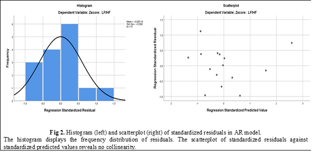
The residuals of the linear regression model satisfied the necessary conditions. The observed distribution exhibited a slight right skewness and a random scatter without any discernible pattern (Fig. 2). These characteristics indicate that the developed model is optimal for predicting LF/HF in individuals with abdominal breathing based on the provided data.

Discussions

The present study documents that PR parameters, measured during both resting breathing and abdominal breathing, can predict sympathovagal modulation in healthy individuals undergoing breathing pattern re-education. Based on the obtained results, we determined that Vt has the greatest predictive power for assessing the balance between sympathetic and parasympathetic activity, as measured by the LF/HF ratio in individuals practicing abdominal breathing. The unstandardized coefficient for Vt is 5.007, followed by Te (B = -3.831) and MVR (B = 4.415), both measured during resting breathing. Consequently, we can predict that decreasing Vt or increasing MVR during resting breathing may lead to a reduction in the LF/HF ratio during abdominal breathing. This can be explained by an accentuation of parasympathetic influences and a decrease in sympathetic influences. However, these findings are not immediately evident due to the general lack of change in HRV. Further studies incorporating longer periods of abdominal breathing may reveal more pronounced alterations in HRV.

The LF/HF ratio observed during the abdominal breathing pattern can also be predicted by the PR parameters measured during abdominal breathing. The most effective predictors are found to be the PR time parameters, including the frequency of breathing in the abdominal breathing pattern (FR) with a coefficient of -5.953, the duration of the inspiratory phase (Ti) with B = -4.037, and the duration of the expiratory phase (Te) with B = -4.194. Increasing FR along with an increase in Ti or increasing FR along with an increase in Te would lead to a reduction in the LF/HF ratio, thereby improving the sympathovagal balance.

Therefore, we can assume that individuals with higher MVR at rest and correspondingly higher frequency in abdominal breathing may experience a decrease in the sympathovagal balance during abdominal breathing.

In conclusion, by modulating these two parameters of the breathing pattern, namely MVR at rest and the total duration of a respiratory cycle (which influences the frequency of breathing), during normal breathing in healthy individuals, we can potentially enhance the sympathovagal balance.

Conclusion

The statistical analysis data presented in this study enable us to propose a hypothesis that certain volume and time parameters of the breathing pattern have the potential to predict changes in the ratio between sympathetic and vagal tone of the heart. Specifically, abdominal breathing has shown the ability to restore or normalize the sympathovagal balance by modulating the duration of inspiration or expiration.

To gain a deeper understanding of the practical applications of breathing pattern parameters in restoring the LF/HF ratio, particularly in disorders characterized by an elevated sympathovagal balance

Competing interests

None declared.

Patient consent 

Obtained

Ethics approval

This study was approved by the Research Ethics Committee of Nicolae Testemițanu State University of Medicine and Pharmacy (minutes no. 15 from 11.01.2016).

Author’s ORCID ID

Andrei Ganenco – https://orcid.org/0000-0002-9835-5461

References

  1. Strauss-Blasche G, Moser M, Voica M, McLeod DR, Klammer N, Marktl, W. Relative timing of inspiration and expiration affects respiratory sinus arrhythmia. Clin Exp Pharmacol Physiol. 2000 Aug;27(8):601-6. doi: 10.1046/j.1440-1681.2000.03306.x.

  2. Lehrer PM, Smetankin A, Potapova T. Respiratory sinus arrhythmia biofeedback therapy for asthma: a report of 20 unmedicated pediatric cases using the Smetankin method. Appl Psychophysiol Biofeedback. 2000 Sep;25(3):193-200. doi: 10.1023/A:1009506909815

  3. Lehrer PM, Vaschillo E, Vaschillo B. Resonant frequency biofeedback training to increase cardiac variability: rationale and manual for training. Appl Psychophysiol Biofeedback. 2000 Sep;25(3):177-91. doi: 10.1023/A:1009554825745.

  4. Draghici AE, Taylor JA. The physiological basis and measurement of heart rate variability in humans. J Physiol Anthropol. 2016 Sep;35(1):22 doi: 10.1186/s40101-016-0113-7.

  5. Berntson GG, Bigger Jr JT, Eckberg DL, Grossman P, Kaufmann PG, Malik M, Nagaraja HN, Porges SW, Saul JP, Stone PH, van der Molen MW. Heart rate variability: origins, methods, and interpretive caveats. Psychophysiology. 1997 Nov;34(6):623-48. doi: 10.1111/j.1469-8986.1997.tb02140.x

  6. Tiwari R, Kumar R, Malik S, Raj T, Kumar P. Analysis of heart rate variability and implication of different factors on heart rate variability. Curr Cardiol Rev. 2021;17(5):e160721189770. doi: 10.2174/1573403X16999201231203854.

  7. Lehrer P. Biofeedback training to increase heart rate variability. In: Lehrer P, Woolfolk RL, Sim WE, editors. Principles and practices of stress management. 3rd ed. New York: Guilford Press; 2007. p. 227-48.

  8. Task Force of the European Society of Cardiology and the North American Society of Pacing and Electrophysiology. Heart rate variability: standards of measurement, physiological interpretation, and clinical use. Circulation. 1996;93(5):1043-65. doi: 10.1161/01.CIR.93.5.1043.

  9. Thomas BL, Claassen N, Becker P, Viljoen M. Validity of commonly used heart rate variability markers of autonomic nervous system function. Neuropsychobiology. 2019;78(1):14-26. doi: 10.1159/000495519

  10. Hopf HB, Skyschally A, Heusch G, Peters J. Low-frequency spectral power of heart rate variability is not a specific marker of cardiac sympathetic modulation. Anesthesiology. 1995 Mar;82(3):609-19. doi: 10.1097/00000542-199503000-00002.

  11. Moak JP, Goldstein DS, Eldadah BA, Saleem A, Holmes C, Pechnik S, Sharabi Y. Supine low-frequency power of heart rate variability reflects baroreflex function, not cardiac sympathetic innervation. Heart Rhythm. 2007 Dec;4(12):1523-9. doi: b10.1016/j.hrthm.2007.07.019.

  12. Billman GE. Heart rate variability: a historical perspective. Front Physiol. 2011 Nov;2:86. doi: 10.3389/fphys.2011.00086.

  13. Billman GE. The effect of heart rate of the heart rate variability response to autonomic interventions. Front Physiol. 2013 Aug 26;4:222. doi: 10.3389/fphys.2013.00222

  14. Reyes del Paso G, Langewitz W, Mulder LJM, van Roon A, Duschek S. The utility of low frequency heart rate variability as an index of sympathetic cardiac tone: a review with emphasis on a reanalysis of previous studies. Psychophysiology. 2013 May;50(5):477-87. doi: 10.1111/psyp.12027

  15. Haensch C-A, Lerch H, Jörg J, Isenmann S. Cardiac denervation occurs independent of orthostatic hypotension and impaired heart rate variability in Parkinson's disease. Parkinsonism Relat Disord. 2009 Feb;15(2):134-7. doi: 10.1016/j.parkreldis.2008.04.031.

  16. RBI Instrumentation. VISURESP RBI-Instrumentation [Internet]. Meylan (France): RBI; 2005 [cited 2022 Apr 25]. Available from: https://rbi-instrumentation.monsite-orange.fr/page6/index.html.

More articles

Research Harmfulness of prooxidants in bronchopulmonary dysplasia in preterm children
Mariana Ceahlău1*, Rodica Selevestru1,2, Olga Tagadiuc3, Svetlana Șciuca1,2
https://doi.org/10.52645/MJHS.2023.3.01
Oxidative stress can be defined as the imbalance of the redox state of a certain system including living one (organelle, cell, organ/tissue), which excessively produces reactive oxygen and/or reactive nitrogen species (ROS/RNS) that exceed the capacity of the antioxidant defense system, which have the ability to slow down or even prevent the oxidative damage of macromolecules. Oxidative stress is a pathogenic mechanism of a large variety of diseases, including pulmonary one.
Research The impact of imuheptin and imupurin on cytokine profile and antioxidant status in rat model of inflammation
Ina Guțu1*, Nicolae Bacinschi1, Valentin Gudumac2
https://doi.org/10.52645/MJHS.2023.3.03
Insects, throughout evolution, have developed a huge arsenal of active compounds, which they use to defend themselves against enemies and diseases, at the same time in recent years insects have shown great interest as a source of food rich in biologically active substances. Research in recent decades has shown that insects produce a variety of proteins and peptides with antibacterial, antifungal, antiviral, immunomodulatory, anti-inflammatory, antioxidant, antitumor, hepatoprotective, antithrombotic, antihypertensive and detoxifying activity during or after contact with the microbial agent or unfavourable factor.
Research Clinical effectiveness study of the new diagnostic score of acute appendicitis in the elderly
Acute appendicitis is among the three most frequent surgical diseases. The lifetime likelihood of developing acute appendicitis is around 7%. The incidence of acute appendicitis reduces with age after adolescence. Several studies evaluated the relevance of the current scores to the general population, mostly children, but a limited number of studies have studied the elderly population. This study aims to assess the clinical effectiveness of the new diagnostic score for the elderly population in comparison to both the Alvarado score and the non-standardized score.
Research Early results of endovascular treatment using percutaneous vacuum-assisted thromboaspiration in acute lower limb ischemia
Alexandru Predenciuc1*, Vasile Culiuc1,2, Dumitru Casian1,2
https://doi.org/10.52645/MJHS.2023.3.05
Open balloon thrombectomy and embolectomy remain the preferred initial option in the management of acute lower limb ischemia (ALI), but various endovascular techniques have become accessible and are growing in popularity. The aim of the study was to assess our early experience with percutaneous vacuum-assisted thromboaspiration using the Penumbra/Indigo® system for non-traumatic ALI.
Review Venous and arterial endothelium: markers of dysfunction and pathophysiological significance
Victor Ojog1*, Svetlana Lozovanu1
https://doi.org/10.52645/MJHS.2023.3.06
Endothelial dysfunction is a result of complex pathogenic interface involving inflammation, oxidative stress, disorders of endothelization and hemostasis etc., in both arteries and veins, leading to a lot of cardiovascular diseases. Identifying markers with high predictive value has an important diagnostic and prognostic significance.
Review Current affairs in the use of medical ozone. Biological effects. Mechanisms of action
Natalia Cernei1*, Serghei Șandru1, Serghei Cobîlețchi1, Ion Grabovschi2, Ivan Cîvîrjîc1, Ruslan Baltaga1,3
https://doi.org/10.52645/MJHS.2023.3.08
Oxygen-ozone therapy stands as a medically endorsed practice confirmed by numerous international clinical studies. Various authors have illustrated the beneficial clinical outcomes of ozone therapy in terms of its capacity to regulate redox balance, cellular inflammatory responses, and adaptation to ischemia/reperfusion processes. Ozone therapy extends to encompass a range of viral infections, inflammatory disorders, and degenerative ailments, used as both monotherapy and as an adjunct to unified conventional therapies.Introduction Ozone (O3), a gas discovered in the mid-19th century and composed of three oxygen atoms, represents a highly reactive allotropic form of oxygen. It exhibits high solubility in plasma, extracellular fluids, and water (approximately 10 times more soluble in water than conventional oxygen). At room temperature, it is unstable, causing rapid decomposition into ordinary diatomic oxygen. Notably, its half-life measures 25 minutes at 30°C, 40 minutes at 20°C, and 140 minutes at 0°C [1-10]. Medical ozone is a blend of oxygen and ozone derived from medical-grade oxygen through the utilization of a medical ozone generator. This medical ozone contains a concentration of 1-5% ozone and 90-95% pure medical oxygen, or 10-80 μg/mL (0.21-1.68 μmol/ml) of ozone per milliliter of blood. Ozone therapy stands as a current and significant avenue of research in contemporary medicine [1, 3-5, 7, 9, 10-15]. Oxygen-ozone therapy is a medically validated practice supported by numerous international clinical studies. Nowadays, many clinical trials have shown its beneficial effects on the modulation of the oxidoreduction balance, cellular inflammation state, and adaptation to ischemia/reperfusion processes. Ozonotherapy is an effective, safe, feasible, and easy-to-perform technique, which finds applications in various inflammatory, infectious, degenerative diseases, as well as in rehabilitation following acute cardiac and cerebral ischemic events. It demonstrates good efficacy both as an independent treatment and, notably, as an adjunct to conventional therapies [3-5, 7, 11, 16-22]. By incorporating this medical practice, patients can attain significant clinical benefits. When combined with standard therapies, it often leads to reduced medication dosages, complication rates, treatment duration, medication toxicity, and medical expenses. It also addresses the issue of bacterial resistance to antibiotics [2, 4, 18, 19, 21, 23]. In the context of the aforementioned, the purpose of this article is to present a synthesis of the most recent findings regarding ozone's mechanisms of action. Materials and methods To achieve the outlined purpose, an initial search of specialized scientific publications was conducted. These were identified through the Google Search engine, namely, PubMed, Hinari, SpringerLink, the National Center of Biotechnology Information, and Medline. The article selection criteria encompassed contemporary data regarding the mechanisms of action of ozone therapy, utilizing the following keywords: “ozone”, “ozone therapy”, “ozone mechanisms of action”, “biological effects of ozone”, “antioxidant effect”, “anti-inflammatory effect”, and “immunomodulatory effect.” These keywords were employed in various combinations to optimize search efficiency. For the advanced selection of bibliographic sources, the following filters were used: full-text articles, articles in English, articles published between 1990 and 2022. After a preliminary analysis of the titles, original articles, editorials, articles of narrative synthesis, taxonomy, and meta-analysis were selected, which contained up-to-date information and contemporary concepts regarding the mechanisms of ozone therapy. Furthermore, a search was conducted within the reference lists of the identified sources to highlight additional relevant publications that were not found during the initial database searches. The information from the publications included in the bibliography was gathered, organized, evaluated, and synthesized, showcasing the key aspects of the contemporary understanding of ozone's mechanisms of action, namely, its antioxidant capacity, vascular and hematological modulation, immune system activation, as well as its anti-inflammatory, bactericidal, virucidal, and fungicidal effects. To minimize the potential systematic errors (bias) in the study, a meticulous search was conducted within databases to identify a maximum number of relevant publications for the study's purpose. Only studies that satisfy validity criteria were evaluated, rigorous exclusion criteria for articles under consideration were applied, and a comprehensive review was conducted of both positive outcome studies and those that did not highlight the treatment's benefits. If necessary, additional sources of information were consulted to clarify some concepts. Duplicate publications and articles that did not meet the purpose of the article and were not available for full viewing were excluded from the list of publications generated by the search engine. Results Following the data processing, as identified by the Google Search engine and from databases such as PubMed, Hinari, SpringerLink, National Center of Biotechnology Information, and Medline, in accordance with the search criteria, a total 475 articles on the topic of ozone therapy were found. After a primary analysis of the titles, 59 articles were eventually deemed relevant for the given synthesis. Upon repeated review of these sources, a final selection of 52 relevant publications was ultimately made in alignment with the intended purpose. The final bibliography of this work comprises 52 articles that have been considered representative of the materials published on the subject of this synthesis article. Publications, the content of which did not reflect the relevant topic, despite being selected by the search program, as well as articles that were not accessible for open viewing through the HINARI (Health Internet Work Access to Research Initiative) database or available in the scientific medical library of the Nicolae Testemițanu State University of Medicine and Pharmacy, were subsequently removed from the list. Although ozone is the most potent natural oxidant, capable of oxidizing a wide range of organic and inorganic substances, and has the potential for cytotoxicity, researchers believe that under controlled conditions, it possesses numerous therapeutic effects. Moreover, the reactivity to ozone can be effectively mitigated by the blood and cellular antioxidant system [1, 2, 15, 24-28]. Initially used as an empirical approach, oxygen-ozone therapy has now evolved to a stage where the majority of ozone's biological mechanisms of action have been extensively studied and elucidated, these findings being found within the fields of biochemistry, physiology, and pharmacology [18, 22, 25, 27-29]. Ozone is not a pharmaceutical medicine but rather a regulatory molecule capable of generating bioactive mediators. The effects of ozone have been demonstrated to be consistent, safe, and associated with minimal preventable side effects [2, 30]. Chronic oxidative stress, chronic inflammatory processes, and immune overactivation are present and highly detrimental in a wide variety of diseases. The effectiveness of ozone therapy is determined by moderate oxidative stress, resulting from the interaction of ozone with the biological components of the body, triggering an endogenous cascade of biochemical reactions [31]. Ozone can function as an oxidant either directly, when it dissolves in plasma and other biological fluids, immediately reacting with polyunsaturated fatty acids, antioxidants, cysteine-rich proteins, and carbohydrates; or indirectly, by generating reactive oxygen species (ROS) and lipid oxidation products (LOP) [25, 28, 32-37]. At the onset of ozone therapy, an endogenous cascade is triggered, releasing bioactive substances in response to transient and moderately induced oxidative stress by ozone ('oxidative eustress'). Ozone can easily induce this oxidative stress due to its plasma solubility. Reacting with polyunsaturated fatty acids and water, ozone forms ROS in human fluids and tissues. The main molecule among ROS is hydrogen peroxide (H2O2) – a non-radical oxidant. Concurrently, ozone also gives rise to LOP – the lipoperoxide radical, hydroperoxides, malondialdehyde, isoprostanes, ozoneides, alkenes, and predominantly, 4-hydroxynonenal. ROS and LOP are the effector molecules responsible for modulating several biological and therapeutic effects in the body following ozone therapy [3, 8, 13, 27, 34, 37-39]. Having reacted with a number of biomolecules, ozone disappears, and hydrogen peroxide, the main molecule of ROS, and other mediators rapidly diffuse into cells, activating various metabolic pathways with numerous biological and therapeutic effects [3, 5, 27, 28, 35, 40]. Therefore, ROS and LOP are “biological messengers of ozone” and are responsible for the biological and therapeutic effects of ozone. ROSs are short-acting early messengers and are responsible for immediate biological effects, while LOPs are important late and long-term messengers [3, 5, 10, 13, 14, 17, 28, 36]. The formation of ROS in plasma occurs extremely quickly (less than a minute), accompanied by a moderate and transient decrease in the antioxidant capacity of the blood (from 5% to 25%). However, this antioxidant capacity returns to normal within 15-20 minutes [3, 9, 17, 28, 31, 40, 41]. Discussion Although not fully comprehended, the present article will delve into the mechanisms underlying the antioxidant, anti-inflammatory, immunomodulatory, antimicrobial, antiviral, and analgesic effects of ozone. The antioxidative capacity is considered one of the key impacts of ozone therapy. Moderate oxidative stress induced by ozone within the therapeutic range (10-80 μg/mL), most commonly 30-45 μg/mL (the 'physiological' dose of ozone), physiologically effective and recommended levels for systemic application, elicits controlled low doses of ROS acting primarily as signaling molecules, thereby stimulating the formation of LOP. ROS triggers the activation of nuclear erythroid factor 2 (Nrf2), well known as a pivotal regulator of manifold cytoprotective responses, responsible for upregulating antioxidant enzyme activity. In response to transient, moderate oxidative stress, the levels of antioxidant enzymes such as superoxide dismutase, glutathione peroxidase, glutathione S-transferase, catalase, and heme oxygenase-1 increase. Thus, moderate, transient, and repetitive oxidative stress causes an intense modulation of antioxidants in the body. A multitude of cells across various organs upregulate the synthesis of antioxidants, which are capable of significantly countering excessive ROS, thereby alleviating chronic oxidative stress, which is present and extremely harmful in a variety of diseases Consequently, ozone, either through oxidative preconditioning or adaptation to chronic oxidative stress, safeguards tissue integrity against ROS-induced damage, fostering a balance between antioxidant and pro-oxidant factors while preserving cellular redox balance [27, 30, 41-45]. Nrf2 and nuclear factor kappa Β (NF-κB) represent the primary signaling pathways through which ozone exerts its effects. Nrf2 activation regulates cell defense and maintains cellular homeostasis [36]. Furthermore, ozone therapy fosters adaptation to oxidative stress by gently triggering the immune system, releasing growth factors, and/or activating metabolic pathways that contribute to maintaining redox balance [38]. By activating Nrf2, LOP induces oxidative stress proteins, including heme-oxygenase-1 (HO-1), another inhibitor of the NF-κB pathway and one of the most crucial antioxidant defense enzymes. Through inhibiting the high expression level of hypoxia-inducible factor-1α (HIF-1α), it contributes to reducing the production of proinflammatory cytokines, directly activating anti-inflammatory cytokines, enhancing antioxidant protection, and consequently, safeguarding cellular integrity [8, 28, 30, 44-46]. Thus, ozone mimics acute oxidative stress which, when properly balanced, is not harmful, but can trigger several beneficial biochemical mechanisms. It can reactivate the intra- and extracellular antioxidant system, thereby reversing chronic oxidative stress in various inflammatory, degenerative processes, etc. During ozone treatment, cells throughout the body receive gradual and subtle impulses of LOP, significant long-term messengers that play a crucial role in up-regulating antioxidant enzymes in multiple cell types while rebalancing the oxidant/antioxidant system [46, 47]. Vascular and Hematological Modulation. Ozone serves as a catalyst for transmembrane oxygen flow. The increase in cellular oxygen levels resulting from ozone therapy enhances the efficiency of the mitochondrial respiratory chain. Moreover, ozone amplifies the production of prostacyclin, a widely acknowledged vasodilator [2, 6, 8, 26]. The effects of ozone on oxygen metabolism can be explained by promoting (1) changes in the rheological properties of blood (reversal of erythrocyte aggregation, increased flexibility and elasticity of red blood cells, favoring the transport and delivery of tissue oxygen), leading to enhanced blood flow in microcirculation; (2) increasing the speed of glycolysis in erythrocytes; and (3) the release of substances (adenosine triphosphate, nitric oxide, and prostaglandins) that may contribute to reducing peripheral vascular resistance and increasing oxygen supply to tissues [18, 25, 35, 37, 40, 48-50]. Hydrogen peroxide (H2O2) diffuses from the plasma into the cellular cytoplasm and serves as the triggering stimulus. Depending on the cell type, various biochemical pathways can be simultaneously activated in red blood cells, white blood cells, and platelets, leading to a multitude of biological effects [10, 28, 32]. The Impact of Ozone on Erythrocytes. Erythrocytes are the focus of ROS. During erythropoiesis, submicromolar concentrations of LOP positively regulate the synthesis of antioxidant enzymes. Consequently, ozone therapy increases the glycolytic rate by enhancing intracellular adenosine triphosphate production. This approach intensifies erythrocyte generation, yielding metabolically enhanced erythrocytes (super-endowed erythrocytes) capable of more effectively transporting and delivering oxygen to tissues, including ischemic tissues, thereby correcting hypoxia in vascular diseases [7, 12, 25, 27, 28, 39, 48]. Coupled with increased nitric oxide synthase activity, there is a significant increase in nitric oxide, an essential element in maintaining optimal levels of vasodilation and blood perfusion [1, 6, 8, 40]. Ozone therapy, through careful regulation of ozone dosage, stimulates the production of antioxidant enzymes within the system (catalase, glutathione peroxidase, and superoxide dismutase) while mitigating excessive formation of ROS, thereby reducing chronic oxidative stress [1, 6, 12, 14, 39, 43, 49]. The impact of ozone on leukocytes. Ozone acts as a mild cytokine and serves as a cytokine inducer by lymphocytes and monocytes, thereby enhancing the immune system's activity. This stimulation fosters intercellular matrix synthesis and contributes to the healing process [1, 12, 32, 35, 37, 39]. The Impact of ozone on platelets. Hydrogen Peroxide (H2O2) and other ROS generated through blood ozonation initiate a cascade of enzymatic reactions. These reactions gradually elevate intracellular Ca levels and trigger the release of prostaglandins (F2a and E2), leading to irreversible platelet aggregation. Increased levels of growth factors released from platelets, mobilization of endogenous stem cells, and stimulation of neoangiogenesis promote tissue regeneration, as well as healing of injuries and wounds [27, 49, 51]. Thus, the impact of ozone on oxygen metabolism is explained by how it alters the blood's rheological properties (reversing red blood cell clumping, enhancing the flexibility and elasticity of red blood cells, promoting the transport and delivery of oxygen to tissues). This, in turn, facilitates blood flow in the microcirculation, increases glycolysis in red blood cells, and triggers the production of substances (such as adenosine triphosphate, nitric oxide, and prostaglandins) that help reduce peripheral vascular resistance. Activation of the immune system. Ozone promotes an increase in the production of interferon-γ (IFN-γ) and some cytokines, with interleukin-2 (IL-2) being the primary one, subsequently triggering a whole cascade of immunological reactions [1, 2]. It has been shown that ROS, including H2O2 and LOP generated by ozone therapy, can easily diffuse into plasma cells and activate NF-κB, inducing the production of immunoactive cytokines in normal cells (IL-2, tumor necrosis factor alpha - TNF-a, IL-6 and IFN-γ), thereby enhancing the immune response [9, 13, 14, 26, 32, 40, 44]. Ozone indirectly activates the innate (non-specific) immune system by enhancing phagocytosis and promoting the synthesis of cytokines and interleukins in neutrophils and leukocytes. It also triggers the components of both cellular and humoral immunity [8, 26, 33, 39]. Within mononuclear cells, ozone stimulates immune responses by modulating the NF-kB transcription factor, thereby reactivating the suppressed immune system [27, 28]. Furthermore, ROSs trigger the activation of the immune system, which acts through monocytes and lymphocytes, promoting the production of a variety of cytokines (IL-1, IL-2, IL-6, IFN-β, IFN-γ, TNFα) [6, 49]. Thus, ozone induces mild immune system activation by stimulating neutrophils and initiating the synthesis of certain cytokines that trigger a whole cascade of immunological responses. Bactericidal, virucidal and fungicidal action of ozone. Ozone used in vitro acts directly on the membrane of bacterial cells (direct oxidative effect), disrupting and damaging the integrity of bacterial cell membranes, oxidizing phospholipids and lipoproteins, thereby impeding their enzymatic function. Additionally, ozone damages the viral capsids, disturbing their structure and interfering with the virus-cell interaction, leading to disruption in the reproductive cycle. When it comes to fungi, ozone inhibits cell growth by perturbing intracellular homeostasis, resulting from the compromised barrier properties of the plasma membrane [2, 6, 12, 16, 26, 44, 48, 50]. Although ozone is one of the most potent disinfectants, used in various ways, it cannot deactivate any pathogens (bacteria, viruses, and fungi) in vivo. This is because pathogens are well protected, especially within cells, by the cell's powerful antioxidant system. Consequently, ozone acts as a gentle enhancer of the immune system by activating neutrophils and stimulating the synthesis of certain cytokines [1, 10, 19, 22, 28, 39, 46]. The anti-inflammatory effect is revealed in ozone's ability to influence the inflammatory cascade by oxidizing biologically active substances (arachidonic acid and its derivatives - prostaglandins), which participate in the development and sustenance of the inflammatory process. Additionally, ozone significantly reduces the levels of pro-inflammatory cytokines (IL-1β, IL-6, IL-8, and TNF-α) without any signs of toxicity or recorded side effects [8, 26, 30, 31]. These cytokines induce the prostaglandin E2 pathway, which causes pain or increases the sensitivity of nerve roots to other algogenic substances (such as bradykinin) [31]. Severe oxidative stress, triggered by high concentrations of ozone, along with proinflammatory cytokines (IL-1β, IL-6, IL-8, TNF-α), activate NF-κB, a key regulator of the inflammatory response and muscle atrophy. This contributes to an increased inflammatory response and tissue damage, including the release of other inflammatory factors that enhance the migration of eosinophils and neutrophils [9, 13, 17, 47, 49]. On the contrary, mild oxidative stress induced by precise and small doses of ozone activates Nrf2. The latter indirectly inhibits the pro-inflammatory mechanism driven by the NF-kB pathway. As a result, there is a reduction in NF-κB activity along with a modification in the expression of inflammatory cytokines associated with NF-kB activity. This triggers an anti-inflammatory effect, leading to a decrease in IL-1, IL-2, IL-6, IL-7, and TNFα, as well as an increase in interleukins such as IL-4, IL-10, IL-13, and the transforming growth factor beta – TGF-β [11, 13, 19, 38, 43, 49, 50]. Nrf2 also plays an important role in intracellular inflammatory signaling pathways. Triggering the Nrf2-antioxidant signal can dampen NF-kB activity, leading to the downregulation of the inflammatory response by suppressing essential inflammatory mediators and cytokines (IL-6, IL-8, and TNF-a) [31, 38, 42, 50]. Moreover, a small amount of H2O2 stimulates the NF-kB pathway, which is typically balanced out by the Nrf2's blocking action, resulting in an immunomodulatory effect [11]. The analgesic effect of ozone is ensured by the oxidation of the byproducts of albuminolysis, known as algopeptides, which act on the nerve endings in the damaged tissue and determine the intensity of the pain response. Additionally, the analgesic effect is attributed to the restoration of the antioxidant system and, subsequently, the reduction of harmful molecular byproducts from lipid peroxidation [26]. Recent preclinical studies have elucidated the role of ROS in hyperalgesia by activating N-methyl-D-aspartate receptors [11]. Following ozone therapy, there has been a demonstrated increase in antioxidant molecules (serotonin and endogenous opioids), which induce pain relief by stimulating antinociceptive pathways [31, 39]. Data from scientific research acknowledge that the mechanisms of action of ozone are due to: (1) a decrease in the production of inflammatory mediators; (2) oxidation (inactivation) of metabolic mediators of pain; (3) improvement of local blood microcirculation leading to improved oxygen delivery to tissues; (4) elimination of toxins and resolution of physiological disorders that generate pain [42, 52]. Therefore, ozone exhibits pleiotropic properties, extending beyond its exclusive role as an antioxidant, anti-inflammatory, or immunomodulatory one. It also encompasses the capacity to employ ROS as a signaling molecule rather than merely as intracellular toxic substances. In existing experimental models and clinical studies, the anti-inflammatory, antioxidant, regenerative and immunomodulatory effects of ozone therapy have been associated with several molecular mechanisms, the main ones being the NF-kB/Nrf2 balance and IL-6 and IL-1β expression. NF-kB and Nrf2 are the most studied and important transcription factors and regulatory proteins that control the expression of a wide range of genes, encoding proteins involved in a multitude of vital biological functions, including those associated with redox status, immunity, and inflammatory responses. Additionally, indirectly through these pathways, LOP initiates the HIF-1α, HO-1, and NO/iNOS pathways. The main pharmacological effects of medical ozone through ozone-produced peroxides are as follows: (1) increased oxygen release by erythrocytes due to activated metabolism; (2) immunomodulation due to leukocyte activation; and (3) regulation of cellular antioxidants via Nrf2 signaling. Ozone therapy can elicit the following biological reactions: (a) improved blood circulation and oxygen delivery to ischemic tissue; (b) optimization of overall metabolism by improving oxygen delivery; (c) regulation of cellular antioxidant enzymes and induction of HO-1; (d) triggering a mild immune system activation and intensified release of growth factors; (e) providing a state of well-being in most patients, probably due to stimulation of the neuroendocrine system. Conclusions 1. Ozone induces both mild and moderate oxidative stress. When appropriately balanced, this stress poses no harm; instead, it can initiate several beneficial biochemical mechanisms. These mechanisms, in turn, reactivate the intracellular and extracellular antioxidant systems, effectively countering long-term oxidative stress in various inflammatory and degenerative processes, etc. Cells throughout the body receive small and gradual bursts of lipid oxidation products, important late and long-term messengers that are responsible for activating antioxidant enzymes in many cell types to rebalance the oxidant/antioxidant system. 2. The impact of ozone on oxygen metabolism is explained by changes in the blood's rheological properties. This involves reversing red blood cell aggregation, enhancing the flexibility and elasticity of hemoglobin, and promoting the efficient transport and delivery of oxygen to tissues. This process also facilitates blood flow within microcirculation, speeds up glycolysis within red blood cells, and triggers the release of substances like adenosine triphosphate, nitric oxide, and prostaglandins, which help to reduce peripheral vascular resistance. 3. Ozone triggers a slight activation of the immune system by up-regulating and activating neutrophils and promoting the synthesis of cytokines (IL-2, TNF-a, IL-6, and IFN-γ), setting off a chain reaction of immune responses. 4. Ozone therapy induces the following biological responses: enhanced blood circulation and oxygen delivery to ischemic tissue, regulation of cellular antioxidant enzymes, mild immune system activation, and intensified release of growth factors. 5. Ozone is an inherently toxic gas that should never be inhaled, cannot be stored, and must be handled with caution. Generally, no toxic effects were reported, and only the respiratory tract was found to be highly sensitive to inhaled ozone since the respiratory mucosal cells contain a minimal amount of antioxidants and are extremely susceptible to oxidation. 6. Although ozone ranks among the most potent disinfectants, being employed in various ways, it cannot neutralize any pathogens (bacteria, viruses, and fungi) in vivo, since pathogens are effectively shielded by the strong blood and cellular antioxidant system. Furthermore, elevated ozone concentrations induce severe oxidative stress, prompting increased inflammatory responses and tissue damage. Competing interests None declared. Authors’ contribution NC and RB conceived the study, participated in the study design and assisted in drafting the manuscript. SȘ and IC performed the analysis and data interpretation. IG drafted the manuscript. SC conceived the significant revision of the manuscript and provided significant intellectual involvement. The authors have read and approved the final version of the manuscript. Authors’ ORCID IDs Natalia Cernei – https://orcid.org/0000-0002-2031-5881 Serghei Șandru – https://orcid.org/0000-0002-2973-9154 Ion Grabovschi – https://orcid.org/0000-0002-7716-9926 Ivan Cîvîrjîc – https://orcid.org/0000-0002-1360-5485 Ruslan Baltaga – https://orcid.org/0000-0003-0659-4877 References 1. Cakir R. General aspects of ozone therapy [Internet]. In: Atroshi F, editor. Pharmacology and nutritional intervention in the treatment of disease. London: IntechOpen, 2014 [cited 2023 Apr 12]. Available from: http://dx.doi.org/10.5772/57470 2. Elvis A.M., Ekta J.S. Ozone therapy: a clinical review. J. Nat. Sci. Biol. Med., 2011 Jan; 2 (1): 66-70. doi: 10.4103/0976-9668.82319. 3. Bocci V. Ozone: a new medical drug. 2nd ed. New York: Springer, 2011; 315p. https://doi.org/10.1007/978-90-481-9234-2. 4. Allorto N. Oxygen-ozone therapy: an extra weapon for the general practitioners and their patients. Ozone Ther., 2019 July; 4 (2): 8424. doi: 10.4081/ozone.2019.8424. 5. Baeza-Noci J., Cabo-Soler J.R., Moraleda-Gómez M. et al. Revisión WFOT sobre Ozonoterapia Basada en Evidencias. Comité Científico Asesor de la WFOT – 2015 [WFOT Review on Evidence-Based Ozone Therapy. WFOT Scientific Advisory Committee – 2015]. Brescia: World Federation of Ozone Therapy, 2015; 117p. Spanish. https://semdor.es/wp-content/uploads/2021/10/WFOT-OZONE-2015-ESP.pdf. 6. Scassellati C., Ciani M., Galoforo A., Zanardini R., Bonvicini C., Geroldi C. Molecular mechanisms in cognitive frailty: potential therapeutic targets for oxygen-ozone treatment. Mech. Ageing Dev., 2020 Mar; 186: 111210. doi: 10.1016/j.mad.2020.111210. 7. Yousefi B., Banihashemian S., Feyzabadi Z., Hasanpour S., Kokhaei P., Abdolshahi A. et al. Potential therapeutic effect of oxygen-ozone in controlling of COVID-19 disease. Med. Gas Res., 2022 Apr-Jun; 12 (2): 33-40. doi: 10.4103/2045-9912.325989. 8. Smith N., Wilson A., Gandhi J., Vatsia S., Khan S. Ozone therapy: an overview of pharmacodynamics, current research, and clinical utility. Med. Gas Res., 2017 Oct 17; 7 (3): 212-9. doi: 10.4103/2045-9912.215752. 9. Sagai M., Bocci V. Mechanisms of action involved in ozone therapy: is healing induced via a mild oxidative stress? Med. Gas Res., 2011 Dec 20; 1:29. doi: 10.1186/2045-9912-1-29. 10. Bocci V., Borrelli E., Travagli V., Zanardi I. The ozone paradox: ozone is a strong oxidant as well as a medical drug. Med. Res. Rev., 2009 Jul; 29 (4): 646-82. doi: 10.1002/med.20150. 11. Hidalgo-Tallón F., Torres-Morera L., Baeza-Noci J., Carrillo-Izquierdo M., Pinto-Bonilla R. Updated review on ozone therapy in pain medicine. Front. Physiol., 2022 Feb 23; 13: 840623. doi: 10.3389/fphys.2022.840623. 12. Manjunath R., Singla D., Singh A. Ozone revisited. J. Adv. Oral Res., 2015 May-Aug; 6 (2): 5-9. https://doi.org/10.1177/222941122015020. 13. Cenci A., Macchia I., La Sorsa V., Sbarigia C., Di Donna V., Pietraforte D. Mechanisms of action of ozone therapy in emerging viral diseases: immunomodulatory effects and therapeutic advantages with reference to SARS-CoV-2. Front Microbiol., 2022 Apr 21; 13: 871645. doi: 10.3389/fmicb.2022.871645. 14. du Plessis L.H., van der Westhuizen F.H., Kotzé H.F. The protective effect of plasma antioxidants during ozone autohemotherapy. Afr. J. Biotechnol., 2008 Aug; 7 (14): 2472-7. doi: 10.4314/ajb.v7i14.59039. 15. Rowen R.J. Ozone and oxidation therapies as a solution to the emerging crisis in infectious disease management: a review of current knowledge and experience. Med. Gas Res., 2019 Oct-Dec; 9 (4): 232-7. doi: 10.4103/2045-9912.273962. 16. Tizaoui C. Ozone: a potential oxidant for COVID-19 virus (SARSCoV-2). Ozone Sci. Eng., 2020 Jul; 42 (5): 378-85. https://doi.org/10.1080/01919512.2020.1795614. 17. Zanardi I., Borrelli E., Valacchi G., Travagli V., Bocci V. Ozone: a multifaceted molecule with unexpected therapeutic activity. Curr. Med. Chem., 2016; 23 (4): 304-14. doi: 10.2174/0929867323666151221150420. 18. Radzimierska M., Śmigielski K. Ozone and products of ozonation in medical use. Biomed J. Sci. Tech. Res., 2019 Apr; 17 (1): 002959. doi: 10.26717/BJSTR.2019.17.002959. 19. de Sire A., Agostini F., Lippi L., Mangone M., Marchese S., Cisari C. et al. Oxygen-Ozone therapy in the rehabilitation field: state of the art on mechanisms of action, safety and effectiveness in patients with musculoskeletal disorders. Biomolecules, 2021 Feb 26; 11 (3): 356. doi: 10.3390/biom11030356. 20. Wen Q., Liu D., Wang X., Zhang Y., Fang S., Qiu X. et al. A systematic review of ozone therapy for treating chronically refractory wounds and ulcers. Int. Wound J., 2022 May; 19 (4): 853-70. doi: 10.1111/iwj.13687. 21. Mendes C., Cunha M., Ferreira A., Santos E. Ozone therapy as a coadjuvant in wound healing and pain reduction. Millenium, 2021 Sep; 2 (9): 131-8. https://doi.org/10.29352/mill029e.25237. 22. Smith A., Oertle J., Warren D., Pratoet D. Ozone therapy: a critical physiological and diverse clinical evaluation with regard to immune modulation, anti-infectious properties, anti-cancer potential, and impact on anti-oxidant enzymes. Open J. Mol. Integr. Physiol., 2015 Aug; 5 (3): 37-48. doi: 10.4236/ojmip.2015.53004. 23. León Fernández O.S., Oru G.T., Hansler R.V., Cabreja G.L., Espinosa I.S., Polo Vega J.P. et al. Medical Ozone: the pharmacological mechanisms accounting for its effectiveness against COVID-19/SARS-COV-2. J. Med. Clin. Res. Rev., 2021 Mar; 5 (3): 1-10. doi: 10.33425/2639-944X.1199. 24. Bocci V. Biological and clinical effects of ozone. Has ozone therapy a future in medicine? Br. J. Biomed. Sci., 1999 Apr; 56 (4): 270-9. 25. Di Mauro R., Cantarella G., Bernardini R., Di Rosa M., Barbagallo I., Distefano A. et al. The biochemical and pharmacological properties of Ozone: the smell of protection in acute and chronic diseases. Int. J. Mol. Sci., 2019 Feb 1; 20 (3): 634. doi: 10.3390/ijms20030634. 26. Maslennikov O.V., Kontorshchikova C.N., Gribkova I.A. Ozone therapy in practice. Health manual [Internet]. Nizhny Novgorod (Russia); 2008 [cited 2023 Feb 23]. 42 p. Available from: https://absoluteozone.com/wp-content/uploads/2022/05/Ozone-Therapy-in-Practice.pdf. 27. Tricarico G., Travagli V. The relationship between Ozone and human blood in the course of a well-controlled, mild, and transitory oxidative eustress. Antioxidants (Basel), 2021 Dec 4; 10 (12): 1946. doi: 10.3390/antiox10121946. 28. Borrelli E., Bocci V. Basic biological and therapeutic effects of ozone therapy in human medicine [Internet]. In: Munter R, editor. Ozone science and technology. Oxford, UK: Encyclopedia of Life Support Systems, 2010 [cited 2022 Apr 18]. Available from: https://www.eolss.net/sample-chapters/c07/E6-192-15-00.pdf 29. Inal M., Dokumacioglu A., Özcelik E., Ucar O. The effects of ozone therapy and coenzyme Q₁₀ combination on oxidative stress markers in healthy subjects. Ir. J. Med. Sci., 2011 Sep; 180 (3): 703-7. doi: 10.1007/s11845-011-0675-7. 30. Chirumbolo S., Valdenassi L., Simonetti V., Bertossi D., Ricevuti G., Franzini M. et al. Insights on the mechanisms of action of ozone in the medical therapy against COVID-19. Int. Immunopharmacol., 2021 Jul; 96: 107777. doi: 10.1016/j.intimp.2021.107777. 31. de Sire A., Marotta N., Ferrillo M., Agostini F., Sconza C., Lippi L. et al. Oxygen-ozone therapy for reducing pro-inflammatory cytokines serum levels in musculoskeletal and temporomandibular disorders: a comprehensive review. Int. J. Mol. Sci., 2022 Feb 25; 23 (5): 2528. doi: 10.3390/ijms23052528. 32. Bocci V., Zanardi I., Travagli V. Has oxygen-ozonetherapy a future in medicine? J. Exp. Integr. Med., 2011 Jan; 1: 5-11. doi: 10.5455/jeim.161210.ir.002. 33. Néri J., Lomba E., Karam A., Reis S., Marchionni A., Medrado A. Ozone therapy influence in the tissue repair process: a literature review. J. Oral Diagn., 2017 Jul; 2: e20170032. doi: 10.5935/2525-5711.20170032. 34. Bocci V., Valacchi G., Corradeschi F., Fanetti G. Studies on the biological effects of ozone: 8. Effects on the total antioxidant status and on interleukin-8 production. Mediators Inflamm., 1998 Jul; 7 (5): 313-7. doi: 10.1080/09629359890820. 35. Bocci V. Oxygen-ozone therapy: a critical evaluation. Dordrect: Kluwer Academic Publishers, 2002. 440 p. http://doi.org/10.1007/978-94-015-9952-8. 36. Viebahn-Haensler R., León Fernández O. Ozone in Medicine. The low-dose ozone concept and its basic biochemical mechanisms of action in chronic inflammatory diseases. Int. J. Mol. Sci., 2021 Jul 23; 22 (15): 7890. doi: 10.3390/ijms22157890. 37. Bocci V. Ozone as Janus: this controversial gas can be either toxic or medically useful. Mediators Inflamm., 2004 Feb; 13 (1): 3-11. doi: 10.1080/0962935062000197083. 38. Scassellati C., Galoforo A., Bonvicini C., Esposito C., Ricevuti G. Ozone: a natural bioactive molecule with antioxidant property as potential new strategy in aging and in neurodegenerative disorders. Ageing Res. Rev., 2020 Nov; 63: 101138. doi: 10.1016/j.arr.2020.101138. 39. Bocci V., Zanardi I., Travagli V. Ozone acting on human blood yields a hormetic dose-response relationship. J. Transl. Med., 2011 May 17; 9: 66. doi: 10.1186/1479-5876-9-66. 40. International Scientific Committee of Ozonetherapy (ISCO3). Madrid Declaration on Ozone Therapy [Internet]. 2nd ed. Madrid: ISCO3; 2012 [cited 2023 Feb 23]. 50 p. Available from: https://bornclinic.com/wp-content/uploads/2020/12/OZONE-THERAPY.pdf. 41. Thorp J., Thorp K., Thorp E., Viglione D. Ozone preconditioning in viral disease. Virol. Curr. Res. [Internet], 2022; 6 (S1): 001 [cited 2023 Feb 23]. Available from: https://www.hilarispublisher.com/open-access/ozone-preconditioning-in-viral-disease.pdf. 42. Chirumbolo S., Varesi A., Franzini M., Valdenassi L., Pandolfi S., Tirelli U. et al. The mito-hormetic mechanisms of ozone in the clearance of SARS-CoV2 and in the COVID-19 therapy. Biomedicines, 2022 Sep 12; 10 (9): 2258. doi: 10.3390/biomedicines10092258. 43. Pivotto A.P., Banhuk F.W., Staffen I.V., Daga M.A., Ayala T.S., Menolli R.A. Clinical uses and molecular aspects of ozone therapy: a review. OnLine J. Biol. Sci., 2020 Feb; 20 (1): 37-49. https://doi.org/10.3844/ojbsci.2020.37.49. 44. Obeid B.M. Ozone autohemotherapy: possible mechanisms of anti-viral action and anti oxidative. J. Infect. Dis. Epidemiol., 2020 Apr; 6: 117. doi: 10.23937/2474-3658/1510117. 45. Galiè M., Covi V., Tabaracci G., Malatesta M. The role of Nrf2 in the antioxidant cellular response to medical ozone exposure. Int. J. Mol. Sci., 2019 Aug 17; 20 (16): 4009. doi: 10.3390/ijms20164009. 46. Martínez-Sánchez G., Schwartz A., Donna V. Potential cytoprotective activity of ozone therapy in SARS-CoV-2/COVID-19. Antioxidants (Basel), 2020 May 6; 9 (5): 389. doi: 10.3390/antiox9050389. 47. Ranaldi G., Villani E., Franza L. Rationale for ozone-therapy as an adjuvant therapy in COVID-19: a narrative review. Med. Gas Res., 2020 Jul-Sep; 10 (3): 134-8. doi: 10.4103/2045-9912.289462. 48. Bodrug N., Barba D., Istrati V., Botezatu A. Eficacitatea terapiei cu ozon în medicină [Effectiveness of ozone therapy in medicine]. Chișinău: Medicina, 2012. 116 p. Romanian. 49. Dias E.N., Andrade K.F., Silveira R.S., Machado R.R.P. [The acting of ozoniotherapy in wounds, neuropathies, infections and inflammations: a systematic review]. Braz. J. Dev., 2021; 7 (5): 48604-29. Portuguese. https://doi.org/10.34117/bjdv.v7i5.29786. 50. Borges F., Meyer P., Jahara R., de Morais Carreiro E., Antonuzzo P., Picariello F. et al. Fundamentals of the use of ozone therapy in the treatment of aesthetic disorders: a review. J. Biosci. Med., 2021 Dec; 9: 40-70. doi: 10.4236/jbm.2021.912005. 51. Fitzpatrick E., Holland O., Vanderlelie J. Ozone therapy for the treatment of chronic wounds: a systematic review. Int. Wound J., 2018 Aug; 15 (4): 633-44. doi: 10.1111/iwj.12907. 52. Franzini M., Valdenassi L., Pandolfi S., Tirelli U., Ricevuti G., Simonetti V. et al. The biological activity of medical ozone in the hormetic range and the role of full expertise professionals. Front Public Health, 2022 Sep 16; 10: 979076. doi: 10.3389/fpubh.2022.979076.
Review The role of the lateral pterygoid muscle in temporomandibular disorders
Vitalie Pântea1*, Felicia Tabără1, Mariana Ceban1, Veronica Burduja1, Lilian Nistor2, Olga Ursu3
https://doi.org/10.52645/MJHS.2023.3.09
The clinical concept that would argue that the activity of the lateral pterygoid muscle, being disturbed, would play an important role as an etiological factor in temporomandibular joint dysfunctions is still widely accepted, being also a decisive factor in the correct choice of the treatment plan. However, because of the fact that very few research and clear evidence were conducted and presented to support completely that concept, it continues to remain a very controversial one.
Case study Laser ureteroscopic endopyelotomy efficacy in pyeloureteral junction stenosis
Vladimir Caraion1*, Eduard Pleșca2, Andrei Mezu2, Corneliu Maximciuc2
https://doi.org/10.52645/MJHS.2023.3.10
Pyeloureteral junction stenosis (PUJS) is a condition that affects urinary drainage at level of the renal pelvis and upper ureter. It is found in approximately 1 in 500 newborns, with a higher prevalence in males (2:1 ratio). PUJS is the main cause of congenital hydronephrosis and can also be caused by other specific pathologies. Endoscopic management is the primary treatment for PUJS, particularly in cases of aperistaltic and <2cm intrinsic ureteral stenosis without aberrant vessels.
Case study Treatment of deep carious lesions with mineral trioxide aggregate: clinical case report
Diana Trifan*, Diana Uncuța
https://doi.org/10.52645/MJHS.2023.3.11
Deep carious lesions are a dental disease widely spread among population of all ages. From clinical point of view, they have little symptoms and go unnoticed by the patients a long time, until they provoke dental pulp inflammations. If diagnosed and treated properly, the tooth can be treated conservatively with certain techniques of pulp vitality preservation. An important role in this process plays the innate capacity of regeneration of the pulp-dentine complex and the enhanced stimulating properties of new biomaterials used in dentistry. The aim of this clinical case report is to describe the clinical manifestations and the diagnostic algorithm used in deep caries and to establish a clinical guideline of treatment of deep carious lesion with a calcium silicate hydraulic cement.