MJHS
Navigation
Journal Navigation
Article Navigation
opened article
Volume 10, Issue 3
September 2023
Cover Image

Article contents

opened journal
Volume 10, Issue 3
September 2023
Cover Image

Abstract

Introduction

Open balloon thrombectomy and embolectomy remain the preferred initial option in the management of acute lower limb ischemia (ALI), but various endovascular techniques have become accessible and are growing in popularity. The aim of the study was to assess our early experience with percutaneous vacuum-assisted thromboaspiration using the Penumbra/Indigo® system for non-traumatic ALI.

Material and methods

The study group comprised 13 patients with ALI who received treatment between September 2022 and June 2023; with 7 (53.8%) being males. The median age was 71 years (25%-75%IQR 62.5-77.5). ALI cases were classified according to the Rutherford scale: grade I – 2 (15.3%), grade IIA – 7 (53.8%), and grade IIB – 4 (30.7%). In 10 (76.9%) cases, ischemia was classified as "acute-on-chronic." The occluded native vascular segment, determined through preoperative computer tomography angiography (n=4; 30.7%), duplex scanning (n=5; 38.4%), or both examinations (n=4; 30.7%), were as follows: superficial femoral artery (n=7) and popliteal artery (n=2). In two patients, thrombosis of the below-knee femoropopliteal bypass with autogenous vein was identified, while two others presented with femoral artery stent thrombosis. An embolic etiology of ALI was observed in 4 (30.7%) cases, and thrombotic etiologyin 9 (69.2%) cases. Endovascular access was established via the ipsilateral common femoral artery (n=10), crossover (n=2), or brachial artery (n=1).  Thromboaspiration was carried out using dedicated CAT6™ and CAT8™ catheters.

Results

The technical success rate of vacuum-assisted thromboaspiration was 92.3%. Subsequent angiography revealed accompanying occlusive-stenotic lesions in all instances, necessitating transluminal angioplasty, and in 8 (61.5%) patients, additional stenting was required. Catheter-directed thrombolysis was utilized as an adjunct in one patient. There were 2 (15.3%) instances of distal embolization, both addressed within the same surgical session. Survival and limb salvage rates at the 30th-day follow-up stood at 100%.

Conclusions

Utilizing the Penumbra/Indigo® system, percutaneous vacuum-assisted thromboaspiration appears to be a safe and effective minimally invasive technique for treating ALI. This method allows for the concurrent correction of coexisting chronic peripheral arterial lesions.

Key Messages

What is not yet known about the issue addressed in the submitted manuscript 

Since open thromb/-embolectomy and catheter-directed thrombolysis remain the first-choice options in the treatment of acute lower limb ischemia (ALI), the role of other endovascular techniques, including percutaneous aspiration thrombectomy, is not yet well defined. 

The research hypothesis 

We assume that percutaneous vacuum-assisted thromboaspiration using Penumbra/Indigo® device and dedicated catheters (CAT™) can be suitable for ALI of multiple etiology. 

The novelty added by manuscript to the already published scientific literature 

In the context of the first experience applying percutaneous vacuum-assisted thromboaspiration using Penumbra/Indigo® system in the Republic of Moldova, we identified that this is a safe and effective minimally invasive technique for the treatment of non-traumatic ALI of various causes: embolism, native arteries or stent thrombosis, acutely occluded femoropopliteal bypass with autogenous vein conduit; as well as for “acute-on-chronic” ischemia. The method allows simultaneous correction of coexisting chronic peripheral arterial lesions. Intraoperative distal embolization occurs rarely and can be solved during the same surgical session.

Introduction

Acute lower limb ischemia (ALI) is characterized by a sudden decrease in arterial perfusion of the pelvic extremity, potentially threatening the viability of the respective anatomical segment and requiring urgent evaluation and treatment [1]. ALI remains one of the most frequent vascular surgical emergencies, being associated with a high rate of amputation of the affected extremity and a mortality rate surpassed only by that recorded in cases of ruptured abdominal aortic aneurysm [2, 3].

Embolism, in-situ thrombosis of the native artery, stent or vascular graft thrombosis, arterial trauma, or a complicated peripheral aneurysm (sac thrombosis or distal embolization) stand among the common etiological factors of ALI [2, 4]. Atrial fibrillation and mural intracardiac thrombosis that develop after myocardial infarction are presently more frequently noted sources of peripheral embolism [4]. Concurrently, a significant proportion is attributed to iatrogenic embolism, which arises intraoperatively during percutaneous transluminal angioplasty (PTA) for peripheral arterial disease of the lower extremities – an endovascular intervention that is frequently performed in the daily practice of vascular surgery services [5]. In the same context, acute stent thrombosis, particularly at the level of the femoropopliteal artery, is diagnosed in over 6% of cases [6].

Restoring the patency of the arterial lumen as swiftly as possible, ideally within the initial 6-8 hours following the onset of ALI, is crucial for preserving the limb and constitutes the primary objective of treatment [4]. Conventionally, this is achieved through open surgery – thromb/embolectomy using the Fogarty balloon catheter. The technique is characterized by its surgical simplicity, cost-effectiveness, speed, and accessibility, and it is largely clinically effective, particularly in cases of embolic ALI where a single arterial segment is obstructed, especially above the knee [3, 7]. However, the same intervention doesn't yield a similar technical success in the presence of organized embolic masses situated within small-caliber arteries or when embolism occurs against the backdrop of peripheral arterial disease – a situation known as "acute-on-chronic" ischemia [7]. By the way, the latter is being increasingly registered in recent studies dedicated to peripheral arterial embolism [1]. Advancing the balloon catheter towards the infrapopliteal vessels, especially in diabetic patients where distal occlusive-stenotic lesions are characteristic, may encounter difficulties [8, 9]. Also, it's important to take into account that the procedure is typically carried out blindly in the vast majority of cases, without the option of separately guiding the catheter towards the lumen of each calf artery (tibial or peroneal arteries). The routine practice of performing fluoroscopic-assisted balloon thrombectomy or intraoperative angiography is still limited [3, 7, 8, 10]. In the clinical circumstances mentioned above, the extraction of thrombotic masses with the Fogarty catheter often remains incomplete, with the documented rate of residual thrombosis in small-caliber (distal) arteries varying between 36% and 86% [3].

Nonetheless, even in the absence of pre-existing atherosclerotic lesions, the indirect surgical embolectomy using a balloon catheter can be linked to the migration of thrombotic masses (resulting in distal embolization or, conversely, propagation in the proximal direction) or harm to the arterial wall (such as dissection or perforation), delayed pseudoaneurysm or arteriovenous fistula formation, and diffuse arterial narrowing due to intimal proliferation [5, 11]. Therefore, in cases of ALI, more intricate surgical interventions for limb revascularization might frequently be necessary: open procedures (like endarterectomy or bypass surgery) or hybrid approaches (combining open surgery with endovascular techniques) [12].

In an effort to address the aforementioned deficiencies in open surgical ALI treatment over the past two decades, specialized medical companies have introduced new technologies and devices for percutaneous revascularization. Consequently, the current array of endovascular treatment methods applicable to ALI patients encompasses: catheter-directed thrombolysis; ultrasound-accelerated thrombolysis; percutaneous mechanical thrombectomy involving rheolytic or fragmentation (rotational) techniques; pharmacomechanical thrombectomy; simultaneous angioplasty combined with thrombolytic irrigation (SATI technique); as well as manual percutaneous aspiration thrombectomy or the utilization of devices offering continuous automatic thromboaspiration [4, 7, 13]. To the latter group is also attributed the Penumbra Indigo® (Penumbra Inc., Alameda, CA, USA) – a device designed for extracting thrombotic masses/emboli from the lumen of peripheral vessels through percutaneous vacuum-assisted aspiration, applicable in cases of ALI and venous thrombosis. The initial data reported up to this point for the utilization of the Penumbra Indigo® system in ALI seem promising, but the overall evidence remains limited. The objective of the current study was to present the preliminary results of our initial experience with the application of percutaneous vacuum-assisted thromboaspiration in patients with non-traumatic ALI resulting from infrainguinal occlusions.

Material and methods

The study was carried out in the University Vascular Surgery Clinic, Chair of General Surgery-Semiology No.3 of the Nicolae Testemițanu State University of Medicine and Pharmacy (Department of Vascular Surgery, Institute of Emergency Medicine, Chișinău), during the period September 2022 – June 2023. Informed consent was acquired from all subjects encompassed by the study. The data from the electronic register of prospective records of patients who underwent revascularization surgery for ALI were evaluated, and cases treated with the application of percutaneous thromboaspiration using the Penumbra Indigo® system were selected for further analysis. The mentioned research received approval from the institutional Ethics Committee, within the project dedicated to the study of acute ischemia of the extremities (No.1 of 16.02.2021). 

Features of Penumbra Indigo® system.As per the manufacturer's specifications, the Indigo® device (Fig. 1A) comprises the subsequent components: Penumbra Engine® aspiration source, Penumbra Engine canister, Indigo aspiration tubing equipped with a valve switch for system activation and deactivation, Indigo Separator™, and Indigo CAT™ mechanical thrombectomy catheters. 

Fig. 1 General aspect of Penumbra Indigo® system (A) and the dedicated CAT6™ and CAT8™ mechanical thrombectomy catheters (B). 

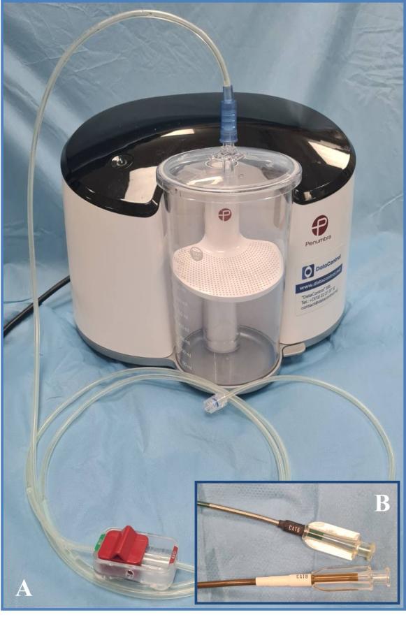
The aspiration source is capable of providing a pure, continuous vacuum (-29 in Hg / 736.6 mm Hg / 98.2 kPa / 0.96 Atm), enabling the elimination of thrombi from the lumen of vessels with various diameters. This capability is also attributed to the availability of an extensive array of catheters with different diameters and lengths, designed to be tapered and resistant to collapsing (Fig. 1B): CAT3, CAT5, CAT6, CAT8 (including various tips: STR / TORQ / XTORQ), CAT D, CAT RX, or CAT7 and CAT12 – the latter two being of a newer generation.

Technical aspects of percutaneous vacuum-assisted thromboaspiration. Following the establishment of endovascular access and the placement of a 6F or 8F sheath, digital subtraction angiography (DSA) was conducted to confirm the location and extent of arterial occlusion. The guide-wire was advanced through the occlusive lesion, and subsequently, the dedicated thromboaspiration catheter was guided towards the proximal end of the lesion. After the catheter tip engaged the thrombus, the suction pump was activated, with a wait time of approximately 90 seconds to enable the creation of negative pressure. Subsequently, the suction tube switch was turned on, and the catheter was gradually withdrawn. Confirmation of thrombi aspiration was visual (by observing the presence of thrombotic masses in the canister after defoaming its contents), and also through DSA. In the presence of co-existing or underlying hemodynamically significant occlusive-stenotic lesions, their simultaneous endovascular treatment was applied, depending on the assessment of the operating surgeons. 

Definitions and data interpretations. As per the guidelines, acute limb ischemia was defined with symptom duration less than 2 weeks [1]. The degree and clinical categories of ischemia were evaluated in line with the widely acknowledged Rutherford ALI classification system, incorporating criteria such as sensory loss, motor deficit, prognosis, and Doppler signals: gr. I (viable), gr. IIA (marginally threatened), gr. IIB (immediately threatened), gr. III (irreversible).

As primary endpoints of the study, the technical and clinical success of thromboaspiration were assessed, with secondary endpoints encompassing the rates of complications, primary patency, limb salvage, and 30-day mortality. Technical success was defined as restoring antegrade blood flow with near/complete aspiration of the embolus/thrombus and maintaining patency in at least one run-off vessel. For interpreting the technical results, the adapted classification for Thrombo-aspiration In Peripheral Ischaemia (TIPI), modified from the Thrombolysis in Myocardial Infarction (TIMI) classification, was employed [14]. Success was indicated by an increase of at least ≥1 point in comparison to the baseline score: 0 (no recanalization of the thrombotic occlusion), 1 (incomplete or partial recanalization of the thrombotic occlusion with no distal flow), 2 (incomplete or partial recanalization of the thrombotic occlusion with any distal flow), and 3 (complete recanalization of the thrombotic occlusion with normal distal flow).

Clinical success was defined as a post-interventional relief of ALI symptoms and an upward shift of at least one grade in the Rutherford classification. Complications/adverse events were categorized using the CIRSE classification system: gr.1 (can be resolved within the same session), gr.2 (requires prolonged observation <48 h), gr.3 (prolonged hospital stay >48 h or additional post-procedure therapy), gr.4 (causes permanent mild sequelae), gr.5 (causes permanent severe sequelae), and gr.6 (results in death) [15]. Primary patency was determined as a target lesion without haemodynamically significant stenosis (>50%) or re-occlusion on duplex scanning, conducted on the first postoperative day and at one month.

Continuous variables are presented as medians with interquartile range (25%-75%IQR), while categorical variables are represented as percentages.

Results 

Patient data and ALI characteristics. The study group comprised 13 patients with ALI resulting from unilateral occlusion of the femoropopliteal arterial segment; 7 (53.8%) were males. Subjects' ages ranged from 43 years to 83 years, with a median value of 71 (25%-75%IQR 62.5-77.5) years. The right lower extremity was affected in 8 (61.5%) cases. All patients exhibited characteristic ALI symptoms (classic "6 P's"), occurring over 72 (25%-75%IQR 24-96) hours after onset (4-302). Thrombosis (n=9; 69.2%) or embolism (n=4; 30.7%) was identified as the etiological factor of ALI. According to the Rutherford classification, cases were distributed as follows: gr. I – 2 (15.3%), gr. IIA – 7 (53.8%), and gr. IIB – 4 (30.7%). In 10 (76.9%) instances, ischemia was categorized as "acute-on-chronic". Among comorbidities, the following were notable: arterial hypertension (n=13), chronic heart failure (n=13), ischemic heart disease (n=9), normo- (n=1) or tachysystolic (n=5) atrial fibrillation, diabetes mellitus (n=3), and chronic obstructive pulmonary disease (n=2). Each patient presented with at least three chronic diseases.

All patients were on chronic anticoagulant/antiplatelet treatment, while 6 (46.1%) were receiving antiarrhythmic medication. In 3 (23%) cases, a recurrent episode of ALI was identified, with the patients having undergone previous open embolectomy at the same limb level. Two (15.3%) patients had a history of superficial femoral artery stenting for chronic occlusive-stenotic lesions, and two others had undergone femoropopliteal below-knee autogenous vein bypass. Laboratory data revealed leukocytosis (n=6), hyperfibrinogenemia (n=6), mild anemia (n=4), and thrombocytopenia (n=2). All patients received therapeutic doses of anticoagulants upon admission: sodium heparin (n=10; 76.9%) or enoxaparin (n=3; 23%).

The occluded arterial segment determined by preoperative computer tomography angiography (n=4; 30.7%), duplex scan (n=5; 38.4%), or both examinations (n=4; 30.7%) included the superficial femoral artery (n=7) and popliteal artery (n=2). Thrombosis of the femoropopliteal below-knee bypass was identified in two patients, while two others experienced acute stent thrombosis of the superficial femoral artery.

Results of the application of thromboaspiration and adjuvant endovascular techniques. 
Patients underwent revascularization through percutaneous mechanical thromboaspiration using the Penumbra/Indigo® system as a primary or salvage intervention within 9 (25%-75%IQR 2.5-48) hours after hospitalization (2-96). In 11 (84.6%) cases, thromboaspiration was performed with local anesthesia, and in two others – under spinal anesthesia. Endovascular access was established via the ipsilateral common femoral artery (n=10), crossover (n=2), or brachial artery (n=1). DSA before thromboaspiration revealed thrombotic occlusion with no distal flow (TIPI score = 0) in all instances. Thromboaspiration was conducted using dedicated CAT6™ (n=3) and CAT8™ (n=10) catheters as the initial choice, depending on the diameter of the targeted vessel. Technical success (TIPI score = 2-3) was achieved in 12 out of 13 (92.3%) cases. In one patient with bypass thrombosis, despite recanalization of the autologous venous conduit and subsequent PTA for infrapopliteal occlusive lesions, restoration of distal flow was not possible. Consequently, a decision was made to opt for catheter-guided thrombolysis.

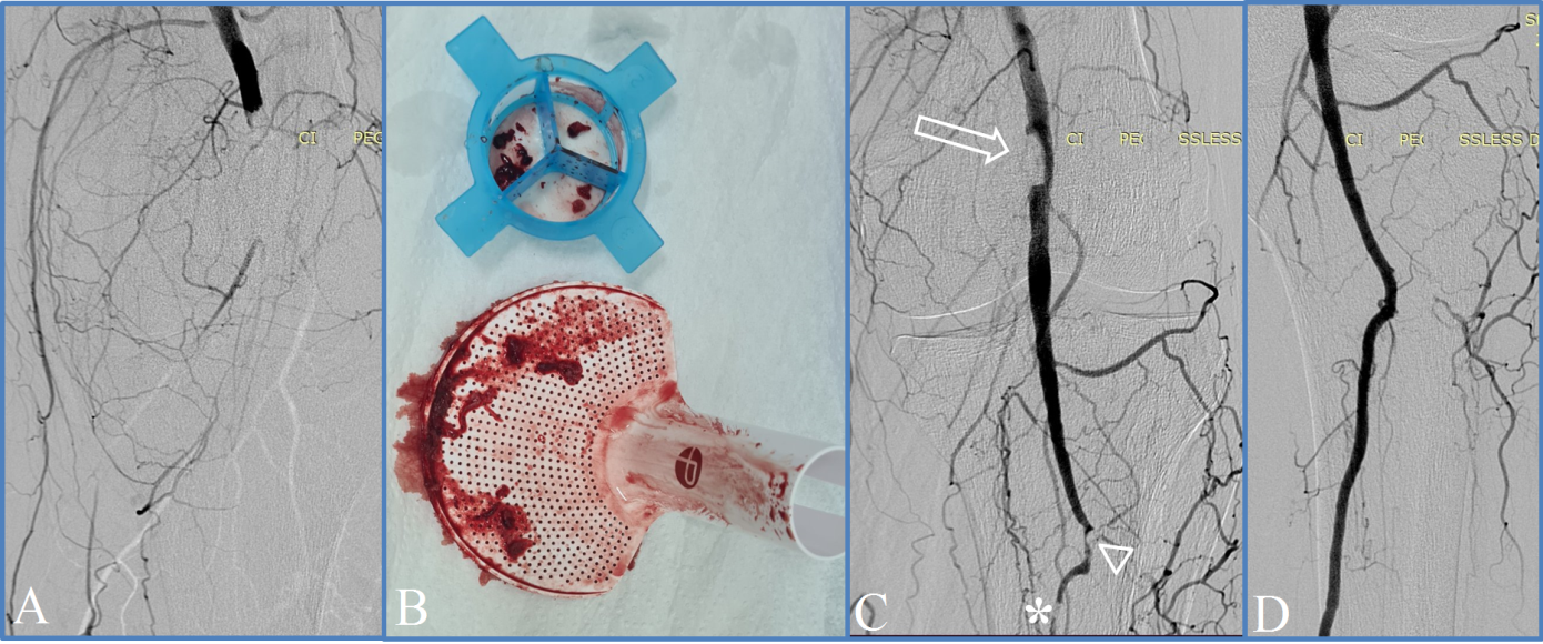
Control angiography following aspiration revealed associated infrainguinal occlusive-stenotic lesions in all cases, necessitating the use of adjunctive techniques: PTA, and in 8 (61.5%) cases – additional stenting of the femoropopliteal segment (Fig. 2). Distal embolization was identified during thromboaspiration in two cases, resulting in a perioperative complication rate of 15.3%. It's important to note that both instances were readily resolved during the same operative session through repeated aspiration (gr.1 CIRSE).

Fig. 2 Angiographic images captured during percutaneous thromboaspiration using the Penumbra/Indigo® system and the extracted thrombotic masses.

Note: (a) – popliteal artery occlusion at diagnostic angiography; (b) – thrombotic masses aspirated into the canister of Indigo® device; (c) – persistent intraluminal embolus (arrow), intraoperative distal embolization (arrowhead) and concomitant chronic arterial lesion (asterisk) identified after initial thromboaspiration; (d) – restoration of blood flow at the completion angiography, after iterative aspiration followed by percutaneous transluminal angioplasty.

The duration of surgical interventions ranged from 60 minutes to 160 minutes, with a median value of 120 (25%-75%IQR 72.5-120) minutes. The volume of intraoperative blood loss (aspirated into the canister) ranged from 260 ml to 480 ml. No patients required postprocedural blood transfusion. The clinical success rate was 92.3%. Follow-up duplex scanning confirmed the preservation of primary patency in all cases. There were no instances of death within the 30-day period, and the limb salvage rate was 100%.

Discussion 

Despite its multiple shortcomings and potential perioperative complications, open thrombectomy with the Fogarty catheter remains the standard approach for ALI caused by embolism [1]. In contrast, the present study reflects the results of the initial experience of percutaneous vacuum-assisted thrombectomy using the Penumbra Indigo® system for ALI in the Republic of Moldova. In all four of our cases considered with embolic etiology, coexisting occlusive-stenotic lesions were identified after percutaneous thromboaspiration, necessitating adjunctive treatment – PTA or stenting. It can be assumed that in these cases, the standard approach might have overlooked the associated lesions with significant hemodynamic impairment, potentially affecting clinical outcomes or necessitating additional surgery. Moreover, three patients had previously undergone surgery for embolism, potentially exposing them to the associated risks of repeated procedures. Generally, considering the substantial percentage of cases presenting with "acute-on-chronic" ischemia (76.9%) and the dominance of arterial thrombosis (69.2%), the conventional open approach within the studied group likely would have necessitated complex revascularization interventions instead of simple balloon thromb-/embolectomy. Given the patients' advanced age and the elevated prevalence of comorbidities, we hold the view that bypass surgery might have been linked to notably higher morbidity rates compared to those observed following the application of the percutaneous technique.

The current guidelines suggest catheter-directed thrombolysis as an alternative to surgery in ALI, with both methods demonstrating comparable clinical outcomes [1, 3, 16]. However, thrombolysis has somewhat more limited indications, being recommended for less severe ALI cases (Rutherford grade IIA); and the rate of complications such as intracranial hemorrhage, major bleeding requiring surgery or transfusion, distal embolization, and compartment syndrome remains high (13%-30%) [11, 14, 17]. Furthermore, the approach demands meticulous monitoring in the intensive care unit with the requirement for subsequent angiography; it is time- and resource-intensive, and the reported high rate of technical success (up to 90%) can be attained only after the accumulation of extensive personal experience [16, 18]. Other previously mentioned endovascular techniques are also associated with a certain percentage of specific complications: hemorrhages, distal embolization (pharmaco-mechanical thrombectomy), hemolysis and renal failure (rheolytic thrombectomy), vessel injury (ultrasound-enhanced thrombolysis) [4, 19].

Among the endovascular techniques potentially associated with reduced periprocedural risks, percutaneous thromboaspiration stands out as a treatment option for ALI patients. In 1978, Horvath et al. first proposed the use of intra-arterial catheter aspiration to address iatrogenic embolism related to PTA [5]. Manual aspiration thrombectomy was subsequently described by Snidermann et al. in 1984 and successfully implemented using sheaths and catheters by Starck et al. in 1985 [11, 14, 20]. Traditionally, this procedure involves the use of a large-bore catheter and manual (50 ml) syringe aspiration, making it a cost-effective and widely available approach with a high technical success rate, ranging between 87% and 96% [21, 22]. Manual thromboaspiration has long been used as a complementary technique to thrombolysis, but current viewpoints suggest that it can also serve as the primary choice, with thrombolysis reserved for cases of treatment failure [10]. However, the method has several limitations. One of the primary concerns is the occurrence of sudden pressure changes during aspiration due to the inability of manual suction to maintain a stable (negative) pressure, which could potentially lead to distal embolization or the movement of the clot in a proximal direction [4, 7]. Additionally, the occurrence of arterial spasm, iatrogenic intimal dissections, or thrombosis is not negligible, mainly due to the necessity for multiple catheter movements [11, 20, 22].

Several devices for automatic thromboaspiration have recently been introduced, among which is the Penumbra Indigo® system. Initially, in 2005, the Penumbra mechanical thrombectomy system, utilizing vacuum aspiration as its primary mechanism of action, became available for revascularizing occluded intracranial vessels in patients with acute ischemic stroke [2, 14]. Following a successful initial experience in treating stroke, acute pulmonary embolism, and renovisceral occlusions, another device was launched in 2014 – the Penumbra Indigo® aspiration thrombectomy system, specifically designed for peripheral applications [5, 14]. The summarized early outcomes, reflecting the experiences of conducting peripheral thromboaspiration for ALI using the Penumbra Indigo® system across various medical centers, in addition to our own data, are outlined in Table 1.

Table 1. Concise synthesis of early outcomes of percutaneous thromboaspiration for ALI using the Penumbra Indigo® system from international experience.

Author (year)

Number of cases

(Assisted) technical success rate

Limb salvage rate at 30 day

Gandini et al. (2015) [8] 

3*

100%

100%

Baumann et al. (2016) [18] 

33

(53.9%)†

-

Saxon et al.; PRISM trial (2017) [11] 

79‡

(96.2%)  

97.5%

Kwok et al. (2018) [19] 

15

53.3%

100%

Lopez et al. (2020) [16] 

43

51%

88.4%

Farhat-Sabet et al. (2020) [5] 

4

100%

100%

de Donato et al.; INDIAN trial (2021) [14] 

150

88.7% (95.3%)

99.3%

Zied et al. (2021) [21] 

19

(94.7%)

100%

Rossi et al. (2021) [12] 

33

70% (90%)

87.8%

Present study (2023) 

13

(92.3%)  

100%  

Note: *treated with Penumbra system (non-dedicated); †for above-the-knee occlusions; ‡52 cases – treated with Penumbra system (non-dedicated), 27 cases – treated with Indigo® system. 

Percutaneous aspiration thrombectomy is suitable for patients with anticipated difficulty during surgical embolectomy (morbidly obese, previous groin surgery) or an anticipated need for adjuvant endovascular procedures [21], as well as in cases when catheter-directed thrombolysis is contraindicated [11, 14]. As in the case of the application of many other techniques, the success of the method is determined by the selection of appropriate cases. More acute thrombus, presumably softer and more malleable, has a higher probability of successful removal [12, 18]. Despite the delayed referral of the patients in the current study (median value of ALI onset–presentation time being 72 hours), we obtained a high clinical success rate, presumably due to the predominance of atherothrombotic cases with well-developed collateralization.

The location of the arterial occlusion is considered a factor that can influence the technical success of the method. In the below-knee segments, the technique has a higher reported rate of success because the lesions are more often iatrogenic, shorter, and there is better concordance between the diameter of the vessel and the catheter [21]. Vice versa, a lower success rate in the above-knee lesions may be explained by a larger mismatch between the vessel size and the thrombectomy catheter size [18]. Therefore, the use of larger catheters, even in smaller vessels, is favored and believed to provide better thrombus removal [16].

Vacuum-assisted thromboaspiration minimizes the risk of endothelium injury and potential iatrogenic distal embolization, which are typical for open surgery [5, 11]. The reported rate of the latter during endovascular interventions is 1-5%, while in some studies it can be as high as 24% [22]. However, the actual frequency of this complication is believed to be much higher, estimated at 30-50%; fortunately, it often remains clinically silent [20, 22]. The open surgical approach in such cases, using the Fogarty balloon catheter in the below-knee arterial segment, could be ineffective due to the difficulty in directing the catheter into crural and foot arteries, and therefore does not appear to be an equivalent alternative to endovascular treatment [8].  In our study group, two (15.3%) cases of distal embolization were identified. However, it should be noted that these occurred during our initial practical experience when the full range of dedicated catheters was not yet available. Fortunately, both complications were ultimately resolved within the same intervention by repeating the aspiration procedure. Jung Guen Cha et al. reported a similar rate of distal embolization (16.7%). It's important to mention that the authors used a Penumbra aspiration catheter and a simple syringe instead of an automated device [3]. Overall, the rate of perioperative complications associated with the use of vacuum-assisted thromboaspiration is acceptable, ranging from 2% to 14%, being basically non-device specific [3, 14, 16]. 

Under the aspect of interventional technique, the use of access sheaths with removable check-flow valves that can be replaced, or rotating hemostatic valves, is considered beneficial. The last one allows for easier introduction of the catheters without damage to the tip, as well as removal of the clot intact when "corked" at the end of the aspiration catheter [11]. Even if its use remains at the discretion of the operating surgeon, the Indigo Separator™ allows thrombus disruption at the tip of the catheter ensuring its patency and makes possible fragmentation of the clot with cleaning the lumen without catheter removal [11, 14]. In general, it is believed that better results are related to a more precise technique, including the 1:1 sizing of the CAT™ catheter to the target vessel diameter in all cases, the application of the Separator™ in almost half of the cases, and the use of more than one catheter per case when necessary [14].

Judicious use of suction control by switching the on/off position of the tube valve and intermittent application of vacuum-assisted aspiration became important tricks for minimizing blood loss [11, 16]. By the way, the volume of hemorrhage in our study was comparable to the amounts described by other authors – on average 240 ml (up to 600 ml) [14].

To the disadvantages of percutaneous aspiration thrombectomy compared to catheter-directed thrombolysis can be attributed a potentially higher risk of traumatic injury to the endothelium and the inability to infuse lytic agents into collaterals or run-off arteries that are too small for an aspiration catheter [3]. However, it is important to mention that in the case of failure of vacuum-assisted thromboaspiration as an initial approach, the possibility of resorting to thrombolysis, embolectomy, or bypass surgery is not ruled out [14, 18]. In a similar context, unsuccessful open thromb-/embolectomy does not exclude the possibility of subsequent application of percutaneous thromboaspiration [3, 23].

Currently, other automated systems for percutaneous aspiration thrombectomy in ALI are also available, such as ClearLumen-II (ClearLumen-II, Groupates), which includes pulse spray thrombolysis; Aspirex (Straub Medical AG), which provides fragmentation of the thrombus by the spinning steel helix; or ThromCat XT (Spectranetics International, Leusden, The Netherlands), which implies rotational thrombectomy [7, 9]. More recently, the Indigo® Lightning™ 7 system, followed by the Lightning Bolt™ 7 and Lightning Flash™, have been implemented, providing intelligent computer-aided mechanical aspiration. Nevertheless, due to the limited evidence regarding the comparative efficacy of the respective methods, at the moment, it cannot be concluded that one of the thromboaspiration techniques is obviously superior in cases of ALI.

In conclusion, the Indigo® system represents a modern, minimally invasive technology with high effectiveness and a low rate of complications, suitable for both primary treatment of ALI and salvage or secondary therapy. The system setup is straightforward and doesn't necessitate the use of adjunctive devices or thrombolytic agents. Advantages of the technique include immediate restoration of blood flow and the absence of the need for protection filters. The availability of dedicated catheters with different diameters allows for reaching clots in very distal arteries, overcoming a limitation present in other techniques [8, 14]. The diverse clinical scenarios suitable for applying this technique to ALI patients, as also confirmed in our study (embolism, atherothrombosis, stent or bypass thrombosis, "acute-on-chronic" ischemia), highlight the wide applicability of the method. These factors could potentially contribute to a shift in the treatment trend for ALI in the near future.

Conclusions 

Percutaneous vacuum-assisted thromboaspiration using the Penumbra/Indigo® system appears to be a safe and effective minimally invasive technique for treating ALI, providing the opportunity for concurrent correction of concomitant chronic peripheral arterial lesions. 

Competing interests

None declared. 

Patient consent 

Obtained

Ethics approval

This study was approved by the Research Ethics Committee of Nicolae Testemițanu State University of Medicine and Pharmacy (minutes no. 1 of 16.02.2021).

Authors’ contributions 

AP performed data collection and drafted the manuscript. VC participated in study design, performed statistical analysis, interpretation of data, and helped drafting the manuscript. DC conceived the study, interpreted the data and helped drafting the manuscript. All the authors reviewed the work critically and approved the final version of the manuscript.

Author’s ORCID IDs

Alexandru Predenciuc - https://orcid.org/0000-0002-2730-8115&nbsp;

Vasile Culiuc - https://orcid.org/0000-0003-3046-3914&nbsp;

Dumitru Casian - https://orcid.org/0000-0002-4823-0804&nbsp;

References 

  1. Björck M, Earnshaw JJ, Acosta S, Bastos Gonçalves F, Cochennec F, Debus ES, et al. Editor's choice - European Society for Vascular Surgery (ESVS) 2020 clinical practice guidelines on the management of acute limb ischaemia. Eur J Vasc Endovasc Surg. 2020;59(2):173-218. doi: 10.1016/j.ejvs.2019.09.006.

  2. Lind B, Morcos O, Ferral H, Chen A, Aquisto T, Lee S, Lee CJ. Endovascular strategies in the management of acute limb ischemia. Vasc Specialist Int. 2019;35(1):4-9. doi: 10.5758/vsi.2019.35.1.4. 

  3. Cha JG, Kim CS, Kim YH, Kim SH. Effectiveness of percutaneous aspiration thrombectomy for acute or subacute thromboembolism in infrainguinal arteries. J Korean Soc Radiol. 2017;76(6):386-94. doi: 10.3348/jksr.2017.76.6.386. 

  4. Araujo ST, Moreno DH, Cacione DG. Percutaneous thrombectomy for initial management of acute limb ischaemia. Cochrane Database Syst Rev. 2019;(11):CD013486. doi: 10.1002/14651858.CD013486. 

  5. Farhat-Sabet AA, Tolaymat B, Voit A, Drucker CB, Santini-Dominguez R, Ucuzian AA, Toursavadkohi SA, Nagarsheth KH. Successful treatment of acute limb ischemia secondary to iatrogenic distal embolization using catheter directed aspiration thrombectomy. Front Surg. 2020;7:22. doi: 10.3389/fsurg.2020.00022. 

  6. Katsanos K, Al-Lamki SA, Parthipun A, Spiliopoulos S, Patel SD, Paraskevopoulos I, Zayed H, Diamantopoulos A. Peripheral stent thrombosis leading to acute limb ischemia and major amputation: incidence and risk factors in the aortoiliac and femoropopliteal arteries. Cardiovasc Intervent Radiol. 2017;40(3):351-9. doi: 10.1007/s00270-016-1513-0. 

  7. de Donato G, Pasqui E, Setacci F, Palasciano G, Nigi L, Fondelli C, Sterpetti A, Dotta F, Weber G, Setacci C. Acute on chronic limb ischemia: from surgical embolectomy and thrombolysis to endovascular options. Semin Vasc Surg. 2018;31(2-4):66-75. doi: 10.1053/j.semvascsurg.2018.12.008. 

  8. Gandini R, Merolla S, Chegai F, Del Giudice C, Stefanini M, Pampana E. Foot embolization during limb salvage procedures in critical limb ischemia patients successfully managed with mechanical thromboaspiration: a technical note. J Endovasc Ther. 2015;22(4):558-63. doi: 10.1177/1526602815589955. 

  9. Canyiğit M, Ateş ÖF, Sağlam MF, Yüce G. Clearlumen-II thrombectomy system for treatment of acute lower limb ischemia with underlying chronic occlusive disease. Diagn Interv Radiol. 2018;24(5):298-301. doi: 10.5152/dir.2018.18122. 

  10. Oğuzkurt L, Özkan U, Gümüş B, Coşkun İ, Koca N, Gülcan Ö. Percutaneous aspiration thrombectomy in the treatment of lower extremity thromboembolic occlusions. Diagn Interv Radiol. 2010;16:79-83. doi: 10.4261/1305-3825.DIR.2654-09.1. 

  11. Saxon RR, Benenati JF, Teigen C, Adams GL, Sewall LE; PRISM Trialists. Utility of a power aspiration-based extraction technique as an initial and secondary approach in the treatment of peripheral arterial thromboembolism: results of the multicenter PRISM trial. J Vasc Interv Radiol. 2018;29(1):92-100. doi: 10.1016/j.jvir.2017.08.019. 

  12. Rossi M, Tipaldi MA, Tagliaferro FB, Pisano A, Ronconi E, Lucertini E, Daffina J, Caruso D, Laghi A, Laurino F. Aspiration thrombectomy with the Indigo system for acute lower limb ischemia: preliminary experience and analysis of parameters affecting the outcome. Ann Vasc Surg. 2021;76:426-35. doi: 10.1016/j.avsg.2021.04.016. 

  13. Gupta R, Hennebry TA. Endovascular therapy for acute limb ischemia. Current and future trends in percutaneous intervention for limb salvage. Endovasc Today. 2010 Sept:90-95. 

  14. de Donato G, Pasqui E, Sponza M, Intrieri F, Spinazzola A, Silingardi R, Guzzardi G, Ruffino MA, Palasciano G, Setacci C; INDIAN trial collaborators. Safety and efficacy of vacuum assisted thrombo-aspiration in patients with acute lower limb ischaemia: the INDIAN trial. Eur J Vasc Endovasc Surg. 2021;61(5):820-8. doi: 10.1016/j.ejvs.2021.01.004. 

  15. Filippiadis DK, Binkert C, Pellerin O, Hoffmann RT, Krajina A, Pereira PL. Cirse quality assurance document and standards for classification of complications: the Cirse classification system. Cardiovasc Intervent Radiol. 2017;40(8):1141-6. doi: 10.1007/s00270-017-1703-4. 

  16. Lopez R, Yamashita TS, Neisen M, Fleming M, Colglazier J, Oderich G, DeMartino R. Single-center experience with Indigo aspiration thrombectomy for acute lower limb ischemia. J Vasc Surg. 2020;72(1):226-32. doi: 10.1016/j.jvs.2019.10.079. 

  17. Rey E, Kaur S, Lee S, Arun G, Risam R, Hochberg K. A hybrid approach to acute limb ischemia utilizing Indigo aspiration thrombectomy in conjunction with catheter-directed thrombolysis. Vasc Dis Manag. 2022;19(2):E29-E33.

  18. Baumann F, Sharpe E 3rd, Peña C, Samuels S, Benenati JF. Technical results of vacuum-assisted thrombectomy for arterial clot removal in patients with acute limb ischemia. J Vasc Interv Radiol. 2016;27(3):330-5. doi: 10.1016/j.jvir.2015.11.061. 

  19. Kwok CHR, Fleming S, Chan KKC, Tibballs J, Samuelson S, Ferguson J, Nadkarni S, Hockley JA, Jansen SJ. Aspiration thrombectomy versus conventional catheter-directed thrombolysis as first-line treatment for noniatrogenic acute lower limb ischemia. J Vasc Interv Radiol. 2018;29(5):607-13. doi: 10.1016/j.jvir.2017.11.030. 

  20. Schleder S, Diekmann M, Manke C, Heiss P. Percutaneous aspiration thrombectomy for the treatment of arterial thromboembolic occlusions following percutaneous transluminal angioplasty. Cardiovasc Intervent Radiol. 2015;38(1):60-4. doi: 10.1007/s00270-014-0857-6. 

  21. Zied S, Desoky H, Eldmarany H. Percutaneous-aspiration thrombectomy for acute and subacute lower-limb ischemia: feasibility and mid-term results. Egypt J Surg. 2021;40(4):1310-20. 

  22. Wei L, Zhu Y, Liu F, Zhang P, Li X, Zhao J, Lu H. Infrainguinal endovascular recanalization: Risk factors for arterial thromboembolic occlusions and efficacy of percutaneous aspiration thrombectomy. J Vasc Interv Radiol. 2016;27(3):322-9. doi: 10.1016/j.jvir.2015.11.025. 

  23. Papadoulas S, Kouri N, Mulita F, Katsanos K. Adjunctive vacuum-assisted aspiration thrombectomy in a patient with acute limb ischaemia and peronea arteria magna. BMJ Case Rep. 2021;14(8):e245490. doi: 10.1136/bcr-2021-245490.

More articles

Research Harmfulness of prooxidants in bronchopulmonary dysplasia in preterm children
Mariana Ceahlău1*, Rodica Selevestru1,2, Olga Tagadiuc3, Svetlana Șciuca1,2
https://doi.org/10.52645/MJHS.2023.3.01
Oxidative stress can be defined as the imbalance of the redox state of a certain system including living one (organelle, cell, organ/tissue), which excessively produces reactive oxygen and/or reactive nitrogen species (ROS/RNS) that exceed the capacity of the antioxidant defense system, which have the ability to slow down or even prevent the oxidative damage of macromolecules. Oxidative stress is a pathogenic mechanism of a large variety of diseases, including pulmonary one.
Research Predicting sympathovagal balance using parameters of breathing patterns in abdominal breathing
Abdominal breathing is utilized as a non-pharmacological treatment method for various stress-related conditions and autonomic dysfunctions. The objective of the study was to determine the predictors in the modulation of sympathovagal balance, as indicated by the ratio of low frequency to high frequency power of heart rate variability, by utilizing the respiratory pattern parameters recorded during the abdominal breathing model.
Research The impact of imuheptin and imupurin on cytokine profile and antioxidant status in rat model of inflammation
Ina Guțu1*, Nicolae Bacinschi1, Valentin Gudumac2
https://doi.org/10.52645/MJHS.2023.3.03
Insects, throughout evolution, have developed a huge arsenal of active compounds, which they use to defend themselves against enemies and diseases, at the same time in recent years insects have shown great interest as a source of food rich in biologically active substances. Research in recent decades has shown that insects produce a variety of proteins and peptides with antibacterial, antifungal, antiviral, immunomodulatory, anti-inflammatory, antioxidant, antitumor, hepatoprotective, antithrombotic, antihypertensive and detoxifying activity during or after contact with the microbial agent or unfavourable factor.
Research Clinical effectiveness study of the new diagnostic score of acute appendicitis in the elderly
Acute appendicitis is among the three most frequent surgical diseases. The lifetime likelihood of developing acute appendicitis is around 7%. The incidence of acute appendicitis reduces with age after adolescence. Several studies evaluated the relevance of the current scores to the general population, mostly children, but a limited number of studies have studied the elderly population. This study aims to assess the clinical effectiveness of the new diagnostic score for the elderly population in comparison to both the Alvarado score and the non-standardized score.
Review Venous and arterial endothelium: markers of dysfunction and pathophysiological significance
Victor Ojog1*, Svetlana Lozovanu1
https://doi.org/10.52645/MJHS.2023.3.06
Endothelial dysfunction is a result of complex pathogenic interface involving inflammation, oxidative stress, disorders of endothelization and hemostasis etc., in both arteries and veins, leading to a lot of cardiovascular diseases. Identifying markers with high predictive value has an important diagnostic and prognostic significance.
Review Current affairs in the use of medical ozone. Biological effects. Mechanisms of action
Natalia Cernei1*, Serghei Șandru1, Serghei Cobîlețchi1, Ion Grabovschi2, Ivan Cîvîrjîc1, Ruslan Baltaga1,3
https://doi.org/10.52645/MJHS.2023.3.08
Oxygen-ozone therapy stands as a medically endorsed practice confirmed by numerous international clinical studies. Various authors have illustrated the beneficial clinical outcomes of ozone therapy in terms of its capacity to regulate redox balance, cellular inflammatory responses, and adaptation to ischemia/reperfusion processes. Ozone therapy extends to encompass a range of viral infections, inflammatory disorders, and degenerative ailments, used as both monotherapy and as an adjunct to unified conventional therapies.Introduction Ozone (O3), a gas discovered in the mid-19th century and composed of three oxygen atoms, represents a highly reactive allotropic form of oxygen. It exhibits high solubility in plasma, extracellular fluids, and water (approximately 10 times more soluble in water than conventional oxygen). At room temperature, it is unstable, causing rapid decomposition into ordinary diatomic oxygen. Notably, its half-life measures 25 minutes at 30°C, 40 minutes at 20°C, and 140 minutes at 0°C [1-10]. Medical ozone is a blend of oxygen and ozone derived from medical-grade oxygen through the utilization of a medical ozone generator. This medical ozone contains a concentration of 1-5% ozone and 90-95% pure medical oxygen, or 10-80 μg/mL (0.21-1.68 μmol/ml) of ozone per milliliter of blood. Ozone therapy stands as a current and significant avenue of research in contemporary medicine [1, 3-5, 7, 9, 10-15]. Oxygen-ozone therapy is a medically validated practice supported by numerous international clinical studies. Nowadays, many clinical trials have shown its beneficial effects on the modulation of the oxidoreduction balance, cellular inflammation state, and adaptation to ischemia/reperfusion processes. Ozonotherapy is an effective, safe, feasible, and easy-to-perform technique, which finds applications in various inflammatory, infectious, degenerative diseases, as well as in rehabilitation following acute cardiac and cerebral ischemic events. It demonstrates good efficacy both as an independent treatment and, notably, as an adjunct to conventional therapies [3-5, 7, 11, 16-22]. By incorporating this medical practice, patients can attain significant clinical benefits. When combined with standard therapies, it often leads to reduced medication dosages, complication rates, treatment duration, medication toxicity, and medical expenses. It also addresses the issue of bacterial resistance to antibiotics [2, 4, 18, 19, 21, 23]. In the context of the aforementioned, the purpose of this article is to present a synthesis of the most recent findings regarding ozone's mechanisms of action. Materials and methods To achieve the outlined purpose, an initial search of specialized scientific publications was conducted. These were identified through the Google Search engine, namely, PubMed, Hinari, SpringerLink, the National Center of Biotechnology Information, and Medline. The article selection criteria encompassed contemporary data regarding the mechanisms of action of ozone therapy, utilizing the following keywords: “ozone”, “ozone therapy”, “ozone mechanisms of action”, “biological effects of ozone”, “antioxidant effect”, “anti-inflammatory effect”, and “immunomodulatory effect.” These keywords were employed in various combinations to optimize search efficiency. For the advanced selection of bibliographic sources, the following filters were used: full-text articles, articles in English, articles published between 1990 and 2022. After a preliminary analysis of the titles, original articles, editorials, articles of narrative synthesis, taxonomy, and meta-analysis were selected, which contained up-to-date information and contemporary concepts regarding the mechanisms of ozone therapy. Furthermore, a search was conducted within the reference lists of the identified sources to highlight additional relevant publications that were not found during the initial database searches. The information from the publications included in the bibliography was gathered, organized, evaluated, and synthesized, showcasing the key aspects of the contemporary understanding of ozone's mechanisms of action, namely, its antioxidant capacity, vascular and hematological modulation, immune system activation, as well as its anti-inflammatory, bactericidal, virucidal, and fungicidal effects. To minimize the potential systematic errors (bias) in the study, a meticulous search was conducted within databases to identify a maximum number of relevant publications for the study's purpose. Only studies that satisfy validity criteria were evaluated, rigorous exclusion criteria for articles under consideration were applied, and a comprehensive review was conducted of both positive outcome studies and those that did not highlight the treatment's benefits. If necessary, additional sources of information were consulted to clarify some concepts. Duplicate publications and articles that did not meet the purpose of the article and were not available for full viewing were excluded from the list of publications generated by the search engine. Results Following the data processing, as identified by the Google Search engine and from databases such as PubMed, Hinari, SpringerLink, National Center of Biotechnology Information, and Medline, in accordance with the search criteria, a total 475 articles on the topic of ozone therapy were found. After a primary analysis of the titles, 59 articles were eventually deemed relevant for the given synthesis. Upon repeated review of these sources, a final selection of 52 relevant publications was ultimately made in alignment with the intended purpose. The final bibliography of this work comprises 52 articles that have been considered representative of the materials published on the subject of this synthesis article. Publications, the content of which did not reflect the relevant topic, despite being selected by the search program, as well as articles that were not accessible for open viewing through the HINARI (Health Internet Work Access to Research Initiative) database or available in the scientific medical library of the Nicolae Testemițanu State University of Medicine and Pharmacy, were subsequently removed from the list. Although ozone is the most potent natural oxidant, capable of oxidizing a wide range of organic and inorganic substances, and has the potential for cytotoxicity, researchers believe that under controlled conditions, it possesses numerous therapeutic effects. Moreover, the reactivity to ozone can be effectively mitigated by the blood and cellular antioxidant system [1, 2, 15, 24-28]. Initially used as an empirical approach, oxygen-ozone therapy has now evolved to a stage where the majority of ozone's biological mechanisms of action have been extensively studied and elucidated, these findings being found within the fields of biochemistry, physiology, and pharmacology [18, 22, 25, 27-29]. Ozone is not a pharmaceutical medicine but rather a regulatory molecule capable of generating bioactive mediators. The effects of ozone have been demonstrated to be consistent, safe, and associated with minimal preventable side effects [2, 30]. Chronic oxidative stress, chronic inflammatory processes, and immune overactivation are present and highly detrimental in a wide variety of diseases. The effectiveness of ozone therapy is determined by moderate oxidative stress, resulting from the interaction of ozone with the biological components of the body, triggering an endogenous cascade of biochemical reactions [31]. Ozone can function as an oxidant either directly, when it dissolves in plasma and other biological fluids, immediately reacting with polyunsaturated fatty acids, antioxidants, cysteine-rich proteins, and carbohydrates; or indirectly, by generating reactive oxygen species (ROS) and lipid oxidation products (LOP) [25, 28, 32-37]. At the onset of ozone therapy, an endogenous cascade is triggered, releasing bioactive substances in response to transient and moderately induced oxidative stress by ozone ('oxidative eustress'). Ozone can easily induce this oxidative stress due to its plasma solubility. Reacting with polyunsaturated fatty acids and water, ozone forms ROS in human fluids and tissues. The main molecule among ROS is hydrogen peroxide (H2O2) – a non-radical oxidant. Concurrently, ozone also gives rise to LOP – the lipoperoxide radical, hydroperoxides, malondialdehyde, isoprostanes, ozoneides, alkenes, and predominantly, 4-hydroxynonenal. ROS and LOP are the effector molecules responsible for modulating several biological and therapeutic effects in the body following ozone therapy [3, 8, 13, 27, 34, 37-39]. Having reacted with a number of biomolecules, ozone disappears, and hydrogen peroxide, the main molecule of ROS, and other mediators rapidly diffuse into cells, activating various metabolic pathways with numerous biological and therapeutic effects [3, 5, 27, 28, 35, 40]. Therefore, ROS and LOP are “biological messengers of ozone” and are responsible for the biological and therapeutic effects of ozone. ROSs are short-acting early messengers and are responsible for immediate biological effects, while LOPs are important late and long-term messengers [3, 5, 10, 13, 14, 17, 28, 36]. The formation of ROS in plasma occurs extremely quickly (less than a minute), accompanied by a moderate and transient decrease in the antioxidant capacity of the blood (from 5% to 25%). However, this antioxidant capacity returns to normal within 15-20 minutes [3, 9, 17, 28, 31, 40, 41]. Discussion Although not fully comprehended, the present article will delve into the mechanisms underlying the antioxidant, anti-inflammatory, immunomodulatory, antimicrobial, antiviral, and analgesic effects of ozone. The antioxidative capacity is considered one of the key impacts of ozone therapy. Moderate oxidative stress induced by ozone within the therapeutic range (10-80 μg/mL), most commonly 30-45 μg/mL (the 'physiological' dose of ozone), physiologically effective and recommended levels for systemic application, elicits controlled low doses of ROS acting primarily as signaling molecules, thereby stimulating the formation of LOP. ROS triggers the activation of nuclear erythroid factor 2 (Nrf2), well known as a pivotal regulator of manifold cytoprotective responses, responsible for upregulating antioxidant enzyme activity. In response to transient, moderate oxidative stress, the levels of antioxidant enzymes such as superoxide dismutase, glutathione peroxidase, glutathione S-transferase, catalase, and heme oxygenase-1 increase. Thus, moderate, transient, and repetitive oxidative stress causes an intense modulation of antioxidants in the body. A multitude of cells across various organs upregulate the synthesis of antioxidants, which are capable of significantly countering excessive ROS, thereby alleviating chronic oxidative stress, which is present and extremely harmful in a variety of diseases Consequently, ozone, either through oxidative preconditioning or adaptation to chronic oxidative stress, safeguards tissue integrity against ROS-induced damage, fostering a balance between antioxidant and pro-oxidant factors while preserving cellular redox balance [27, 30, 41-45]. Nrf2 and nuclear factor kappa Β (NF-κB) represent the primary signaling pathways through which ozone exerts its effects. Nrf2 activation regulates cell defense and maintains cellular homeostasis [36]. Furthermore, ozone therapy fosters adaptation to oxidative stress by gently triggering the immune system, releasing growth factors, and/or activating metabolic pathways that contribute to maintaining redox balance [38]. By activating Nrf2, LOP induces oxidative stress proteins, including heme-oxygenase-1 (HO-1), another inhibitor of the NF-κB pathway and one of the most crucial antioxidant defense enzymes. Through inhibiting the high expression level of hypoxia-inducible factor-1α (HIF-1α), it contributes to reducing the production of proinflammatory cytokines, directly activating anti-inflammatory cytokines, enhancing antioxidant protection, and consequently, safeguarding cellular integrity [8, 28, 30, 44-46]. Thus, ozone mimics acute oxidative stress which, when properly balanced, is not harmful, but can trigger several beneficial biochemical mechanisms. It can reactivate the intra- and extracellular antioxidant system, thereby reversing chronic oxidative stress in various inflammatory, degenerative processes, etc. During ozone treatment, cells throughout the body receive gradual and subtle impulses of LOP, significant long-term messengers that play a crucial role in up-regulating antioxidant enzymes in multiple cell types while rebalancing the oxidant/antioxidant system [46, 47]. Vascular and Hematological Modulation. Ozone serves as a catalyst for transmembrane oxygen flow. The increase in cellular oxygen levels resulting from ozone therapy enhances the efficiency of the mitochondrial respiratory chain. Moreover, ozone amplifies the production of prostacyclin, a widely acknowledged vasodilator [2, 6, 8, 26]. The effects of ozone on oxygen metabolism can be explained by promoting (1) changes in the rheological properties of blood (reversal of erythrocyte aggregation, increased flexibility and elasticity of red blood cells, favoring the transport and delivery of tissue oxygen), leading to enhanced blood flow in microcirculation; (2) increasing the speed of glycolysis in erythrocytes; and (3) the release of substances (adenosine triphosphate, nitric oxide, and prostaglandins) that may contribute to reducing peripheral vascular resistance and increasing oxygen supply to tissues [18, 25, 35, 37, 40, 48-50]. Hydrogen peroxide (H2O2) diffuses from the plasma into the cellular cytoplasm and serves as the triggering stimulus. Depending on the cell type, various biochemical pathways can be simultaneously activated in red blood cells, white blood cells, and platelets, leading to a multitude of biological effects [10, 28, 32]. The Impact of Ozone on Erythrocytes. Erythrocytes are the focus of ROS. During erythropoiesis, submicromolar concentrations of LOP positively regulate the synthesis of antioxidant enzymes. Consequently, ozone therapy increases the glycolytic rate by enhancing intracellular adenosine triphosphate production. This approach intensifies erythrocyte generation, yielding metabolically enhanced erythrocytes (super-endowed erythrocytes) capable of more effectively transporting and delivering oxygen to tissues, including ischemic tissues, thereby correcting hypoxia in vascular diseases [7, 12, 25, 27, 28, 39, 48]. Coupled with increased nitric oxide synthase activity, there is a significant increase in nitric oxide, an essential element in maintaining optimal levels of vasodilation and blood perfusion [1, 6, 8, 40]. Ozone therapy, through careful regulation of ozone dosage, stimulates the production of antioxidant enzymes within the system (catalase, glutathione peroxidase, and superoxide dismutase) while mitigating excessive formation of ROS, thereby reducing chronic oxidative stress [1, 6, 12, 14, 39, 43, 49]. The impact of ozone on leukocytes. Ozone acts as a mild cytokine and serves as a cytokine inducer by lymphocytes and monocytes, thereby enhancing the immune system's activity. This stimulation fosters intercellular matrix synthesis and contributes to the healing process [1, 12, 32, 35, 37, 39]. The Impact of ozone on platelets. Hydrogen Peroxide (H2O2) and other ROS generated through blood ozonation initiate a cascade of enzymatic reactions. These reactions gradually elevate intracellular Ca levels and trigger the release of prostaglandins (F2a and E2), leading to irreversible platelet aggregation. Increased levels of growth factors released from platelets, mobilization of endogenous stem cells, and stimulation of neoangiogenesis promote tissue regeneration, as well as healing of injuries and wounds [27, 49, 51]. Thus, the impact of ozone on oxygen metabolism is explained by how it alters the blood's rheological properties (reversing red blood cell clumping, enhancing the flexibility and elasticity of red blood cells, promoting the transport and delivery of oxygen to tissues). This, in turn, facilitates blood flow in the microcirculation, increases glycolysis in red blood cells, and triggers the production of substances (such as adenosine triphosphate, nitric oxide, and prostaglandins) that help reduce peripheral vascular resistance. Activation of the immune system. Ozone promotes an increase in the production of interferon-γ (IFN-γ) and some cytokines, with interleukin-2 (IL-2) being the primary one, subsequently triggering a whole cascade of immunological reactions [1, 2]. It has been shown that ROS, including H2O2 and LOP generated by ozone therapy, can easily diffuse into plasma cells and activate NF-κB, inducing the production of immunoactive cytokines in normal cells (IL-2, tumor necrosis factor alpha - TNF-a, IL-6 and IFN-γ), thereby enhancing the immune response [9, 13, 14, 26, 32, 40, 44]. Ozone indirectly activates the innate (non-specific) immune system by enhancing phagocytosis and promoting the synthesis of cytokines and interleukins in neutrophils and leukocytes. It also triggers the components of both cellular and humoral immunity [8, 26, 33, 39]. Within mononuclear cells, ozone stimulates immune responses by modulating the NF-kB transcription factor, thereby reactivating the suppressed immune system [27, 28]. Furthermore, ROSs trigger the activation of the immune system, which acts through monocytes and lymphocytes, promoting the production of a variety of cytokines (IL-1, IL-2, IL-6, IFN-β, IFN-γ, TNFα) [6, 49]. Thus, ozone induces mild immune system activation by stimulating neutrophils and initiating the synthesis of certain cytokines that trigger a whole cascade of immunological responses. Bactericidal, virucidal and fungicidal action of ozone. Ozone used in vitro acts directly on the membrane of bacterial cells (direct oxidative effect), disrupting and damaging the integrity of bacterial cell membranes, oxidizing phospholipids and lipoproteins, thereby impeding their enzymatic function. Additionally, ozone damages the viral capsids, disturbing their structure and interfering with the virus-cell interaction, leading to disruption in the reproductive cycle. When it comes to fungi, ozone inhibits cell growth by perturbing intracellular homeostasis, resulting from the compromised barrier properties of the plasma membrane [2, 6, 12, 16, 26, 44, 48, 50]. Although ozone is one of the most potent disinfectants, used in various ways, it cannot deactivate any pathogens (bacteria, viruses, and fungi) in vivo. This is because pathogens are well protected, especially within cells, by the cell's powerful antioxidant system. Consequently, ozone acts as a gentle enhancer of the immune system by activating neutrophils and stimulating the synthesis of certain cytokines [1, 10, 19, 22, 28, 39, 46]. The anti-inflammatory effect is revealed in ozone's ability to influence the inflammatory cascade by oxidizing biologically active substances (arachidonic acid and its derivatives - prostaglandins), which participate in the development and sustenance of the inflammatory process. Additionally, ozone significantly reduces the levels of pro-inflammatory cytokines (IL-1β, IL-6, IL-8, and TNF-α) without any signs of toxicity or recorded side effects [8, 26, 30, 31]. These cytokines induce the prostaglandin E2 pathway, which causes pain or increases the sensitivity of nerve roots to other algogenic substances (such as bradykinin) [31]. Severe oxidative stress, triggered by high concentrations of ozone, along with proinflammatory cytokines (IL-1β, IL-6, IL-8, TNF-α), activate NF-κB, a key regulator of the inflammatory response and muscle atrophy. This contributes to an increased inflammatory response and tissue damage, including the release of other inflammatory factors that enhance the migration of eosinophils and neutrophils [9, 13, 17, 47, 49]. On the contrary, mild oxidative stress induced by precise and small doses of ozone activates Nrf2. The latter indirectly inhibits the pro-inflammatory mechanism driven by the NF-kB pathway. As a result, there is a reduction in NF-κB activity along with a modification in the expression of inflammatory cytokines associated with NF-kB activity. This triggers an anti-inflammatory effect, leading to a decrease in IL-1, IL-2, IL-6, IL-7, and TNFα, as well as an increase in interleukins such as IL-4, IL-10, IL-13, and the transforming growth factor beta – TGF-β [11, 13, 19, 38, 43, 49, 50]. Nrf2 also plays an important role in intracellular inflammatory signaling pathways. Triggering the Nrf2-antioxidant signal can dampen NF-kB activity, leading to the downregulation of the inflammatory response by suppressing essential inflammatory mediators and cytokines (IL-6, IL-8, and TNF-a) [31, 38, 42, 50]. Moreover, a small amount of H2O2 stimulates the NF-kB pathway, which is typically balanced out by the Nrf2's blocking action, resulting in an immunomodulatory effect [11]. The analgesic effect of ozone is ensured by the oxidation of the byproducts of albuminolysis, known as algopeptides, which act on the nerve endings in the damaged tissue and determine the intensity of the pain response. Additionally, the analgesic effect is attributed to the restoration of the antioxidant system and, subsequently, the reduction of harmful molecular byproducts from lipid peroxidation [26]. Recent preclinical studies have elucidated the role of ROS in hyperalgesia by activating N-methyl-D-aspartate receptors [11]. Following ozone therapy, there has been a demonstrated increase in antioxidant molecules (serotonin and endogenous opioids), which induce pain relief by stimulating antinociceptive pathways [31, 39]. Data from scientific research acknowledge that the mechanisms of action of ozone are due to: (1) a decrease in the production of inflammatory mediators; (2) oxidation (inactivation) of metabolic mediators of pain; (3) improvement of local blood microcirculation leading to improved oxygen delivery to tissues; (4) elimination of toxins and resolution of physiological disorders that generate pain [42, 52]. Therefore, ozone exhibits pleiotropic properties, extending beyond its exclusive role as an antioxidant, anti-inflammatory, or immunomodulatory one. It also encompasses the capacity to employ ROS as a signaling molecule rather than merely as intracellular toxic substances. In existing experimental models and clinical studies, the anti-inflammatory, antioxidant, regenerative and immunomodulatory effects of ozone therapy have been associated with several molecular mechanisms, the main ones being the NF-kB/Nrf2 balance and IL-6 and IL-1β expression. NF-kB and Nrf2 are the most studied and important transcription factors and regulatory proteins that control the expression of a wide range of genes, encoding proteins involved in a multitude of vital biological functions, including those associated with redox status, immunity, and inflammatory responses. Additionally, indirectly through these pathways, LOP initiates the HIF-1α, HO-1, and NO/iNOS pathways. The main pharmacological effects of medical ozone through ozone-produced peroxides are as follows: (1) increased oxygen release by erythrocytes due to activated metabolism; (2) immunomodulation due to leukocyte activation; and (3) regulation of cellular antioxidants via Nrf2 signaling. Ozone therapy can elicit the following biological reactions: (a) improved blood circulation and oxygen delivery to ischemic tissue; (b) optimization of overall metabolism by improving oxygen delivery; (c) regulation of cellular antioxidant enzymes and induction of HO-1; (d) triggering a mild immune system activation and intensified release of growth factors; (e) providing a state of well-being in most patients, probably due to stimulation of the neuroendocrine system. Conclusions 1. Ozone induces both mild and moderate oxidative stress. When appropriately balanced, this stress poses no harm; instead, it can initiate several beneficial biochemical mechanisms. These mechanisms, in turn, reactivate the intracellular and extracellular antioxidant systems, effectively countering long-term oxidative stress in various inflammatory and degenerative processes, etc. Cells throughout the body receive small and gradual bursts of lipid oxidation products, important late and long-term messengers that are responsible for activating antioxidant enzymes in many cell types to rebalance the oxidant/antioxidant system. 2. The impact of ozone on oxygen metabolism is explained by changes in the blood's rheological properties. This involves reversing red blood cell aggregation, enhancing the flexibility and elasticity of hemoglobin, and promoting the efficient transport and delivery of oxygen to tissues. This process also facilitates blood flow within microcirculation, speeds up glycolysis within red blood cells, and triggers the release of substances like adenosine triphosphate, nitric oxide, and prostaglandins, which help to reduce peripheral vascular resistance. 3. Ozone triggers a slight activation of the immune system by up-regulating and activating neutrophils and promoting the synthesis of cytokines (IL-2, TNF-a, IL-6, and IFN-γ), setting off a chain reaction of immune responses. 4. Ozone therapy induces the following biological responses: enhanced blood circulation and oxygen delivery to ischemic tissue, regulation of cellular antioxidant enzymes, mild immune system activation, and intensified release of growth factors. 5. Ozone is an inherently toxic gas that should never be inhaled, cannot be stored, and must be handled with caution. Generally, no toxic effects were reported, and only the respiratory tract was found to be highly sensitive to inhaled ozone since the respiratory mucosal cells contain a minimal amount of antioxidants and are extremely susceptible to oxidation. 6. Although ozone ranks among the most potent disinfectants, being employed in various ways, it cannot neutralize any pathogens (bacteria, viruses, and fungi) in vivo, since pathogens are effectively shielded by the strong blood and cellular antioxidant system. Furthermore, elevated ozone concentrations induce severe oxidative stress, prompting increased inflammatory responses and tissue damage. Competing interests None declared. Authors’ contribution NC and RB conceived the study, participated in the study design and assisted in drafting the manuscript. SȘ and IC performed the analysis and data interpretation. IG drafted the manuscript. SC conceived the significant revision of the manuscript and provided significant intellectual involvement. The authors have read and approved the final version of the manuscript. Authors’ ORCID IDs Natalia Cernei – https://orcid.org/0000-0002-2031-5881 Serghei Șandru – https://orcid.org/0000-0002-2973-9154 Ion Grabovschi – https://orcid.org/0000-0002-7716-9926 Ivan Cîvîrjîc – https://orcid.org/0000-0002-1360-5485 Ruslan Baltaga – https://orcid.org/0000-0003-0659-4877 References 1. Cakir R. General aspects of ozone therapy [Internet]. In: Atroshi F, editor. Pharmacology and nutritional intervention in the treatment of disease. London: IntechOpen, 2014 [cited 2023 Apr 12]. Available from: http://dx.doi.org/10.5772/57470 2. Elvis A.M., Ekta J.S. Ozone therapy: a clinical review. J. Nat. Sci. Biol. Med., 2011 Jan; 2 (1): 66-70. doi: 10.4103/0976-9668.82319. 3. Bocci V. Ozone: a new medical drug. 2nd ed. New York: Springer, 2011; 315p. https://doi.org/10.1007/978-90-481-9234-2. 4. Allorto N. Oxygen-ozone therapy: an extra weapon for the general practitioners and their patients. Ozone Ther., 2019 July; 4 (2): 8424. doi: 10.4081/ozone.2019.8424. 5. Baeza-Noci J., Cabo-Soler J.R., Moraleda-Gómez M. et al. Revisión WFOT sobre Ozonoterapia Basada en Evidencias. Comité Científico Asesor de la WFOT – 2015 [WFOT Review on Evidence-Based Ozone Therapy. WFOT Scientific Advisory Committee – 2015]. Brescia: World Federation of Ozone Therapy, 2015; 117p. Spanish. https://semdor.es/wp-content/uploads/2021/10/WFOT-OZONE-2015-ESP.pdf. 6. Scassellati C., Ciani M., Galoforo A., Zanardini R., Bonvicini C., Geroldi C. Molecular mechanisms in cognitive frailty: potential therapeutic targets for oxygen-ozone treatment. Mech. Ageing Dev., 2020 Mar; 186: 111210. doi: 10.1016/j.mad.2020.111210. 7. Yousefi B., Banihashemian S., Feyzabadi Z., Hasanpour S., Kokhaei P., Abdolshahi A. et al. Potential therapeutic effect of oxygen-ozone in controlling of COVID-19 disease. Med. Gas Res., 2022 Apr-Jun; 12 (2): 33-40. doi: 10.4103/2045-9912.325989. 8. Smith N., Wilson A., Gandhi J., Vatsia S., Khan S. Ozone therapy: an overview of pharmacodynamics, current research, and clinical utility. Med. Gas Res., 2017 Oct 17; 7 (3): 212-9. doi: 10.4103/2045-9912.215752. 9. Sagai M., Bocci V. Mechanisms of action involved in ozone therapy: is healing induced via a mild oxidative stress? Med. Gas Res., 2011 Dec 20; 1:29. doi: 10.1186/2045-9912-1-29. 10. Bocci V., Borrelli E., Travagli V., Zanardi I. The ozone paradox: ozone is a strong oxidant as well as a medical drug. Med. Res. Rev., 2009 Jul; 29 (4): 646-82. doi: 10.1002/med.20150. 11. Hidalgo-Tallón F., Torres-Morera L., Baeza-Noci J., Carrillo-Izquierdo M., Pinto-Bonilla R. Updated review on ozone therapy in pain medicine. Front. Physiol., 2022 Feb 23; 13: 840623. doi: 10.3389/fphys.2022.840623. 12. Manjunath R., Singla D., Singh A. Ozone revisited. J. Adv. Oral Res., 2015 May-Aug; 6 (2): 5-9. https://doi.org/10.1177/222941122015020. 13. Cenci A., Macchia I., La Sorsa V., Sbarigia C., Di Donna V., Pietraforte D. Mechanisms of action of ozone therapy in emerging viral diseases: immunomodulatory effects and therapeutic advantages with reference to SARS-CoV-2. Front Microbiol., 2022 Apr 21; 13: 871645. doi: 10.3389/fmicb.2022.871645. 14. du Plessis L.H., van der Westhuizen F.H., Kotzé H.F. The protective effect of plasma antioxidants during ozone autohemotherapy. Afr. J. Biotechnol., 2008 Aug; 7 (14): 2472-7. doi: 10.4314/ajb.v7i14.59039. 15. Rowen R.J. Ozone and oxidation therapies as a solution to the emerging crisis in infectious disease management: a review of current knowledge and experience. Med. Gas Res., 2019 Oct-Dec; 9 (4): 232-7. doi: 10.4103/2045-9912.273962. 16. Tizaoui C. Ozone: a potential oxidant for COVID-19 virus (SARSCoV-2). Ozone Sci. Eng., 2020 Jul; 42 (5): 378-85. https://doi.org/10.1080/01919512.2020.1795614. 17. Zanardi I., Borrelli E., Valacchi G., Travagli V., Bocci V. Ozone: a multifaceted molecule with unexpected therapeutic activity. Curr. Med. Chem., 2016; 23 (4): 304-14. doi: 10.2174/0929867323666151221150420. 18. Radzimierska M., Śmigielski K. Ozone and products of ozonation in medical use. Biomed J. Sci. Tech. Res., 2019 Apr; 17 (1): 002959. doi: 10.26717/BJSTR.2019.17.002959. 19. de Sire A., Agostini F., Lippi L., Mangone M., Marchese S., Cisari C. et al. Oxygen-Ozone therapy in the rehabilitation field: state of the art on mechanisms of action, safety and effectiveness in patients with musculoskeletal disorders. Biomolecules, 2021 Feb 26; 11 (3): 356. doi: 10.3390/biom11030356. 20. Wen Q., Liu D., Wang X., Zhang Y., Fang S., Qiu X. et al. A systematic review of ozone therapy for treating chronically refractory wounds and ulcers. Int. Wound J., 2022 May; 19 (4): 853-70. doi: 10.1111/iwj.13687. 21. Mendes C., Cunha M., Ferreira A., Santos E. Ozone therapy as a coadjuvant in wound healing and pain reduction. Millenium, 2021 Sep; 2 (9): 131-8. https://doi.org/10.29352/mill029e.25237. 22. Smith A., Oertle J., Warren D., Pratoet D. Ozone therapy: a critical physiological and diverse clinical evaluation with regard to immune modulation, anti-infectious properties, anti-cancer potential, and impact on anti-oxidant enzymes. Open J. Mol. Integr. Physiol., 2015 Aug; 5 (3): 37-48. doi: 10.4236/ojmip.2015.53004. 23. León Fernández O.S., Oru G.T., Hansler R.V., Cabreja G.L., Espinosa I.S., Polo Vega J.P. et al. Medical Ozone: the pharmacological mechanisms accounting for its effectiveness against COVID-19/SARS-COV-2. J. Med. Clin. Res. Rev., 2021 Mar; 5 (3): 1-10. doi: 10.33425/2639-944X.1199. 24. Bocci V. Biological and clinical effects of ozone. Has ozone therapy a future in medicine? Br. J. Biomed. Sci., 1999 Apr; 56 (4): 270-9. 25. Di Mauro R., Cantarella G., Bernardini R., Di Rosa M., Barbagallo I., Distefano A. et al. The biochemical and pharmacological properties of Ozone: the smell of protection in acute and chronic diseases. Int. J. Mol. Sci., 2019 Feb 1; 20 (3): 634. doi: 10.3390/ijms20030634. 26. Maslennikov O.V., Kontorshchikova C.N., Gribkova I.A. Ozone therapy in practice. Health manual [Internet]. Nizhny Novgorod (Russia); 2008 [cited 2023 Feb 23]. 42 p. Available from: https://absoluteozone.com/wp-content/uploads/2022/05/Ozone-Therapy-in-Practice.pdf. 27. Tricarico G., Travagli V. The relationship between Ozone and human blood in the course of a well-controlled, mild, and transitory oxidative eustress. Antioxidants (Basel), 2021 Dec 4; 10 (12): 1946. doi: 10.3390/antiox10121946. 28. Borrelli E., Bocci V. Basic biological and therapeutic effects of ozone therapy in human medicine [Internet]. In: Munter R, editor. Ozone science and technology. Oxford, UK: Encyclopedia of Life Support Systems, 2010 [cited 2022 Apr 18]. Available from: https://www.eolss.net/sample-chapters/c07/E6-192-15-00.pdf 29. Inal M., Dokumacioglu A., Özcelik E., Ucar O. The effects of ozone therapy and coenzyme Q₁₀ combination on oxidative stress markers in healthy subjects. Ir. J. Med. Sci., 2011 Sep; 180 (3): 703-7. doi: 10.1007/s11845-011-0675-7. 30. Chirumbolo S., Valdenassi L., Simonetti V., Bertossi D., Ricevuti G., Franzini M. et al. Insights on the mechanisms of action of ozone in the medical therapy against COVID-19. Int. Immunopharmacol., 2021 Jul; 96: 107777. doi: 10.1016/j.intimp.2021.107777. 31. de Sire A., Marotta N., Ferrillo M., Agostini F., Sconza C., Lippi L. et al. Oxygen-ozone therapy for reducing pro-inflammatory cytokines serum levels in musculoskeletal and temporomandibular disorders: a comprehensive review. Int. J. Mol. Sci., 2022 Feb 25; 23 (5): 2528. doi: 10.3390/ijms23052528. 32. Bocci V., Zanardi I., Travagli V. Has oxygen-ozonetherapy a future in medicine? J. Exp. Integr. Med., 2011 Jan; 1: 5-11. doi: 10.5455/jeim.161210.ir.002. 33. Néri J., Lomba E., Karam A., Reis S., Marchionni A., Medrado A. Ozone therapy influence in the tissue repair process: a literature review. J. Oral Diagn., 2017 Jul; 2: e20170032. doi: 10.5935/2525-5711.20170032. 34. Bocci V., Valacchi G., Corradeschi F., Fanetti G. Studies on the biological effects of ozone: 8. Effects on the total antioxidant status and on interleukin-8 production. Mediators Inflamm., 1998 Jul; 7 (5): 313-7. doi: 10.1080/09629359890820. 35. Bocci V. Oxygen-ozone therapy: a critical evaluation. Dordrect: Kluwer Academic Publishers, 2002. 440 p. http://doi.org/10.1007/978-94-015-9952-8. 36. Viebahn-Haensler R., León Fernández O. Ozone in Medicine. The low-dose ozone concept and its basic biochemical mechanisms of action in chronic inflammatory diseases. Int. J. Mol. Sci., 2021 Jul 23; 22 (15): 7890. doi: 10.3390/ijms22157890. 37. Bocci V. Ozone as Janus: this controversial gas can be either toxic or medically useful. Mediators Inflamm., 2004 Feb; 13 (1): 3-11. doi: 10.1080/0962935062000197083. 38. Scassellati C., Galoforo A., Bonvicini C., Esposito C., Ricevuti G. Ozone: a natural bioactive molecule with antioxidant property as potential new strategy in aging and in neurodegenerative disorders. Ageing Res. Rev., 2020 Nov; 63: 101138. doi: 10.1016/j.arr.2020.101138. 39. Bocci V., Zanardi I., Travagli V. Ozone acting on human blood yields a hormetic dose-response relationship. J. Transl. Med., 2011 May 17; 9: 66. doi: 10.1186/1479-5876-9-66. 40. International Scientific Committee of Ozonetherapy (ISCO3). Madrid Declaration on Ozone Therapy [Internet]. 2nd ed. Madrid: ISCO3; 2012 [cited 2023 Feb 23]. 50 p. Available from: https://bornclinic.com/wp-content/uploads/2020/12/OZONE-THERAPY.pdf. 41. Thorp J., Thorp K., Thorp E., Viglione D. Ozone preconditioning in viral disease. Virol. Curr. Res. [Internet], 2022; 6 (S1): 001 [cited 2023 Feb 23]. Available from: https://www.hilarispublisher.com/open-access/ozone-preconditioning-in-viral-disease.pdf. 42. Chirumbolo S., Varesi A., Franzini M., Valdenassi L., Pandolfi S., Tirelli U. et al. The mito-hormetic mechanisms of ozone in the clearance of SARS-CoV2 and in the COVID-19 therapy. Biomedicines, 2022 Sep 12; 10 (9): 2258. doi: 10.3390/biomedicines10092258. 43. Pivotto A.P., Banhuk F.W., Staffen I.V., Daga M.A., Ayala T.S., Menolli R.A. Clinical uses and molecular aspects of ozone therapy: a review. OnLine J. Biol. Sci., 2020 Feb; 20 (1): 37-49. https://doi.org/10.3844/ojbsci.2020.37.49. 44. Obeid B.M. Ozone autohemotherapy: possible mechanisms of anti-viral action and anti oxidative. J. Infect. Dis. Epidemiol., 2020 Apr; 6: 117. doi: 10.23937/2474-3658/1510117. 45. Galiè M., Covi V., Tabaracci G., Malatesta M. The role of Nrf2 in the antioxidant cellular response to medical ozone exposure. Int. J. Mol. Sci., 2019 Aug 17; 20 (16): 4009. doi: 10.3390/ijms20164009. 46. Martínez-Sánchez G., Schwartz A., Donna V. Potential cytoprotective activity of ozone therapy in SARS-CoV-2/COVID-19. Antioxidants (Basel), 2020 May 6; 9 (5): 389. doi: 10.3390/antiox9050389. 47. Ranaldi G., Villani E., Franza L. Rationale for ozone-therapy as an adjuvant therapy in COVID-19: a narrative review. Med. Gas Res., 2020 Jul-Sep; 10 (3): 134-8. doi: 10.4103/2045-9912.289462. 48. Bodrug N., Barba D., Istrati V., Botezatu A. Eficacitatea terapiei cu ozon în medicină [Effectiveness of ozone therapy in medicine]. Chișinău: Medicina, 2012. 116 p. Romanian. 49. Dias E.N., Andrade K.F., Silveira R.S., Machado R.R.P. [The acting of ozoniotherapy in wounds, neuropathies, infections and inflammations: a systematic review]. Braz. J. Dev., 2021; 7 (5): 48604-29. Portuguese. https://doi.org/10.34117/bjdv.v7i5.29786. 50. Borges F., Meyer P., Jahara R., de Morais Carreiro E., Antonuzzo P., Picariello F. et al. Fundamentals of the use of ozone therapy in the treatment of aesthetic disorders: a review. J. Biosci. Med., 2021 Dec; 9: 40-70. doi: 10.4236/jbm.2021.912005. 51. Fitzpatrick E., Holland O., Vanderlelie J. Ozone therapy for the treatment of chronic wounds: a systematic review. Int. Wound J., 2018 Aug; 15 (4): 633-44. doi: 10.1111/iwj.12907. 52. Franzini M., Valdenassi L., Pandolfi S., Tirelli U., Ricevuti G., Simonetti V. et al. The biological activity of medical ozone in the hormetic range and the role of full expertise professionals. Front Public Health, 2022 Sep 16; 10: 979076. doi: 10.3389/fpubh.2022.979076.
Review The role of the lateral pterygoid muscle in temporomandibular disorders
Vitalie Pântea1*, Felicia Tabără1, Mariana Ceban1, Veronica Burduja1, Lilian Nistor2, Olga Ursu3
https://doi.org/10.52645/MJHS.2023.3.09
The clinical concept that would argue that the activity of the lateral pterygoid muscle, being disturbed, would play an important role as an etiological factor in temporomandibular joint dysfunctions is still widely accepted, being also a decisive factor in the correct choice of the treatment plan. However, because of the fact that very few research and clear evidence were conducted and presented to support completely that concept, it continues to remain a very controversial one.
Case study Laser ureteroscopic endopyelotomy efficacy in pyeloureteral junction stenosis
Vladimir Caraion1*, Eduard Pleșca2, Andrei Mezu2, Corneliu Maximciuc2
https://doi.org/10.52645/MJHS.2023.3.10
Pyeloureteral junction stenosis (PUJS) is a condition that affects urinary drainage at level of the renal pelvis and upper ureter. It is found in approximately 1 in 500 newborns, with a higher prevalence in males (2:1 ratio). PUJS is the main cause of congenital hydronephrosis and can also be caused by other specific pathologies. Endoscopic management is the primary treatment for PUJS, particularly in cases of aperistaltic and <2cm intrinsic ureteral stenosis without aberrant vessels.
Case study Treatment of deep carious lesions with mineral trioxide aggregate: clinical case report
Diana Trifan*, Diana Uncuța
https://doi.org/10.52645/MJHS.2023.3.11
Deep carious lesions are a dental disease widely spread among population of all ages. From clinical point of view, they have little symptoms and go unnoticed by the patients a long time, until they provoke dental pulp inflammations. If diagnosed and treated properly, the tooth can be treated conservatively with certain techniques of pulp vitality preservation. An important role in this process plays the innate capacity of regeneration of the pulp-dentine complex and the enhanced stimulating properties of new biomaterials used in dentistry. The aim of this clinical case report is to describe the clinical manifestations and the diagnostic algorithm used in deep caries and to establish a clinical guideline of treatment of deep carious lesion with a calcium silicate hydraulic cement.