MJHS
Navigation
Journal Navigation
Article Navigation
opened article
Volume 10, Issue 3
September 2023
Cover Image

Article contents

opened journal
Volume 10, Issue 3
September 2023
Cover Image

Abstract

Introduction

Pyeloureteral junction stenosis (PUJS) is a condition that affects urinary drainage at level of the renal pelvis and upper ureter. It is found in approximately 1 in 500 newborns, with a higher prevalence in males (2:1 ratio). PUJS is the main cause of congenital hydronephrosis and can also be caused by other specific pathologies. Endoscopic management is the primary treatment for PUJS, particularly in cases of aperistaltic and <2cm intrinsic ureteral stenosis without aberrant vessels. 

Aim of the study

Efficacy assessment of endoscopic retrograde incision of PUJS for urinary drainage recovery and duration of postoperatory effect.

Materials and methods

5 patients were operated, from November 2022 to February 2023. Each patient has been operated by using retrograde LASER endopyelotomy method. There were excluded patients with extrinsic ureteral obstruction, defected segment more than 2 cm, massive hydronephrosis, split renal function <20%, tumor in the obstruction area, high ureteral insertion, patients <18 years of age. Mean follow-up time of patients is 8 weeks. 

Results

One month after intervention patients were recalled for investigations. There were observed way more better results in the patients with grade 1 hydronephrosis than those with grade 2 (p = 0.002). All patients at 3-month postoperative follow-up reported resolution of symptoms.

Conclusions. Efficacy of LASER endopyelotomy is 99.9% in first months of the follow-up, after double J stent extraction. More follow-up time and patients are required to present more statistically significant results.

Key Messages

What is not yet known about the issue addressed in the submitted manuscript

There is no specific data about the PUJ patency years after the intervention. Our research is a prospective study of patients, and relapses that may occur after laser endopyelotomy.

The research hypothesis

In a defined patient subgroup with PUJ defects <2 cm, hydronephrosis grade 1 or 2, and renal function >20%, our study suggests that laser endopyelotomy significantly reduces relapse rates, highlighting its potential efficacy for sustaining PUJ patency.

The novelty added by the manuscript to the already published scientific literature

This study uniquely investigates the prolonged efficacy of laser endopyelotomy in a specific subgroup with PUJ defects <2 cm, hydronephrosis grade 1 or 2, and renal function >20%. The manuscript fills a research gap in this context, enhancing our understanding of laser endopyelotomy's potential in maintaining PUJ patency.

Introduction

Stenosis of the pyeloureteral junction (PUJ) is a condition that disrupts the urinary passage at the level of the renal pelvis and the upper third of the ureter. It occurs in 1 in 500 newborns, with a higher prevalence in males than females, with a ratio of 2:1 [1, 2]. Stenosis of the pyeloureteral junction is considered the most common cause of congenital hydronephrosis, and it is determined by anomalies in the anatomical structure of the renal-urinary tract, such as ureteral hypoplasia, high insertion of the ureter, aberrant vessels, and adhesions that compress and irritate the ureter, leading to urodynamic disorders and subsequent morphological changes. However, PUJ stenosis can also occur later in life as a secondary condition, with etiologies including: (a) extrinsic factors such as compression of the PUJ due to retroperitoneal fibrosis, retroperitoneal lymphadenopathy, retroperitoneal tumors; (b) intrinsic factors such as fibrosis of the PUJ caused by stones, chronic inflammation, exposure to radiation, radiotherapy, transitional cell and urothelial tumors of the ureter, and iatrogenic causes [3-5]. 

There is a variety of minimally invasive interventions available for the treatment of stenosis and strictures of the pyeloureteral junction (PUJ): balloon dilation, laparoscopic pyeloplasty, antegrade (percutaneous) endopyelotomy, and retrograde endopyelotomy. Although laparoscopic pyeloplasty is considered the gold standard for all causes of PUJ stenosis, endoscopic management can be considered as a first-line treatment for aperistaltic ureteral segment and intrinsic ureteral stenosis (<2 cm) in the absence of aberrant renal vessels [6]. Methods with maximal precision and direct ureteroscopic visualization of the lesion are preferred over blind incisions with Acucise, as they carry a higher risk of intraoperative bleeding [7, 8]. 

Ureteroscopic endopyelotomy with LASER was first described by Inglis and Tolley in 1986 [9]. Retrograde incision of PUJ offers several advantages: shorter procedure and recovery duration, reduced hospitalization time, minimal use of postoperative analgesics, and avoidance of external urinary drainage. Although it does not show more significant results compared to open surgeries, studies on the success rate of LASER endopyelotomy in patients with primary or secondary PUJ continue to this day. Literature results show a success rate of retrograde LASER endopyelotomy ranging from 39% to 100% in primary or secondary PUJ [10, 11]. Other studies present ureteroscopic LASER endopyelotomy as a treatment method for PUJ stenosis with a success rate starting at 89% [12, 13]. The primary objective of any intervention, whether open or minimally invasive, is to preserve renal function.

The purpose of presenting these cases is to evaluate the effectiveness and morbidity of retrograde endoscopic incision for restoring urinary passage in PUJ, as well as the duration of maintaining the postoperative result.

Case presentation

Inclusion and exclusion criteria, patients’ description, case history, investigation results. 

Patient selection was based on specific indications for retrograde endopyelotomy, aiming to achieve a higher success rate in cases without significant hydronephrosis, with a ureteral segment affected by stenosis less than 2 cm in length, and lacking aberrant vasculature. The study data was collected by recording the clinical and paraclinical data of the enrolled subjects. Patients with extrinsic ureteral obstruction, stenotic segments exceeding 2 cm, severe hydronephrosis, renal function less than 20% of the affected kidney by PUJ stenosis, tumor involvement at the site of obstruction, high ureteral insertion, pediatric patients, and individuals whose treatment modality did not involve the employed method were excluded from the study.

From November 2022 to February 2023, a cohort of 5 female patients of 35 to 45 years of age with ureteropelvic junction stenosis were admitted for retrograde laser pyelotomy. All participants presented with ureteral stenosis and secondary PUJ obstruction resulting from chronic inflammation due to renal calculi that periodically lodges in PUJ. The primary symptoms reported by patients included lumbago, recurrent urinary tract infections, and, in one case, hematuria. Among the five patients, three exhibited grade 2 hydronephrosis, while the remaining two showed grade 1 hydronephrosis, as confirmed by ultrasonography. The renal function of the affected kidney was evaluated using scintigraphy, revealing a function greater than 30% in all candidates. The diagnosis was established based on computed tomography and intravenous pyelography, with the identification of contrast medium restriction at the junction without visualization of the ureter or encountering delayed passage (image of the distal portion of the ureter appear after 30 minutes). The length of the defect and the degree of hydronephrosis were determined using the aforementioned imaging methods. Renal function was assessed through dynamic scintigraphy. CT angiography was performed to exclude the presence of aberrant vessels.

The patients were prepared for the intervention by treating urinary tract infection, if present, performing ureteral stenting to facilitate drainage of infected urine, and resolving the renal calculi.

Treatment plan, surgical technique, and follow-ups

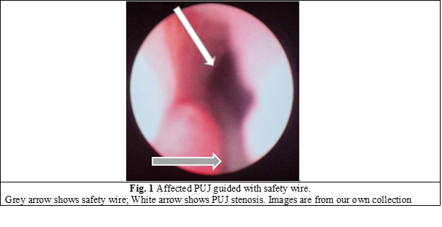
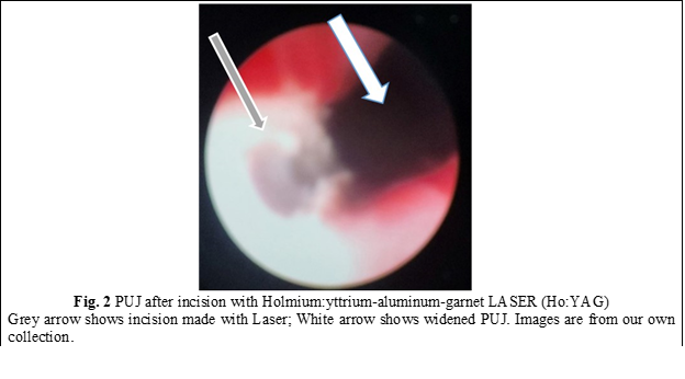
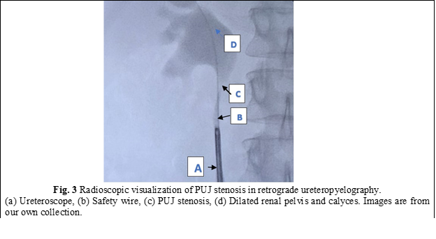
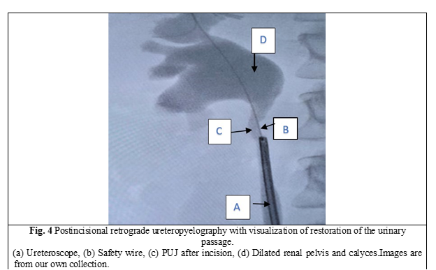
In all cases, a semirigid ureteroscope was employed for the procedure. In instances where the ureter presented excessive narrowness, impeding access to the upper third, a 5-Fr JJ stent was inserted for a duration of 2 weeks to facilitate ureteral dilation and establish an adequate working space. Subsequently, the patients were scheduled for stent extraction and the continuation of the previously determined treatment protocol. All patients underwent spinal anesthesia. Initially, intraoperatively, retrograde ureteropyelography was performed by injecting contrast medium into the ureter via the ureteroscope, allowing for repeated assessment of the anatomy of the PUJ stenosis. A semirigid ureteroscope, specifically the OES Pro 6.4/7.8 Fr x 430 mm model with a 4.2 Fr working channel, was utilized. The safety guide was used to reach the level of the pelviureteric junction stenosis as we can see in Fig. 1. A 365-µm laser fiber was employed during the procedure. Using real-time imaging guidance, the incision was made in a postero-lateral, caudo-cranial direction until visualization of the normal urothelium was achieved (Fig. 2), utilizing Ho:YAG LASER with an energy range of 0.5-3.5J and a frequency of 5-20. A control retrograde ureteropyelography was performed to determine the estimated length and level of the stenosis (Fig. 3). Post-incisional retrograde ureteropyelography was performed to assess the outcome (Fig. 4). Following the procedure, patients received the placement of 7-8 Fr JJ stents, which remained in place for a period of 4 weeks. After the completion of the procedure, patients were catheterized with a 16 Fr Foley catheter for 24 hours, following which they were discharged for outpatient follow-up and continuation of medical treatment at home. After a period of 3-4 weeks, patients were called back for reevaluation and analysis of postoperative results. During the first month, ultrasound, urine culture, and intravenous urography (IVU) were performed to assess urinary passage. If the positive result was maintained, the patient would be scheduled for a follow-up examination after 6 months. Successful treatment was defined by the resolution of symptoms, restoration of urinary passage, shortened visualization time of contrast in the ureter as observed through imaging studies, absence of obstructive patterns in dynamic scintigraphy, and preserved renal function. The half-life (T ½) of the radiopharmaceutical preparation (RFP) was less than 20 minutes. If these criteria were not met, the treatment was considered unsuccessful. Postoperative complications were classified according to the Clavien-Dindo classification system [14]. All patients have been informed about the surgical technique, purpose, and complications of the procedure. Consent for the surgical intervention and the use of personal data for the study has been obtained from each patient.


Results

Subjects involved in the present study were all detected with grade 2 (3 patients) and grade 1 (2 patients). When assessing pre-procedural (PUJ stenosis (Fig. 3)) and post-procedural results in retrograde ureteropyelography, we observe the restoration of urinary passage in all cases (Fig. 4). The average duration of the surgical intervention was 42.8 ± 2,58 minutes, and the mean duration of hospitalization was 4 ± 1.73 days. The patients were followed up for an average period of 8 weeks. One patient experienced a postoperative complication in the form of a urinary tract infection on the second day, which prolonged the hospital stay by an additional 4 days. During the one-month follow-up, a statistically significant difference was observed between patients with grade 1 hydronephrosis (complete restoration of urinary flow) and those with grade 2 hydronephrosis (faster contrast visualization in UIV) (p < 0.005). Furthermore, all patients reported the resolution of symptoms within three months of postoperative clinical monitoring.

Discussion

Open pyeloplasty represents the gold standard in the treatment of ureteropelvic junction (UPJ) stenosis, with a success rate ranging from 80% to 97% [15, 16]. However, due to its drawbacks, such as the need for a large surgical approach, lengthy recovery period, and high cost, alternative minimally invasive methods have been explored. Despite the lower postoperative success rate, these alternative approaches offer advantages in terms of cost, intraoperative complications, recovery time, and length of hospital stay.           

Thorough preoperative evaluation and investigation of the patient allow for the selection of suitable candidates for minimally invasive interventions, which offer a high postoperative success rate. The European Association of Urology guidelines recommend laser endopyelotomy as the first-line treatment with a grade C recommendation. It is primarily applied in cases of intrinsic stenosis with a defect length of less than 2 cm, absence of a dilated pelvis, high ureteral insertion, renal split function less than 20%, and ipsilateral renal calculi. The level of evidence for this recommendation is 4 [17]. Some studies have reported a higher efficacy of laser endopyelotomy in cases of primary UPJ obstruction. Other authors did not observe a difference, while some reported a higher success rate in patients with secondary etiology of UPJ obstruction [11, 18]. Gupta and colleagues have reported on the success rate of retrograde endopyelotomy based on the degree of hydronephrosis and the length of the stenotic segment. In both cases, as the degree of hydronephrosis and the length of the stenotic segment increase, the success rate decreases. However, the difference in results between the two cases is not statistically significant [19]. Similarly, Rassweiler and colleagues have also addressed the correlation between multiple factors that influence the success rate, with one of the factors being hydronephrosis. They found that as the severity of hydronephrosis increases, the postoperative outcomes become less favorable [20], which is consistent with the findings of the current study. The complication rate is estimated to be between 5% and 35%, with the most common complication being urinary tract infection (UTI) [21]. In our case, the complication rate was 20%, with a UTI being identified in a patient on the second day postoperatively during a urine analysis. The duration of stent placement for ureteral integrity restoration post-endopyelotomy varies among different authors. Anil Mandhani and colleagues conducted an animal study that showed positive results with complete recovery after 2 weeks [22]. Geavlete reported a necessary period of 6 weeks, as the urothelium regenerates in 5 days and the muscular tissue in 6 weeks [23]. In the current study, the stent was left in place for 4 weeks. The success rate in our study, at an 8-week patient follow-up, is currently 99.9%. However, patients require ongoing monitoring to assess the maintenance of JPU patency. Nevertheless, although the effectiveness of the intervention is continuously studied, JPU patency can decrease over time, as mentioned by Shalhav Al and colleagues in 1989 [24].

Conclusion

Compared to other minimally invasive methods such as antegrade endopyelotomy, retrograde incision with balloon (Acucise), laparoscopic, and robot-assisted procedures, the retrograde technique has several advantages, with cost-effectiveness being the most significant one: comparatively affordable equipment, shorter recovery period, and hospital stay. To achieve a clear success rate, certain factors need to be considered: defect length <2 cm, insignificant hydronephrosis, absence of a large pelvis, and high ureteral insertion, with preserved ipsilateral renal function. The current study results demonstrate an efficacy rate of 99.9% in the early months of monitoring. However, longer monitoring with a larger patient sample is necessary to provide more conclusive data in the future.

Competing interests

The authors report no conflicts of interest in this work.

Patient consent 

Obtained.

Funding Statement

The authors report no financial support.

Authors’ contributions

VC conceived and designed the study, performed surgical interventions, collected and analyzed data, and significantly contributed to manuscript writing and revision. EP assisted in study design, participated as an assistant in surgical interventions, contributed to data analysis, and played a substantial role in manuscript drafting and critical review. AM assisted in study design, participated as an assistant in surgical interventions, provided clinical insights, and contributed to manuscript revision. CM assisted with data collection, participated as an assistant in surgical interventions, and participated in manuscript revision. Final manuscript was read and approved by all authors.

Authors’ ORCID IDs

Vladimir Caraion - https://orcid.org/0009-0003-2917-9665

Eduard Pleșca - https://orcid.org/0000-0002-0021-0396

Andrei Mezu - https://orcid.org/0009-0006-7355-9982

Corneliu Maximciuc - https://orcid.org/0009-0000-3163-8822

References

1.  

  1. European Association of Urology. EAU Guidelines on paediatric urology [Internet]. Arnhem: EAU; 2023 [cited 2023 May 12]. Available from: https://uroweb.org/guidelines/paediatric-urology/chapter/the-guideline.

  2. Termos S, AlKabbani M, Ulinski T, Sanjad S, Kotobi H, Chalard F, et al. Ureteropelvic junction obstruction and parathyroid adenoma: coincidence or link? Case Rep Nephrol. 2017;2017:9852912. doi: 10.1155/2017/9852912.

  3. Borin JF. Ureteropelvic junction obstruction in adults. Rev Urol. 2017;19(4):261-4. doi: 10.3909/riu0781.

  4. Grasso M, Caruso RP, Phillips CK. UPJ obstruction in the adult population: are crossing vessels significant? Rev Urol. 2001;3(1):42-51.

  5. Jackson L, Woodward M, Coward RJ. The molecular biology of pelvi-ureteric junction obstruction. Pediatr Nephrol. 2018;33(4):553-71. doi: 10.1007/s00467-017-3629-0.

  6. Sutherland DE, Jarrett TW. Surgical options in the management of ureteropelvic junction obstruction. Curr Urol Rep. 2009;10(1):23-8. doi: 10.1007/s11934-009-0006-y.

  7. Gerber GS, Kim JC. Ureteroscopic endopyelotomy in the treatment of patients with ureteropelvic junction obstruction. Urology. 2000;55(2):198-202; discussion 202-3. doi: 10.1016/s0090-4295(99)00530-0.

  8. Kartal I, Cimen S, Karakoyunlu N, Sandikci F, Eraslan A, Yalcinkaya F. Factors affecting the effectiveness and success of retrograde holmium laser endopyelotomy as the primary treatment of ureteropelvic junction obstruction in adults. Urologia. 2021;88(1):34-40.doi: 10.1177/0391560320904259.

  9. Sen H, Bayrak O, Erdem Yilmaz A, Turgut O, Ozturk M, Erturhan S, et al. Evaluation of the results of laser endopyelotomy with two different technique in ureteropelvic junction obstruction. J Pediatr Urol. 2021;17(3):397e1-e6. doi: 10.1016/j.jpurol.2021.01.035.

  10. Braga LH, Lorenzo AJ, Skeldon S, Dave S, Bagli DJ, Khoury AE, et al. Failed pyeloplasty in children: comparative analysis of retrograde endopyelotomy versus redo pyeloplasty. J Urol. 2007;178(6):2571-5; discussion 2575. doi: 10.1016/j.juro.2007.08.050.

  11. Stilling NM, Jung H, Norby B, Osther SS, Osther PJ. Retrograde ureteroscopic holmium laser endopyelotomy in a selected population of patients with ureteropelvic junction obstruction. Scand J Urol Nephrol. 2009;43(1):68-72. doi: 10.1080/00365590802473164.

  12. Gnessin E, Yossepowitch O, Holland R, Livne PM, Lifshitz DA. Holmium laser endoureterotomy for benign ureteral stricture: a single center experience. J Urol. 2009;182(6):2775-9. doi: 10.1016/j.juro.2009.08.051.

  13. Hibi H, Ohori T, Taki T, Yamada Y, Honda N. Long-term results of endoureterotomy using a holmium laser. Int J Urol. 2007;14(9):872-4. doi: 10.1111/j.1442-2042.2007.01835.x.

  14. Dindo D, Demartines N, Clavien PA. Classification of surgical complications: a new proposal with evaluation in a cohort of 6336 patients and results of a survey. Ann Surg. 2004;240(2):205-13.doi: 10.1097/01.sla.0000133083.54934.ae.

  15. Rassweiler JJ, Gozen AS, Erdogru T, Sugiono M, Teber D. Ureteral reimplantation for management of ureteral strictures: a retrospective comparison of laparoscopic and open techniques. Eur Urol. 2007;51(2):512-22; discussion 522-3. doi: 10.1016/j.eururo.2006.08.004.

  16. Simforoosh N, Basiri A, Tabibi A, Danesh AK, Sharifi-Aghdas F, Ziaee SA, et al. A comparison between laparoscopic and open pyeloplasty in patients with ureteropelvic junction obstruction. Urol J. 2004;1(3):165-9.

  17. Herrmann TR, Liatsikos EN, Nagele U, Traxer O, Merseburger AS; Eau Guidelines Panel on Lasers, Technologies. EAU guidelines on laser technologies. Eur Urol. 2012;61(4):783-95. doi: 10.1016/j.eururo.2012.01.010.

  18. Seveso M, Giusti G, Taverna G, Piccinelli A, Benetti A, Maugeri O, et al. Retrograde endopyelotomy using the holmium laser: technical aspects and functional results. Arch Ital Urol Androl. 2005;77(1):10-2.

  19. Gupta M, Tuncay OL, Smith AD. Open surgical exploration after failed endopyelotomy: a 12-year perspective. J Urol. 1997;157(5):1613-8; discussion 1618-9.

  20. Rassweiler JJ, Subotic S, Feist-Schwenk M, Sugiono M, Schulze M, Teber D, et al. Minimally invasive treatment of ureteropelvic junction obstruction: long-term experience with an algorithm for laser endopyelotomy and laparoscopic retroperitoneal pyeloplasty. J Urol. 2007;177(3):1000-5. doi: 10.1016/j.juro.2006.10.049.

  21. Elmussareh M, Traxer O, Somani BK, Biyani CS. Laser endopyelotomy in the management of pelviureteric junction obstruction in adults: a systematic review of the literature. Urology. 2017;107:11-22. doi: 10.1016/j.urology.2017.04.018.

  22. Mandhani A, Kapoor R, Zaman W, Kumar A, Bhandari M, Gambhir S. Is a 2-week duration sufficient for stenting in endopyelotomy? J Urol. 2003;169(3):886-9. doi: 10.1097/01.ju.0000051341.22163.21.

  23. Geavlete P, Georgescu D, Mirciulescu V, Nita G. Ureteroscopic laser approach in recurrent ureteropelvic junction stenosis. Eur Urol. 2007;51(6):1542-8. doi: 10.1016/j.eururo.2006.08.035. 

  24. Shalhav AL, Giusti G, Elbahnasy AM, Hoenig DM, McDougall EM, Smith DS, et al. Adult endopyelotomy: impact of etiology and antegrade versus retrograde approach on outcome. J Urol. 1998;160(3 Pt 1):685-9. doi: 10.1016/S0022-5347(01)62755-1


More articles

Research Harmfulness of prooxidants in bronchopulmonary dysplasia in preterm children
Mariana Ceahlău1*, Rodica Selevestru1,2, Olga Tagadiuc3, Svetlana Șciuca1,2
https://doi.org/10.52645/MJHS.2023.3.01
Oxidative stress can be defined as the imbalance of the redox state of a certain system including living one (organelle, cell, organ/tissue), which excessively produces reactive oxygen and/or reactive nitrogen species (ROS/RNS) that exceed the capacity of the antioxidant defense system, which have the ability to slow down or even prevent the oxidative damage of macromolecules. Oxidative stress is a pathogenic mechanism of a large variety of diseases, including pulmonary one.
Research Predicting sympathovagal balance using parameters of breathing patterns in abdominal breathing
Abdominal breathing is utilized as a non-pharmacological treatment method for various stress-related conditions and autonomic dysfunctions. The objective of the study was to determine the predictors in the modulation of sympathovagal balance, as indicated by the ratio of low frequency to high frequency power of heart rate variability, by utilizing the respiratory pattern parameters recorded during the abdominal breathing model.
Research The impact of imuheptin and imupurin on cytokine profile and antioxidant status in rat model of inflammation
Ina Guțu1*, Nicolae Bacinschi1, Valentin Gudumac2
https://doi.org/10.52645/MJHS.2023.3.03
Insects, throughout evolution, have developed a huge arsenal of active compounds, which they use to defend themselves against enemies and diseases, at the same time in recent years insects have shown great interest as a source of food rich in biologically active substances. Research in recent decades has shown that insects produce a variety of proteins and peptides with antibacterial, antifungal, antiviral, immunomodulatory, anti-inflammatory, antioxidant, antitumor, hepatoprotective, antithrombotic, antihypertensive and detoxifying activity during or after contact with the microbial agent or unfavourable factor.
Research Clinical effectiveness study of the new diagnostic score of acute appendicitis in the elderly
Acute appendicitis is among the three most frequent surgical diseases. The lifetime likelihood of developing acute appendicitis is around 7%. The incidence of acute appendicitis reduces with age after adolescence. Several studies evaluated the relevance of the current scores to the general population, mostly children, but a limited number of studies have studied the elderly population. This study aims to assess the clinical effectiveness of the new diagnostic score for the elderly population in comparison to both the Alvarado score and the non-standardized score.
Research Early results of endovascular treatment using percutaneous vacuum-assisted thromboaspiration in acute lower limb ischemia
Alexandru Predenciuc1*, Vasile Culiuc1,2, Dumitru Casian1,2
https://doi.org/10.52645/MJHS.2023.3.05
Open balloon thrombectomy and embolectomy remain the preferred initial option in the management of acute lower limb ischemia (ALI), but various endovascular techniques have become accessible and are growing in popularity. The aim of the study was to assess our early experience with percutaneous vacuum-assisted thromboaspiration using the Penumbra/Indigo® system for non-traumatic ALI.
Review Venous and arterial endothelium: markers of dysfunction and pathophysiological significance
Victor Ojog1*, Svetlana Lozovanu1
https://doi.org/10.52645/MJHS.2023.3.06
Endothelial dysfunction is a result of complex pathogenic interface involving inflammation, oxidative stress, disorders of endothelization and hemostasis etc., in both arteries and veins, leading to a lot of cardiovascular diseases. Identifying markers with high predictive value has an important diagnostic and prognostic significance.
Review Current affairs in the use of medical ozone. Biological effects. Mechanisms of action
Natalia Cernei1*, Serghei Șandru1, Serghei Cobîlețchi1, Ion Grabovschi2, Ivan Cîvîrjîc1, Ruslan Baltaga1,3
https://doi.org/10.52645/MJHS.2023.3.08
Oxygen-ozone therapy stands as a medically endorsed practice confirmed by numerous international clinical studies. Various authors have illustrated the beneficial clinical outcomes of ozone therapy in terms of its capacity to regulate redox balance, cellular inflammatory responses, and adaptation to ischemia/reperfusion processes. Ozone therapy extends to encompass a range of viral infections, inflammatory disorders, and degenerative ailments, used as both monotherapy and as an adjunct to unified conventional therapies.Introduction Ozone (O3), a gas discovered in the mid-19th century and composed of three oxygen atoms, represents a highly reactive allotropic form of oxygen. It exhibits high solubility in plasma, extracellular fluids, and water (approximately 10 times more soluble in water than conventional oxygen). At room temperature, it is unstable, causing rapid decomposition into ordinary diatomic oxygen. Notably, its half-life measures 25 minutes at 30°C, 40 minutes at 20°C, and 140 minutes at 0°C [1-10]. Medical ozone is a blend of oxygen and ozone derived from medical-grade oxygen through the utilization of a medical ozone generator. This medical ozone contains a concentration of 1-5% ozone and 90-95% pure medical oxygen, or 10-80 μg/mL (0.21-1.68 μmol/ml) of ozone per milliliter of blood. Ozone therapy stands as a current and significant avenue of research in contemporary medicine [1, 3-5, 7, 9, 10-15]. Oxygen-ozone therapy is a medically validated practice supported by numerous international clinical studies. Nowadays, many clinical trials have shown its beneficial effects on the modulation of the oxidoreduction balance, cellular inflammation state, and adaptation to ischemia/reperfusion processes. Ozonotherapy is an effective, safe, feasible, and easy-to-perform technique, which finds applications in various inflammatory, infectious, degenerative diseases, as well as in rehabilitation following acute cardiac and cerebral ischemic events. It demonstrates good efficacy both as an independent treatment and, notably, as an adjunct to conventional therapies [3-5, 7, 11, 16-22]. By incorporating this medical practice, patients can attain significant clinical benefits. When combined with standard therapies, it often leads to reduced medication dosages, complication rates, treatment duration, medication toxicity, and medical expenses. It also addresses the issue of bacterial resistance to antibiotics [2, 4, 18, 19, 21, 23]. In the context of the aforementioned, the purpose of this article is to present a synthesis of the most recent findings regarding ozone's mechanisms of action. Materials and methods To achieve the outlined purpose, an initial search of specialized scientific publications was conducted. These were identified through the Google Search engine, namely, PubMed, Hinari, SpringerLink, the National Center of Biotechnology Information, and Medline. The article selection criteria encompassed contemporary data regarding the mechanisms of action of ozone therapy, utilizing the following keywords: “ozone”, “ozone therapy”, “ozone mechanisms of action”, “biological effects of ozone”, “antioxidant effect”, “anti-inflammatory effect”, and “immunomodulatory effect.” These keywords were employed in various combinations to optimize search efficiency. For the advanced selection of bibliographic sources, the following filters were used: full-text articles, articles in English, articles published between 1990 and 2022. After a preliminary analysis of the titles, original articles, editorials, articles of narrative synthesis, taxonomy, and meta-analysis were selected, which contained up-to-date information and contemporary concepts regarding the mechanisms of ozone therapy. Furthermore, a search was conducted within the reference lists of the identified sources to highlight additional relevant publications that were not found during the initial database searches. The information from the publications included in the bibliography was gathered, organized, evaluated, and synthesized, showcasing the key aspects of the contemporary understanding of ozone's mechanisms of action, namely, its antioxidant capacity, vascular and hematological modulation, immune system activation, as well as its anti-inflammatory, bactericidal, virucidal, and fungicidal effects. To minimize the potential systematic errors (bias) in the study, a meticulous search was conducted within databases to identify a maximum number of relevant publications for the study's purpose. Only studies that satisfy validity criteria were evaluated, rigorous exclusion criteria for articles under consideration were applied, and a comprehensive review was conducted of both positive outcome studies and those that did not highlight the treatment's benefits. If necessary, additional sources of information were consulted to clarify some concepts. Duplicate publications and articles that did not meet the purpose of the article and were not available for full viewing were excluded from the list of publications generated by the search engine. Results Following the data processing, as identified by the Google Search engine and from databases such as PubMed, Hinari, SpringerLink, National Center of Biotechnology Information, and Medline, in accordance with the search criteria, a total 475 articles on the topic of ozone therapy were found. After a primary analysis of the titles, 59 articles were eventually deemed relevant for the given synthesis. Upon repeated review of these sources, a final selection of 52 relevant publications was ultimately made in alignment with the intended purpose. The final bibliography of this work comprises 52 articles that have been considered representative of the materials published on the subject of this synthesis article. Publications, the content of which did not reflect the relevant topic, despite being selected by the search program, as well as articles that were not accessible for open viewing through the HINARI (Health Internet Work Access to Research Initiative) database or available in the scientific medical library of the Nicolae Testemițanu State University of Medicine and Pharmacy, were subsequently removed from the list. Although ozone is the most potent natural oxidant, capable of oxidizing a wide range of organic and inorganic substances, and has the potential for cytotoxicity, researchers believe that under controlled conditions, it possesses numerous therapeutic effects. Moreover, the reactivity to ozone can be effectively mitigated by the blood and cellular antioxidant system [1, 2, 15, 24-28]. Initially used as an empirical approach, oxygen-ozone therapy has now evolved to a stage where the majority of ozone's biological mechanisms of action have been extensively studied and elucidated, these findings being found within the fields of biochemistry, physiology, and pharmacology [18, 22, 25, 27-29]. Ozone is not a pharmaceutical medicine but rather a regulatory molecule capable of generating bioactive mediators. The effects of ozone have been demonstrated to be consistent, safe, and associated with minimal preventable side effects [2, 30]. Chronic oxidative stress, chronic inflammatory processes, and immune overactivation are present and highly detrimental in a wide variety of diseases. The effectiveness of ozone therapy is determined by moderate oxidative stress, resulting from the interaction of ozone with the biological components of the body, triggering an endogenous cascade of biochemical reactions [31]. Ozone can function as an oxidant either directly, when it dissolves in plasma and other biological fluids, immediately reacting with polyunsaturated fatty acids, antioxidants, cysteine-rich proteins, and carbohydrates; or indirectly, by generating reactive oxygen species (ROS) and lipid oxidation products (LOP) [25, 28, 32-37]. At the onset of ozone therapy, an endogenous cascade is triggered, releasing bioactive substances in response to transient and moderately induced oxidative stress by ozone ('oxidative eustress'). Ozone can easily induce this oxidative stress due to its plasma solubility. Reacting with polyunsaturated fatty acids and water, ozone forms ROS in human fluids and tissues. The main molecule among ROS is hydrogen peroxide (H2O2) – a non-radical oxidant. Concurrently, ozone also gives rise to LOP – the lipoperoxide radical, hydroperoxides, malondialdehyde, isoprostanes, ozoneides, alkenes, and predominantly, 4-hydroxynonenal. ROS and LOP are the effector molecules responsible for modulating several biological and therapeutic effects in the body following ozone therapy [3, 8, 13, 27, 34, 37-39]. Having reacted with a number of biomolecules, ozone disappears, and hydrogen peroxide, the main molecule of ROS, and other mediators rapidly diffuse into cells, activating various metabolic pathways with numerous biological and therapeutic effects [3, 5, 27, 28, 35, 40]. Therefore, ROS and LOP are “biological messengers of ozone” and are responsible for the biological and therapeutic effects of ozone. ROSs are short-acting early messengers and are responsible for immediate biological effects, while LOPs are important late and long-term messengers [3, 5, 10, 13, 14, 17, 28, 36]. The formation of ROS in plasma occurs extremely quickly (less than a minute), accompanied by a moderate and transient decrease in the antioxidant capacity of the blood (from 5% to 25%). However, this antioxidant capacity returns to normal within 15-20 minutes [3, 9, 17, 28, 31, 40, 41]. Discussion Although not fully comprehended, the present article will delve into the mechanisms underlying the antioxidant, anti-inflammatory, immunomodulatory, antimicrobial, antiviral, and analgesic effects of ozone. The antioxidative capacity is considered one of the key impacts of ozone therapy. Moderate oxidative stress induced by ozone within the therapeutic range (10-80 μg/mL), most commonly 30-45 μg/mL (the 'physiological' dose of ozone), physiologically effective and recommended levels for systemic application, elicits controlled low doses of ROS acting primarily as signaling molecules, thereby stimulating the formation of LOP. ROS triggers the activation of nuclear erythroid factor 2 (Nrf2), well known as a pivotal regulator of manifold cytoprotective responses, responsible for upregulating antioxidant enzyme activity. In response to transient, moderate oxidative stress, the levels of antioxidant enzymes such as superoxide dismutase, glutathione peroxidase, glutathione S-transferase, catalase, and heme oxygenase-1 increase. Thus, moderate, transient, and repetitive oxidative stress causes an intense modulation of antioxidants in the body. A multitude of cells across various organs upregulate the synthesis of antioxidants, which are capable of significantly countering excessive ROS, thereby alleviating chronic oxidative stress, which is present and extremely harmful in a variety of diseases Consequently, ozone, either through oxidative preconditioning or adaptation to chronic oxidative stress, safeguards tissue integrity against ROS-induced damage, fostering a balance between antioxidant and pro-oxidant factors while preserving cellular redox balance [27, 30, 41-45]. Nrf2 and nuclear factor kappa Β (NF-κB) represent the primary signaling pathways through which ozone exerts its effects. Nrf2 activation regulates cell defense and maintains cellular homeostasis [36]. Furthermore, ozone therapy fosters adaptation to oxidative stress by gently triggering the immune system, releasing growth factors, and/or activating metabolic pathways that contribute to maintaining redox balance [38]. By activating Nrf2, LOP induces oxidative stress proteins, including heme-oxygenase-1 (HO-1), another inhibitor of the NF-κB pathway and one of the most crucial antioxidant defense enzymes. Through inhibiting the high expression level of hypoxia-inducible factor-1α (HIF-1α), it contributes to reducing the production of proinflammatory cytokines, directly activating anti-inflammatory cytokines, enhancing antioxidant protection, and consequently, safeguarding cellular integrity [8, 28, 30, 44-46]. Thus, ozone mimics acute oxidative stress which, when properly balanced, is not harmful, but can trigger several beneficial biochemical mechanisms. It can reactivate the intra- and extracellular antioxidant system, thereby reversing chronic oxidative stress in various inflammatory, degenerative processes, etc. During ozone treatment, cells throughout the body receive gradual and subtle impulses of LOP, significant long-term messengers that play a crucial role in up-regulating antioxidant enzymes in multiple cell types while rebalancing the oxidant/antioxidant system [46, 47]. Vascular and Hematological Modulation. Ozone serves as a catalyst for transmembrane oxygen flow. The increase in cellular oxygen levels resulting from ozone therapy enhances the efficiency of the mitochondrial respiratory chain. Moreover, ozone amplifies the production of prostacyclin, a widely acknowledged vasodilator [2, 6, 8, 26]. The effects of ozone on oxygen metabolism can be explained by promoting (1) changes in the rheological properties of blood (reversal of erythrocyte aggregation, increased flexibility and elasticity of red blood cells, favoring the transport and delivery of tissue oxygen), leading to enhanced blood flow in microcirculation; (2) increasing the speed of glycolysis in erythrocytes; and (3) the release of substances (adenosine triphosphate, nitric oxide, and prostaglandins) that may contribute to reducing peripheral vascular resistance and increasing oxygen supply to tissues [18, 25, 35, 37, 40, 48-50]. Hydrogen peroxide (H2O2) diffuses from the plasma into the cellular cytoplasm and serves as the triggering stimulus. Depending on the cell type, various biochemical pathways can be simultaneously activated in red blood cells, white blood cells, and platelets, leading to a multitude of biological effects [10, 28, 32]. The Impact of Ozone on Erythrocytes. Erythrocytes are the focus of ROS. During erythropoiesis, submicromolar concentrations of LOP positively regulate the synthesis of antioxidant enzymes. Consequently, ozone therapy increases the glycolytic rate by enhancing intracellular adenosine triphosphate production. This approach intensifies erythrocyte generation, yielding metabolically enhanced erythrocytes (super-endowed erythrocytes) capable of more effectively transporting and delivering oxygen to tissues, including ischemic tissues, thereby correcting hypoxia in vascular diseases [7, 12, 25, 27, 28, 39, 48]. Coupled with increased nitric oxide synthase activity, there is a significant increase in nitric oxide, an essential element in maintaining optimal levels of vasodilation and blood perfusion [1, 6, 8, 40]. Ozone therapy, through careful regulation of ozone dosage, stimulates the production of antioxidant enzymes within the system (catalase, glutathione peroxidase, and superoxide dismutase) while mitigating excessive formation of ROS, thereby reducing chronic oxidative stress [1, 6, 12, 14, 39, 43, 49]. The impact of ozone on leukocytes. Ozone acts as a mild cytokine and serves as a cytokine inducer by lymphocytes and monocytes, thereby enhancing the immune system's activity. This stimulation fosters intercellular matrix synthesis and contributes to the healing process [1, 12, 32, 35, 37, 39]. The Impact of ozone on platelets. Hydrogen Peroxide (H2O2) and other ROS generated through blood ozonation initiate a cascade of enzymatic reactions. These reactions gradually elevate intracellular Ca levels and trigger the release of prostaglandins (F2a and E2), leading to irreversible platelet aggregation. Increased levels of growth factors released from platelets, mobilization of endogenous stem cells, and stimulation of neoangiogenesis promote tissue regeneration, as well as healing of injuries and wounds [27, 49, 51]. Thus, the impact of ozone on oxygen metabolism is explained by how it alters the blood's rheological properties (reversing red blood cell clumping, enhancing the flexibility and elasticity of red blood cells, promoting the transport and delivery of oxygen to tissues). This, in turn, facilitates blood flow in the microcirculation, increases glycolysis in red blood cells, and triggers the production of substances (such as adenosine triphosphate, nitric oxide, and prostaglandins) that help reduce peripheral vascular resistance. Activation of the immune system. Ozone promotes an increase in the production of interferon-γ (IFN-γ) and some cytokines, with interleukin-2 (IL-2) being the primary one, subsequently triggering a whole cascade of immunological reactions [1, 2]. It has been shown that ROS, including H2O2 and LOP generated by ozone therapy, can easily diffuse into plasma cells and activate NF-κB, inducing the production of immunoactive cytokines in normal cells (IL-2, tumor necrosis factor alpha - TNF-a, IL-6 and IFN-γ), thereby enhancing the immune response [9, 13, 14, 26, 32, 40, 44]. Ozone indirectly activates the innate (non-specific) immune system by enhancing phagocytosis and promoting the synthesis of cytokines and interleukins in neutrophils and leukocytes. It also triggers the components of both cellular and humoral immunity [8, 26, 33, 39]. Within mononuclear cells, ozone stimulates immune responses by modulating the NF-kB transcription factor, thereby reactivating the suppressed immune system [27, 28]. Furthermore, ROSs trigger the activation of the immune system, which acts through monocytes and lymphocytes, promoting the production of a variety of cytokines (IL-1, IL-2, IL-6, IFN-β, IFN-γ, TNFα) [6, 49]. Thus, ozone induces mild immune system activation by stimulating neutrophils and initiating the synthesis of certain cytokines that trigger a whole cascade of immunological responses. Bactericidal, virucidal and fungicidal action of ozone. Ozone used in vitro acts directly on the membrane of bacterial cells (direct oxidative effect), disrupting and damaging the integrity of bacterial cell membranes, oxidizing phospholipids and lipoproteins, thereby impeding their enzymatic function. Additionally, ozone damages the viral capsids, disturbing their structure and interfering with the virus-cell interaction, leading to disruption in the reproductive cycle. When it comes to fungi, ozone inhibits cell growth by perturbing intracellular homeostasis, resulting from the compromised barrier properties of the plasma membrane [2, 6, 12, 16, 26, 44, 48, 50]. Although ozone is one of the most potent disinfectants, used in various ways, it cannot deactivate any pathogens (bacteria, viruses, and fungi) in vivo. This is because pathogens are well protected, especially within cells, by the cell's powerful antioxidant system. Consequently, ozone acts as a gentle enhancer of the immune system by activating neutrophils and stimulating the synthesis of certain cytokines [1, 10, 19, 22, 28, 39, 46]. The anti-inflammatory effect is revealed in ozone's ability to influence the inflammatory cascade by oxidizing biologically active substances (arachidonic acid and its derivatives - prostaglandins), which participate in the development and sustenance of the inflammatory process. Additionally, ozone significantly reduces the levels of pro-inflammatory cytokines (IL-1β, IL-6, IL-8, and TNF-α) without any signs of toxicity or recorded side effects [8, 26, 30, 31]. These cytokines induce the prostaglandin E2 pathway, which causes pain or increases the sensitivity of nerve roots to other algogenic substances (such as bradykinin) [31]. Severe oxidative stress, triggered by high concentrations of ozone, along with proinflammatory cytokines (IL-1β, IL-6, IL-8, TNF-α), activate NF-κB, a key regulator of the inflammatory response and muscle atrophy. This contributes to an increased inflammatory response and tissue damage, including the release of other inflammatory factors that enhance the migration of eosinophils and neutrophils [9, 13, 17, 47, 49]. On the contrary, mild oxidative stress induced by precise and small doses of ozone activates Nrf2. The latter indirectly inhibits the pro-inflammatory mechanism driven by the NF-kB pathway. As a result, there is a reduction in NF-κB activity along with a modification in the expression of inflammatory cytokines associated with NF-kB activity. This triggers an anti-inflammatory effect, leading to a decrease in IL-1, IL-2, IL-6, IL-7, and TNFα, as well as an increase in interleukins such as IL-4, IL-10, IL-13, and the transforming growth factor beta – TGF-β [11, 13, 19, 38, 43, 49, 50]. Nrf2 also plays an important role in intracellular inflammatory signaling pathways. Triggering the Nrf2-antioxidant signal can dampen NF-kB activity, leading to the downregulation of the inflammatory response by suppressing essential inflammatory mediators and cytokines (IL-6, IL-8, and TNF-a) [31, 38, 42, 50]. Moreover, a small amount of H2O2 stimulates the NF-kB pathway, which is typically balanced out by the Nrf2's blocking action, resulting in an immunomodulatory effect [11]. The analgesic effect of ozone is ensured by the oxidation of the byproducts of albuminolysis, known as algopeptides, which act on the nerve endings in the damaged tissue and determine the intensity of the pain response. Additionally, the analgesic effect is attributed to the restoration of the antioxidant system and, subsequently, the reduction of harmful molecular byproducts from lipid peroxidation [26]. Recent preclinical studies have elucidated the role of ROS in hyperalgesia by activating N-methyl-D-aspartate receptors [11]. Following ozone therapy, there has been a demonstrated increase in antioxidant molecules (serotonin and endogenous opioids), which induce pain relief by stimulating antinociceptive pathways [31, 39]. Data from scientific research acknowledge that the mechanisms of action of ozone are due to: (1) a decrease in the production of inflammatory mediators; (2) oxidation (inactivation) of metabolic mediators of pain; (3) improvement of local blood microcirculation leading to improved oxygen delivery to tissues; (4) elimination of toxins and resolution of physiological disorders that generate pain [42, 52]. Therefore, ozone exhibits pleiotropic properties, extending beyond its exclusive role as an antioxidant, anti-inflammatory, or immunomodulatory one. It also encompasses the capacity to employ ROS as a signaling molecule rather than merely as intracellular toxic substances. In existing experimental models and clinical studies, the anti-inflammatory, antioxidant, regenerative and immunomodulatory effects of ozone therapy have been associated with several molecular mechanisms, the main ones being the NF-kB/Nrf2 balance and IL-6 and IL-1β expression. NF-kB and Nrf2 are the most studied and important transcription factors and regulatory proteins that control the expression of a wide range of genes, encoding proteins involved in a multitude of vital biological functions, including those associated with redox status, immunity, and inflammatory responses. Additionally, indirectly through these pathways, LOP initiates the HIF-1α, HO-1, and NO/iNOS pathways. The main pharmacological effects of medical ozone through ozone-produced peroxides are as follows: (1) increased oxygen release by erythrocytes due to activated metabolism; (2) immunomodulation due to leukocyte activation; and (3) regulation of cellular antioxidants via Nrf2 signaling. Ozone therapy can elicit the following biological reactions: (a) improved blood circulation and oxygen delivery to ischemic tissue; (b) optimization of overall metabolism by improving oxygen delivery; (c) regulation of cellular antioxidant enzymes and induction of HO-1; (d) triggering a mild immune system activation and intensified release of growth factors; (e) providing a state of well-being in most patients, probably due to stimulation of the neuroendocrine system. Conclusions 1. Ozone induces both mild and moderate oxidative stress. When appropriately balanced, this stress poses no harm; instead, it can initiate several beneficial biochemical mechanisms. These mechanisms, in turn, reactivate the intracellular and extracellular antioxidant systems, effectively countering long-term oxidative stress in various inflammatory and degenerative processes, etc. Cells throughout the body receive small and gradual bursts of lipid oxidation products, important late and long-term messengers that are responsible for activating antioxidant enzymes in many cell types to rebalance the oxidant/antioxidant system. 2. The impact of ozone on oxygen metabolism is explained by changes in the blood's rheological properties. This involves reversing red blood cell aggregation, enhancing the flexibility and elasticity of hemoglobin, and promoting the efficient transport and delivery of oxygen to tissues. This process also facilitates blood flow within microcirculation, speeds up glycolysis within red blood cells, and triggers the release of substances like adenosine triphosphate, nitric oxide, and prostaglandins, which help to reduce peripheral vascular resistance. 3. Ozone triggers a slight activation of the immune system by up-regulating and activating neutrophils and promoting the synthesis of cytokines (IL-2, TNF-a, IL-6, and IFN-γ), setting off a chain reaction of immune responses. 4. Ozone therapy induces the following biological responses: enhanced blood circulation and oxygen delivery to ischemic tissue, regulation of cellular antioxidant enzymes, mild immune system activation, and intensified release of growth factors. 5. Ozone is an inherently toxic gas that should never be inhaled, cannot be stored, and must be handled with caution. Generally, no toxic effects were reported, and only the respiratory tract was found to be highly sensitive to inhaled ozone since the respiratory mucosal cells contain a minimal amount of antioxidants and are extremely susceptible to oxidation. 6. Although ozone ranks among the most potent disinfectants, being employed in various ways, it cannot neutralize any pathogens (bacteria, viruses, and fungi) in vivo, since pathogens are effectively shielded by the strong blood and cellular antioxidant system. Furthermore, elevated ozone concentrations induce severe oxidative stress, prompting increased inflammatory responses and tissue damage. Competing interests None declared. Authors’ contribution NC and RB conceived the study, participated in the study design and assisted in drafting the manuscript. SȘ and IC performed the analysis and data interpretation. IG drafted the manuscript. SC conceived the significant revision of the manuscript and provided significant intellectual involvement. The authors have read and approved the final version of the manuscript. Authors’ ORCID IDs Natalia Cernei – https://orcid.org/0000-0002-2031-5881 Serghei Șandru – https://orcid.org/0000-0002-2973-9154 Ion Grabovschi – https://orcid.org/0000-0002-7716-9926 Ivan Cîvîrjîc – https://orcid.org/0000-0002-1360-5485 Ruslan Baltaga – https://orcid.org/0000-0003-0659-4877 References 1. Cakir R. General aspects of ozone therapy [Internet]. In: Atroshi F, editor. Pharmacology and nutritional intervention in the treatment of disease. London: IntechOpen, 2014 [cited 2023 Apr 12]. Available from: http://dx.doi.org/10.5772/57470 2. Elvis A.M., Ekta J.S. Ozone therapy: a clinical review. J. Nat. Sci. Biol. Med., 2011 Jan; 2 (1): 66-70. doi: 10.4103/0976-9668.82319. 3. Bocci V. Ozone: a new medical drug. 2nd ed. New York: Springer, 2011; 315p. https://doi.org/10.1007/978-90-481-9234-2. 4. Allorto N. Oxygen-ozone therapy: an extra weapon for the general practitioners and their patients. Ozone Ther., 2019 July; 4 (2): 8424. doi: 10.4081/ozone.2019.8424. 5. Baeza-Noci J., Cabo-Soler J.R., Moraleda-Gómez M. et al. Revisión WFOT sobre Ozonoterapia Basada en Evidencias. Comité Científico Asesor de la WFOT – 2015 [WFOT Review on Evidence-Based Ozone Therapy. WFOT Scientific Advisory Committee – 2015]. Brescia: World Federation of Ozone Therapy, 2015; 117p. Spanish. https://semdor.es/wp-content/uploads/2021/10/WFOT-OZONE-2015-ESP.pdf. 6. Scassellati C., Ciani M., Galoforo A., Zanardini R., Bonvicini C., Geroldi C. Molecular mechanisms in cognitive frailty: potential therapeutic targets for oxygen-ozone treatment. Mech. Ageing Dev., 2020 Mar; 186: 111210. doi: 10.1016/j.mad.2020.111210. 7. Yousefi B., Banihashemian S., Feyzabadi Z., Hasanpour S., Kokhaei P., Abdolshahi A. et al. Potential therapeutic effect of oxygen-ozone in controlling of COVID-19 disease. Med. Gas Res., 2022 Apr-Jun; 12 (2): 33-40. doi: 10.4103/2045-9912.325989. 8. Smith N., Wilson A., Gandhi J., Vatsia S., Khan S. Ozone therapy: an overview of pharmacodynamics, current research, and clinical utility. Med. Gas Res., 2017 Oct 17; 7 (3): 212-9. doi: 10.4103/2045-9912.215752. 9. Sagai M., Bocci V. Mechanisms of action involved in ozone therapy: is healing induced via a mild oxidative stress? Med. Gas Res., 2011 Dec 20; 1:29. doi: 10.1186/2045-9912-1-29. 10. Bocci V., Borrelli E., Travagli V., Zanardi I. The ozone paradox: ozone is a strong oxidant as well as a medical drug. Med. Res. Rev., 2009 Jul; 29 (4): 646-82. doi: 10.1002/med.20150. 11. Hidalgo-Tallón F., Torres-Morera L., Baeza-Noci J., Carrillo-Izquierdo M., Pinto-Bonilla R. Updated review on ozone therapy in pain medicine. Front. Physiol., 2022 Feb 23; 13: 840623. doi: 10.3389/fphys.2022.840623. 12. Manjunath R., Singla D., Singh A. Ozone revisited. J. Adv. Oral Res., 2015 May-Aug; 6 (2): 5-9. https://doi.org/10.1177/222941122015020. 13. Cenci A., Macchia I., La Sorsa V., Sbarigia C., Di Donna V., Pietraforte D. Mechanisms of action of ozone therapy in emerging viral diseases: immunomodulatory effects and therapeutic advantages with reference to SARS-CoV-2. Front Microbiol., 2022 Apr 21; 13: 871645. doi: 10.3389/fmicb.2022.871645. 14. du Plessis L.H., van der Westhuizen F.H., Kotzé H.F. The protective effect of plasma antioxidants during ozone autohemotherapy. Afr. J. Biotechnol., 2008 Aug; 7 (14): 2472-7. doi: 10.4314/ajb.v7i14.59039. 15. Rowen R.J. Ozone and oxidation therapies as a solution to the emerging crisis in infectious disease management: a review of current knowledge and experience. Med. Gas Res., 2019 Oct-Dec; 9 (4): 232-7. doi: 10.4103/2045-9912.273962. 16. Tizaoui C. Ozone: a potential oxidant for COVID-19 virus (SARSCoV-2). Ozone Sci. Eng., 2020 Jul; 42 (5): 378-85. https://doi.org/10.1080/01919512.2020.1795614. 17. Zanardi I., Borrelli E., Valacchi G., Travagli V., Bocci V. Ozone: a multifaceted molecule with unexpected therapeutic activity. Curr. Med. Chem., 2016; 23 (4): 304-14. doi: 10.2174/0929867323666151221150420. 18. Radzimierska M., Śmigielski K. Ozone and products of ozonation in medical use. Biomed J. Sci. Tech. Res., 2019 Apr; 17 (1): 002959. doi: 10.26717/BJSTR.2019.17.002959. 19. de Sire A., Agostini F., Lippi L., Mangone M., Marchese S., Cisari C. et al. Oxygen-Ozone therapy in the rehabilitation field: state of the art on mechanisms of action, safety and effectiveness in patients with musculoskeletal disorders. Biomolecules, 2021 Feb 26; 11 (3): 356. doi: 10.3390/biom11030356. 20. Wen Q., Liu D., Wang X., Zhang Y., Fang S., Qiu X. et al. A systematic review of ozone therapy for treating chronically refractory wounds and ulcers. Int. Wound J., 2022 May; 19 (4): 853-70. doi: 10.1111/iwj.13687. 21. Mendes C., Cunha M., Ferreira A., Santos E. Ozone therapy as a coadjuvant in wound healing and pain reduction. Millenium, 2021 Sep; 2 (9): 131-8. https://doi.org/10.29352/mill029e.25237. 22. Smith A., Oertle J., Warren D., Pratoet D. Ozone therapy: a critical physiological and diverse clinical evaluation with regard to immune modulation, anti-infectious properties, anti-cancer potential, and impact on anti-oxidant enzymes. Open J. Mol. Integr. Physiol., 2015 Aug; 5 (3): 37-48. doi: 10.4236/ojmip.2015.53004. 23. León Fernández O.S., Oru G.T., Hansler R.V., Cabreja G.L., Espinosa I.S., Polo Vega J.P. et al. Medical Ozone: the pharmacological mechanisms accounting for its effectiveness against COVID-19/SARS-COV-2. J. Med. Clin. Res. Rev., 2021 Mar; 5 (3): 1-10. doi: 10.33425/2639-944X.1199. 24. Bocci V. Biological and clinical effects of ozone. Has ozone therapy a future in medicine? Br. J. Biomed. Sci., 1999 Apr; 56 (4): 270-9. 25. Di Mauro R., Cantarella G., Bernardini R., Di Rosa M., Barbagallo I., Distefano A. et al. The biochemical and pharmacological properties of Ozone: the smell of protection in acute and chronic diseases. Int. J. Mol. Sci., 2019 Feb 1; 20 (3): 634. doi: 10.3390/ijms20030634. 26. Maslennikov O.V., Kontorshchikova C.N., Gribkova I.A. Ozone therapy in practice. Health manual [Internet]. Nizhny Novgorod (Russia); 2008 [cited 2023 Feb 23]. 42 p. Available from: https://absoluteozone.com/wp-content/uploads/2022/05/Ozone-Therapy-in-Practice.pdf. 27. Tricarico G., Travagli V. The relationship between Ozone and human blood in the course of a well-controlled, mild, and transitory oxidative eustress. Antioxidants (Basel), 2021 Dec 4; 10 (12): 1946. doi: 10.3390/antiox10121946. 28. Borrelli E., Bocci V. Basic biological and therapeutic effects of ozone therapy in human medicine [Internet]. In: Munter R, editor. Ozone science and technology. Oxford, UK: Encyclopedia of Life Support Systems, 2010 [cited 2022 Apr 18]. Available from: https://www.eolss.net/sample-chapters/c07/E6-192-15-00.pdf 29. Inal M., Dokumacioglu A., Özcelik E., Ucar O. The effects of ozone therapy and coenzyme Q₁₀ combination on oxidative stress markers in healthy subjects. Ir. J. Med. Sci., 2011 Sep; 180 (3): 703-7. doi: 10.1007/s11845-011-0675-7. 30. Chirumbolo S., Valdenassi L., Simonetti V., Bertossi D., Ricevuti G., Franzini M. et al. Insights on the mechanisms of action of ozone in the medical therapy against COVID-19. Int. Immunopharmacol., 2021 Jul; 96: 107777. doi: 10.1016/j.intimp.2021.107777. 31. de Sire A., Marotta N., Ferrillo M., Agostini F., Sconza C., Lippi L. et al. Oxygen-ozone therapy for reducing pro-inflammatory cytokines serum levels in musculoskeletal and temporomandibular disorders: a comprehensive review. Int. J. Mol. Sci., 2022 Feb 25; 23 (5): 2528. doi: 10.3390/ijms23052528. 32. Bocci V., Zanardi I., Travagli V. Has oxygen-ozonetherapy a future in medicine? J. Exp. Integr. Med., 2011 Jan; 1: 5-11. doi: 10.5455/jeim.161210.ir.002. 33. Néri J., Lomba E., Karam A., Reis S., Marchionni A., Medrado A. Ozone therapy influence in the tissue repair process: a literature review. J. Oral Diagn., 2017 Jul; 2: e20170032. doi: 10.5935/2525-5711.20170032. 34. Bocci V., Valacchi G., Corradeschi F., Fanetti G. Studies on the biological effects of ozone: 8. Effects on the total antioxidant status and on interleukin-8 production. Mediators Inflamm., 1998 Jul; 7 (5): 313-7. doi: 10.1080/09629359890820. 35. Bocci V. Oxygen-ozone therapy: a critical evaluation. Dordrect: Kluwer Academic Publishers, 2002. 440 p. http://doi.org/10.1007/978-94-015-9952-8. 36. Viebahn-Haensler R., León Fernández O. Ozone in Medicine. The low-dose ozone concept and its basic biochemical mechanisms of action in chronic inflammatory diseases. Int. J. Mol. Sci., 2021 Jul 23; 22 (15): 7890. doi: 10.3390/ijms22157890. 37. Bocci V. Ozone as Janus: this controversial gas can be either toxic or medically useful. Mediators Inflamm., 2004 Feb; 13 (1): 3-11. doi: 10.1080/0962935062000197083. 38. Scassellati C., Galoforo A., Bonvicini C., Esposito C., Ricevuti G. Ozone: a natural bioactive molecule with antioxidant property as potential new strategy in aging and in neurodegenerative disorders. Ageing Res. Rev., 2020 Nov; 63: 101138. doi: 10.1016/j.arr.2020.101138. 39. Bocci V., Zanardi I., Travagli V. Ozone acting on human blood yields a hormetic dose-response relationship. J. Transl. Med., 2011 May 17; 9: 66. doi: 10.1186/1479-5876-9-66. 40. International Scientific Committee of Ozonetherapy (ISCO3). Madrid Declaration on Ozone Therapy [Internet]. 2nd ed. Madrid: ISCO3; 2012 [cited 2023 Feb 23]. 50 p. Available from: https://bornclinic.com/wp-content/uploads/2020/12/OZONE-THERAPY.pdf. 41. Thorp J., Thorp K., Thorp E., Viglione D. Ozone preconditioning in viral disease. Virol. Curr. Res. [Internet], 2022; 6 (S1): 001 [cited 2023 Feb 23]. Available from: https://www.hilarispublisher.com/open-access/ozone-preconditioning-in-viral-disease.pdf. 42. Chirumbolo S., Varesi A., Franzini M., Valdenassi L., Pandolfi S., Tirelli U. et al. The mito-hormetic mechanisms of ozone in the clearance of SARS-CoV2 and in the COVID-19 therapy. Biomedicines, 2022 Sep 12; 10 (9): 2258. doi: 10.3390/biomedicines10092258. 43. Pivotto A.P., Banhuk F.W., Staffen I.V., Daga M.A., Ayala T.S., Menolli R.A. Clinical uses and molecular aspects of ozone therapy: a review. OnLine J. Biol. Sci., 2020 Feb; 20 (1): 37-49. https://doi.org/10.3844/ojbsci.2020.37.49. 44. Obeid B.M. Ozone autohemotherapy: possible mechanisms of anti-viral action and anti oxidative. J. Infect. Dis. Epidemiol., 2020 Apr; 6: 117. doi: 10.23937/2474-3658/1510117. 45. Galiè M., Covi V., Tabaracci G., Malatesta M. The role of Nrf2 in the antioxidant cellular response to medical ozone exposure. Int. J. Mol. Sci., 2019 Aug 17; 20 (16): 4009. doi: 10.3390/ijms20164009. 46. Martínez-Sánchez G., Schwartz A., Donna V. Potential cytoprotective activity of ozone therapy in SARS-CoV-2/COVID-19. Antioxidants (Basel), 2020 May 6; 9 (5): 389. doi: 10.3390/antiox9050389. 47. Ranaldi G., Villani E., Franza L. Rationale for ozone-therapy as an adjuvant therapy in COVID-19: a narrative review. Med. Gas Res., 2020 Jul-Sep; 10 (3): 134-8. doi: 10.4103/2045-9912.289462. 48. Bodrug N., Barba D., Istrati V., Botezatu A. Eficacitatea terapiei cu ozon în medicină [Effectiveness of ozone therapy in medicine]. Chișinău: Medicina, 2012. 116 p. Romanian. 49. Dias E.N., Andrade K.F., Silveira R.S., Machado R.R.P. [The acting of ozoniotherapy in wounds, neuropathies, infections and inflammations: a systematic review]. Braz. J. Dev., 2021; 7 (5): 48604-29. Portuguese. https://doi.org/10.34117/bjdv.v7i5.29786. 50. Borges F., Meyer P., Jahara R., de Morais Carreiro E., Antonuzzo P., Picariello F. et al. Fundamentals of the use of ozone therapy in the treatment of aesthetic disorders: a review. J. Biosci. Med., 2021 Dec; 9: 40-70. doi: 10.4236/jbm.2021.912005. 51. Fitzpatrick E., Holland O., Vanderlelie J. Ozone therapy for the treatment of chronic wounds: a systematic review. Int. Wound J., 2018 Aug; 15 (4): 633-44. doi: 10.1111/iwj.12907. 52. Franzini M., Valdenassi L., Pandolfi S., Tirelli U., Ricevuti G., Simonetti V. et al. The biological activity of medical ozone in the hormetic range and the role of full expertise professionals. Front Public Health, 2022 Sep 16; 10: 979076. doi: 10.3389/fpubh.2022.979076.
Review The role of the lateral pterygoid muscle in temporomandibular disorders
Vitalie Pântea1*, Felicia Tabără1, Mariana Ceban1, Veronica Burduja1, Lilian Nistor2, Olga Ursu3
https://doi.org/10.52645/MJHS.2023.3.09
The clinical concept that would argue that the activity of the lateral pterygoid muscle, being disturbed, would play an important role as an etiological factor in temporomandibular joint dysfunctions is still widely accepted, being also a decisive factor in the correct choice of the treatment plan. However, because of the fact that very few research and clear evidence were conducted and presented to support completely that concept, it continues to remain a very controversial one.
Case study Treatment of deep carious lesions with mineral trioxide aggregate: clinical case report
Diana Trifan*, Diana Uncuța
https://doi.org/10.52645/MJHS.2023.3.11
Deep carious lesions are a dental disease widely spread among population of all ages. From clinical point of view, they have little symptoms and go unnoticed by the patients a long time, until they provoke dental pulp inflammations. If diagnosed and treated properly, the tooth can be treated conservatively with certain techniques of pulp vitality preservation. An important role in this process plays the innate capacity of regeneration of the pulp-dentine complex and the enhanced stimulating properties of new biomaterials used in dentistry. The aim of this clinical case report is to describe the clinical manifestations and the diagnostic algorithm used in deep caries and to establish a clinical guideline of treatment of deep carious lesion with a calcium silicate hydraulic cement.